Lab 3: Osmosis and Diffusion
advertisement
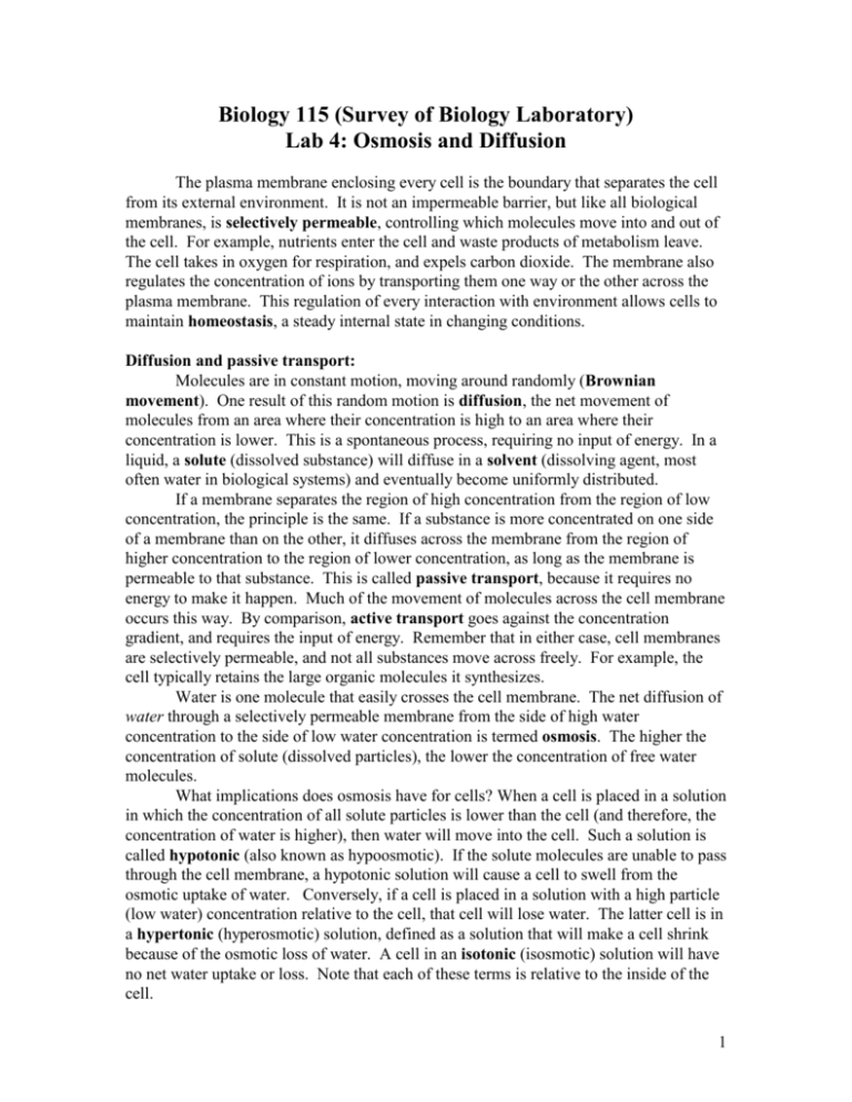
Biology 115 (Survey of Biology Laboratory) Lab 4: Osmosis and Diffusion The plasma membrane enclosing every cell is the boundary that separates the cell from its external environment. It is not an impermeable barrier, but like all biological membranes, is selectively permeable, controlling which molecules move into and out of the cell. For example, nutrients enter the cell and waste products of metabolism leave. The cell takes in oxygen for respiration, and expels carbon dioxide. The membrane also regulates the concentration of ions by transporting them one way or the other across the plasma membrane. This regulation of every interaction with environment allows cells to maintain homeostasis, a steady internal state in changing conditions. Diffusion and passive transport: Molecules are in constant motion, moving around randomly (Brownian movement). One result of this random motion is diffusion, the net movement of molecules from an area where their concentration is high to an area where their concentration is lower. This is a spontaneous process, requiring no input of energy. In a liquid, a solute (dissolved substance) will diffuse in a solvent (dissolving agent, most often water in biological systems) and eventually become uniformly distributed. If a membrane separates the region of high concentration from the region of low concentration, the principle is the same. If a substance is more concentrated on one side of a membrane than on the other, it diffuses across the membrane from the region of higher concentration to the region of lower concentration, as long as the membrane is permeable to that substance. This is called passive transport, because it requires no energy to make it happen. Much of the movement of molecules across the cell membrane occurs this way. By comparison, active transport goes against the concentration gradient, and requires the input of energy. Remember that in either case, cell membranes are selectively permeable, and not all substances move across freely. For example, the cell typically retains the large organic molecules it synthesizes. Water is one molecule that easily crosses the cell membrane. The net diffusion of water through a selectively permeable membrane from the side of high water concentration to the side of low water concentration is termed osmosis. The higher the concentration of solute (dissolved particles), the lower the concentration of free water molecules. What implications does osmosis have for cells? When a cell is placed in a solution in which the concentration of all solute particles is lower than the cell (and therefore, the concentration of water is higher), then water will move into the cell. Such a solution is called hypotonic (also known as hypoosmotic). If the solute molecules are unable to pass through the cell membrane, a hypotonic solution will cause a cell to swell from the osmotic uptake of water. Conversely, if a cell is placed in a solution with a high particle (low water) concentration relative to the cell, that cell will lose water. The latter cell is in a hypertonic (hyperosmotic) solution, defined as a solution that will make a cell shrink because of the osmotic loss of water. A cell in an isotonic (isosmotic) solution will have no net water uptake or loss. Note that each of these terms is relative to the inside of the cell. 1 Brownian Movement Place a drop of diluted India ink (a suspension of particles) on a clean slide, add a coverslip, and examine with your microscope using high power and reduced illumination. Focus carefully until you see the entire field of jiggling particles. The India ink particles “bombard” each other and are being “bombarded” by water molecules. The same energy that produces the movement of the ink particles causes diffusion of molecules. When a gradient exists, if there is no membrane blocking particle movement, diffusion will ultimately result in a uniform distribution. Place about 50 ml of water in a small beaker. Set the beaker on a white piece of paper at your table for a few minutes, until you are convinced that there are no more ‘currents’ in the beaker. Drop ONE drop of Methylene blue into the beaker. Immediately record the appearance of the beaker, with respect to the concentration of Methylene blue (the blue color). After 3 minutes, record the appearance of the beaker again, with respect to the concentration of Methylene blue. Has there been a change? What might have caused the change? Diffusion of Water Across Cell Membranes: Osmosis Consider a hypothetical animal cell with a composition of 10% protein and 90% water in an environment of 100% water (pure water). Remember the definition of diffusion. Water is more concentrated outside the cell, so it will move into the cell (from 100% concentration to 90% concentration). In this case, the protein molecules are too large to pass out of the cell membrane. If this movement of water (osmosis) continues unchecked, the cell may burst like a balloon. Plant cells have a rigid cell wall that prevents them from bursting. They become firm or turgid under the above conditions (that’s why plants wilt from a lack of water). You looked at plant cells in under different saline concentrations two weeks ago. Osmosis Experiment Sometimes, we use non-living models to study living systems. In this experiment, dialysis tubing is used to represent a differentially permeable membrane. When you make up a dialysis bag, think of it as a simplified cell. 2 Obtain three pieces of equal length of dialysis tubing, and several lengths of string. Fold over one end of the tubing and tie it closed with the string (you may also simply tie a knot in one end, but be careful not to put small rips in the tubing) or a clasp. To each tube, add 5 ml of 15% sucrose solution. Then, squeeze the bag gently to remove excess air, fold over the top, and tie it off with string. The bag should not be tight or turgid. Leave some slack in the bag as room for expansion, but get the air out. Briefly rinse the bags in running tap water, and then gently dry them on paper towels. Carefully weigh each bag to the nearest 0.1 g and record the results below. BE SURE TO KEEP TRACK OF WHICH BAG IS WHICH! After weighing, place one bag in each of three labeled beakers containing: (1) tap water, (2) 30% sucrose, (3) 60% sucrose. Allow the bags to remain undisturbed for thirty minutes. Write up this experiment in the form of the scientific method below. What is your question, hypothesis, prediction(s)? After thirty minutes, remove the bags, quickly rinse and dry and reweigh. Again, record the results below. Table 1. Weight (g) of dialysis bags Tap Water 15% Sucrose 30% Sucrose Weight at Time 0 min Weight at Time 30 min Change in Weight Which solution is hypotonic to the ‘cell’? ___________________________ Which solution is hypertonic to the ‘cell’? ___________________________ 3 What would have happened in the following three beakers if the cell were filled with tap water instead of 30% sucrose solution? Tap Water 15% Sucrose 30% Sucrose Diffusion across a differentially permeable membrane: Dialysis Dialysis is the diffusion of solute molecules across a differentially permeable membrane. The cell membrane is differentially permeable. Thus, through dialysis, certain substances may enter a cell, and certain metabolic products, including wastes, may leave. Depending on the permeability of a membrane, small solute molecules may pass through, while larger molecules are held back. Utilizing this principle, it is through dialysis that artificial kidney machines remove the smaller waste particles from the human bloodstream. The following experiment demonstrates the separation of different-sized molecules by dialysis. The two molecules used are starch (a large molecule) and sodium chloride (salt, a small molecule). In order to determine the presence of each of these molecules, we must be able to test for them. Sodium chloride (NaCl) plus silver nitrate (AgNO3) produces a dense white precipitate. So, if we add silver nitrate to a solution and a precipitate forms, we may conclude that sodium chloride is present. Starch plus iodine produces a blue-black color. So, if we add iodine to a solution which then turns blue-black, we may conclude that starch is present. Add 2 drops of sodium chloride and 2 drops of silver nitrate to a culture tube. Record your results. As a control, in a second tube, add 2 drops of tap water and 2 drops of silver nitrate. Record your results. Add 2 drops of 1% starch solution and 2 drops of iodine to a culture tube. Record your results. 4 Again, as a control, add 2 drops of tap water to another tube, and 2 drops of iodine. Record your results. Now you have seen what positive and negative tests for both sodium chloride and starch look like, so you are ready to proceed with an experiment to test the permeability of a membrane. Obtain another length of dialysis tubing and tie off one end like you did in the osmosis experiment. Fill the bag with 2 ml of starch solution, and 2 ml of sodium chloride solution. Gently squeeze the air out, and tie off the top. Rinse the cell off in tap water, and briefly place it on a paper towel while you perform the control test below. Control Fill a beaker with tap water, and then place 2 drops from the beaker into each of two wells on a spot plate. Test one well with 2 drops of silver nitrate, and the other with 2 drops of iodine. Record your results below. (You could just write the results you got above here – there is no need to do it again.) Beaker water (time 0 min) silver nitrate test iodine test Experiment Now, place the ‘cell’ in the beaker, and record the time __________:_________ After 30 minutes, remove the cell and place it on a paper towel. Place two drops from the beaker into each of two wells on a spot plate. Test one well with 2 drops of silver nitrate, and the other with two drops of iodine. Record your results below (+/-). Beaker water - silver nitrate test iodine test What can you conclude from this experiment? Why did you do the control? 5 Osmotic Potential of Plant Cells As you saw two weeks ago with the Elodea leaf, living cells have some amount of water inside them, and some amount of dissolved substance (solute: sugars, salts, proteins). In this part of the lab, you will measure the amount of water either taken up or lost from living plant cells (cells of potato tubers), and infer the proportion of the cytoplasm that is water, and the proportion that is solute. Procedure: Skin a potato and cut the tuber into small cubes (approximately 1cm each). You will need approximately 40 cubes. Divide the cubes into 4 groups of 10. Each group will be immersed for 30 minutes in a different solution. The four solutions you will use are: A. distilled water (water concentration 100%) B. 1% saline solution (water concentration 99%) C. 2% saline solution (water concentration 98%) D. 4% saline solution (water concentration 96%) Decide which of your four groups will go into each solution. Blot the cubes dry with a paper towel, and place them on a weighing boat (dry it first too if it is moist) and weigh them to the nearest 0.01g. Record the initial weight (t=0) on Table 2 below. Place each group of cubes in their appropriate solutions, and wait 30 minutes (you will be collecting data from the other two experiments during this time). At the end of 30 minutes, remove the groups and blot them dry again (being sure to keep track of which group is which). Record the weight after 30 minutes in the table below. In order to account for different initial weights, you need to calculate the percent change in mass for each of your four groups. To calculate the percent change, use the formula: % change = (weight after 30 minutes – initial weight)/initial weight X 100 Record the % change in the table. Table 2. Change in weight in potato tissue Solution Initial Weight (t=0) 0% salt 1% salt 2% salt 4% salt Final Weight (t=30) % Change Which group had the least amount of change in mass? Which group had the greatest amount of gain in mass? Which group had the greatest loss of mass? 6 Which group was in the most hypertonic solution? The most hypotonic? What can you infer about the internal tonicity of potato tuber cells? Approximately how much solute and how much water are present? When you are done with the lab, open all the ‘cells’, pour the contents down the drain, dispose of the tubing and potato cubes, rinse and dry all your equipment, and wipe off your lab bench. 7

