CRITICAL CARE
advertisement
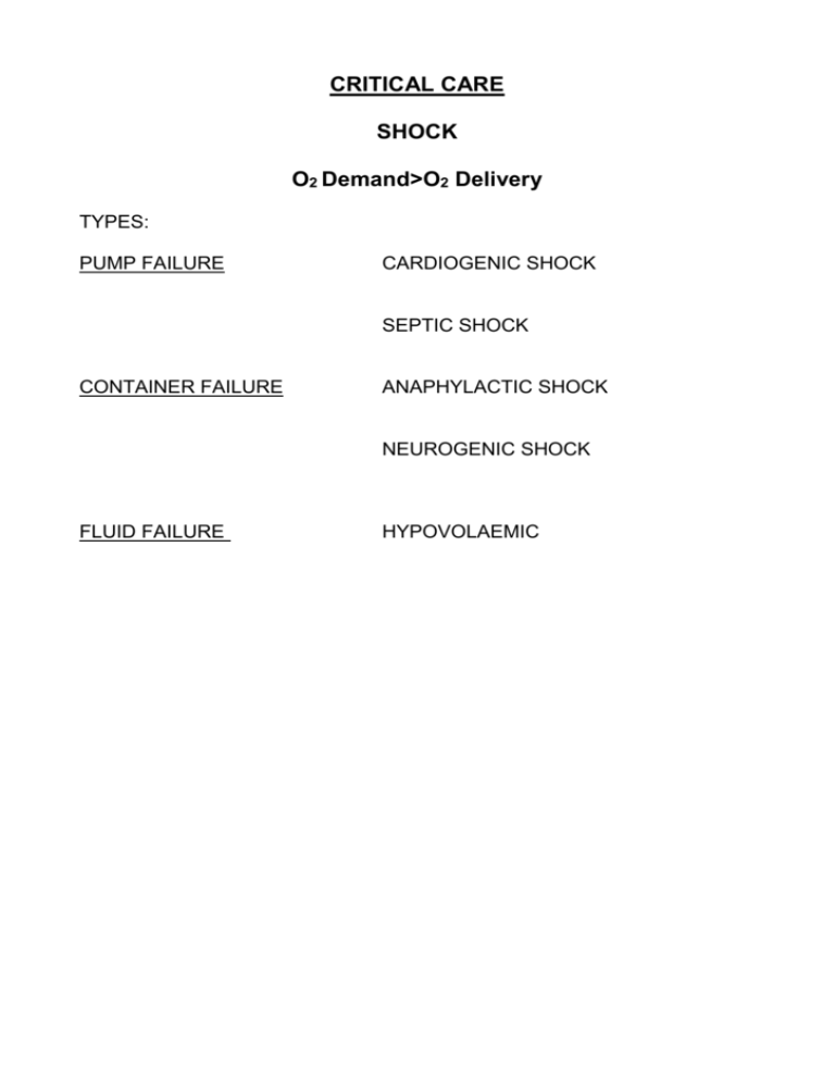
CRITICAL CARE SHOCK O2 Demand>O2 Delivery TYPES: PUMP FAILURE CARDIOGENIC SHOCK SEPTIC SHOCK CONTAINER FAILURE ANAPHYLACTIC SHOCK NEUROGENIC SHOCK FLUID FAILURE HYPOVOLAEMIC PATHOLOGY: MANAGEMENT: Depends on type of cause Treat underlying cause Aim to restore oxygen delivery to tissues SPECIFIC TREATMENTS (SURGICAL) 1. HYPOVOLAEMIC SHOCK Haemorrhage: trauma, GI bleed, ruptured aneurysm Dehydration: bowel obstruction, d+v Classification of haemorrhage Class I Hemorrhage involves up to 15% of blood volume. There is typically no change in vital signs and fluid resuscitation is not usually necessary. Class II Hemorrhage involves 15-30% of total blood volume. A patient is often tachycardic (rapid heart beat) with a narrowing of the difference between the systolic and diastolic blood pressures. The body attempts to compensate with peripheral vasoconstriction. Skin may start to look pale and be cool to the touch. The patient might start acting differently. Volume resuscitation with crystaloids (Normal Saline or Lactated Ringer's Solution) is all that is typically required. Blood transfusion is not typically required. Class III Hemorrhage involves loss of 30-40% of circulating blood volume. The patient's blood pressure drops, the heart rate increases, peripheral perfusion, such as capillary refill worsens, and the mental status worsens. Fluid resuscitation with crystaloid and blood transfusion are usually necessary. Class IV Hemorrhage involves loss of >40% of circulating blood volume. The limit of the body's compensation are reached and aggressive resuscitation is required to prevent death. Principles of Trauma Management (ATLS) PRIMARY SURVEY A: Airway and C-spine control (it does not matter how much blood there is if patient cannot get oxygen into lungs !) B: Breathing C: Circulation D: Disability (nervous system) E: Exposure SECONDARY SURVEY General Management of Hypovolaemic Shock Stop the bleeding ! Oxygen (increases oxygen delivery to tissues) Oxygen delivery=cardiac output X Hb concn X O2 saturation Fluid resuscitation: colloid vs crystalloid (NOT 5% dextrose EVER) Catheterise (0.5ml/kg/hr urine output) NG tube (not in suspected basal skull fractures) Invasive monitoring (maybe ?) Specific Causes and Management Bleeding Visceral injury, blunt or penetrating Hepatic: Spleen: conservative vs surgical (packing) conservative vs splenic preservation vs splenectomy (remember pneumococcal prophylaxis) Pancreas: conservative vs stent vs resection GIT: primary repair vs faecal diversion Cardiac: primary repair Pulmonary: conservative vs thoracotomy (>1.5L or more 200mL/hr for 2 hrs) Great vessel:often fatal RADIOLOGICAL METHODS (USA) DAMAGE LIMITATION CONCEPT GI Bleed Upper GI: Lower GI: Aortoenteric: Endoscopic vs surgical Often conservative, radiological, surgical rare, surgery possible but often fatal Aneursym Abdominal: careful BP management (increase pressure, increase bleed) Surgery Endovascular stent (if you are very lucky, right time, right place and right size stent !) Thoracic: Stent vs surgery Require bypass Dehydration Bowel obstruction Small bowel: Hernia: Adhesions: Others: Large bowel: Volvulus: Malignant: Pseudo-obstruction: “drip and suck”, resuscitate, reduce, repair conservative vs surgical (rising WCC, peritonism) often surgical endoscopic, surgery if fails or intestinal ischaemia surgery (defunction or resect) colonic stent medical (prokinetics, enemas, neostigmine) endoscopic (colonoscopy) surgery (caecostomy) 2. SEPTIC SHOCK SIRS: SIRS can be diagnosed when two or more of the following are present Heart rate > 90 beats per minute Body temperature < 36 or > 38°C Hyperventilation (high respiratory rate) > 20 breaths per minute or, on blood gas, a PaCO2 < 4.3 kPa (32 mm Hg) White blood cell count < 4000 cells/mm 3 or > 12000 cells/mm 3 (< 4 x 10⁹ or > 12 x 109 cells/L), or the presence of greater than 10% immature neutrophils. SEPSIS: SIRS with infection SEVERE SEPSIS: Sepsis with hypoperfusion ie. Septic shock Causes Visceral perforation Urosepsis Pelvic sepsis Respiratory General Management Same as shock, only treat cause of sepsis Fluid resuscitation Antibiotic therapy (BLOOD CULTURES PRIOR TO THIS) Involve critical care early: invasive monitoring inotropic support reducing mortality from sepsis now a national priority
