Trauma Resuscitation Part 2- interventions (Word Format)
advertisement
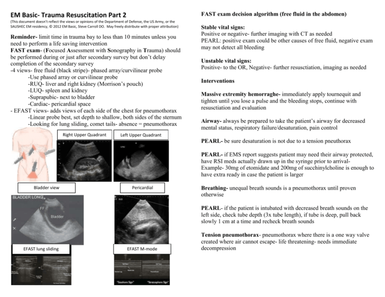
FAST exam decision algorithm (free fluid in the abdomen) EM Basic- Trauma Resuscitation Part 2 (This document doesn’t reflect the views or opinions of the Department of Defense, the US Army, or the SAUSHEC EM residency, © 2012 EM Basic, Steve Carroll DO. May freely distribute with proper attribution) Reminder- limit time in trauma bay to less than 10 minutes unless you need to perform a life saving intervention FAST exam- (Focused Assessment with Sonography in Trauma) should be performed during or just after secondary survey but don’t delay completion of the secondary survey -4 views- free fluid (black stripe)- phased array/curvilinear probe -Use phased array or curvilinear probe -RUQ- liver and right kidney (Morrison’s pouch) -LUQ- spleen and kidney -Suprapubic- next to bladder -Cardiac- pericardial space - EFAST views- adds views of each side of the chest for pneumothorax -Linear probe best, set depth to shallow, both sides of the sternum -Looking for lung sliding, comet tails- absence = pneumothorax Right Upper Quadrant Stable vital signs: Positive or negative- further imaging with CT as needed PEARL: positive exam could be other causes of free fluid, negative exam may not detect all bleeding Unstable vital signs: Positive- to the OR, Negative- further resusctiation, imaging as needed Interventions Massive extremity hemorraghe- immediately apply tournequit and tighten until you lose a pulse and the bleeding stops, continue with resusctiation and evaluation Airway- always be prepared to take the patient’s airway for decreased mental status, respiratory failure/desaturation, pain control Left Upper Quadrant PEARL- be sure desaturation is not due to a tension pneuthorax PEARL- if EMS report suggests patient may need their airway protected, have RSI meds actually drawn up in the syringe prior to arrivalExample- 30mg of etomidate and 200mg of succhinylcholine is enough to have extra ready in case the patient is larger Bladder view Pericardial Breathing- unequal breath sounds is a pneumothorax until proven otherwise PEARL- if the patient is intubated with decreased breath sounds on the left side, check tube depth (3x tube length), if tube is deep, pull back slowly 1 cm at a time and recheck breath sounds EFAST lung sliding EFAST M-mode Tension pneumothorax- pneumothorax where there is a one way valve created where air cannot escape- life threatening- needs immediate decompression ATLS- advocates needle decompression- 14 gague needle at the 2nd intercostal, midclavicular line -Problems- available needles are usually too short, patients with thick chest walls are hard to penetrate with needle Current practice- rapidly insert a chest tube but with an emphasis on rapidly cutting through the chest wall- most important thing is to relieve the tension pneumothorax, not actually inserting the tube- that can come later Chest tube insertion in tension pneumothorax -Prep the skin with betadine/chlorohexadine -Palpate for the 4th/5th intercostal space (lateral to the nipple) -Cut just above the rib and dissect down to muscle -Firmly hold curved kellys, placing finger one inch from end -Apply strong pressure to puncture through chest wall -Should get a rush of air/improvement in vital signs -Insert your finger into chest wall and twist 360 degrees -Place chest tube in a controlled manner Circulation-First step is to establish IV or IO access PEARL- If you can’t get an IV within 1 minute or 2 attempts in a critically ill trauma patient, go immediately to an intraosseous line (IO) IO locations- medial tibia (preferred), proximal humerus, distal femur Medial tibia insertion- feel for the tibial tuberosity (bump just below the patellar ligament insertion on the tibia), go one finger breadth below and medial on the flat part of the tibia, insert IO needle perpendicular until there is a lack of resistance, flush 5cc of saline to ensure placement (look for fluid in surrounding skin) Central access- 2 large bore peripheral IVs can have greater flow than a central line, if central access is desired, usually want a cordis central line with a large single lumen for rapid volume delivery PEARL- one exception- spinal shock- patient with a spinal injury with hypotension in which bleeding has been ruled out, caused by lack of vascular tone- in that case, put a triple lumen (regular) central line in for vasopressors (norepinephrine probably best Sedation and pain control for chest tubes In case of a tension pneumothorax, won’t have time to sedate since patient is close to cardiac arrest However- most pneumothoaxes aren’t tension physiology and can wait 30-60 seconds for sedation/pain control Best option- ketamine- 1-2 mg/kg IV/IO or 2-4 mg/kg IM- in 30-60 seconds you’ll have a calm and disscoatied patient that won’t move during chest tube insertion Fluids- ATLS advocates 2 liters of warmed normal saline but current practice is to go straight to blood products in trauma patients- saline doesn’t carry oxygen or glucose, dilutes clotting factors and hemoglobin Blood products- practice is to give blood products- pRBCs, plasma, platelets in a 1:1:1 ratio to approximate whole blood Blood pressure goal- “permissive hypotension”- not trying to normalize blood pressure- give enough volume to maintain mental status- rougly systolic of 90 but younger patients will tolerate lower systolics Contact- steve@embasic.org Twitter- @embasic
