transaxillary subpectoral endoscopic breast augmentation
advertisement
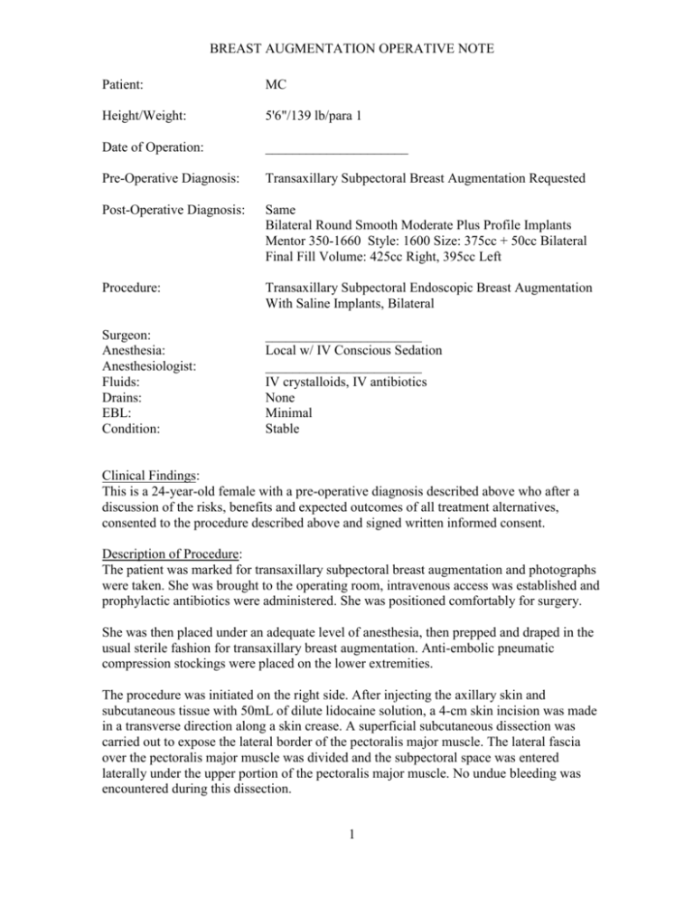
BREAST AUGMENTATION OPERATIVE NOTE Patient: MC Height/Weight: 5'6"/139 lb/para 1 Date of Operation: _____________________ Pre-Operative Diagnosis: Transaxillary Subpectoral Breast Augmentation Requested Post-Operative Diagnosis: Same Bilateral Round Smooth Moderate Plus Profile Implants Mentor 350-1660 Style: 1600 Size: 375cc + 50cc Bilateral Final Fill Volume: 425cc Right, 395cc Left Procedure: Transaxillary Subpectoral Endoscopic Breast Augmentation With Saline Implants, Bilateral Surgeon: Anesthesia: Anesthesiologist: Fluids: Drains: EBL: Condition: _______________________ Local w/ IV Conscious Sedation _______________________ IV crystalloids, IV antibiotics None Minimal Stable Clinical Findings: This is a 24-year-old female with a pre-operative diagnosis described above who after a discussion of the risks, benefits and expected outcomes of all treatment alternatives, consented to the procedure described above and signed written informed consent. Description of Procedure: The patient was marked for transaxillary subpectoral breast augmentation and photographs were taken. She was brought to the operating room, intravenous access was established and prophylactic antibiotics were administered. She was positioned comfortably for surgery. She was then placed under an adequate level of anesthesia, then prepped and draped in the usual sterile fashion for transaxillary breast augmentation. Anti-embolic pneumatic compression stockings were placed on the lower extremities. The procedure was initiated on the right side. After injecting the axillary skin and subcutaneous tissue with 50mL of dilute lidocaine solution, a 4-cm skin incision was made in a transverse direction along a skin crease. A superficial subcutaneous dissection was carried out to expose the lateral border of the pectoralis major muscle. The lateral fascia over the pectoralis major muscle was divided and the subpectoral space was entered laterally under the upper portion of the pectoralis major muscle. No undue bleeding was encountered during this dissection. 1 BREAST AUGMENTATION OPERATIVE NOTE Gentle, blunt finger dissection beneath the pectoralis major muscle was made extending as far laterally and inferiorly as possible to create a space for the endoscope. The space was further dissected with a blunt breast dissector with a gentle sweeping motion. The endoscope was attached to the endoscopic retractor and advanced through the incision into the subpectoral space under direct video surveillance without complications. The undersurface of the pectoralis major, the juncture of the muscles origins, and the chest wall and ribs were clearly identified. The unipolar spatula was advanced into the subpectoral space and employed to divide the pectoralis major muscle from the level of the areola extending inferiorly and laterally to the lateral extent of the muscle staying approximately 1 cm above the chest wall, avoiding damage to the undersurface of the breast skin, and maintaining absolute hemostasis. The endoscope was removed and the Agris-Dingman dissector was inserted into the subpectoral space. With a gentle, blunt sweeping motion, the lower pocket was advanced to the level of the new inframammary fold. The lateral aspect of the pocket was dissected bluntly in a similar fashion with care taken to avoid damage to the antero-lateral sensory nerves. Hemostasis was confirmed endoscopically following this maneuver. A breast sizer was inserted through the incision into the subpectoral space, inflated with air to the volume of the proposed implant and the adequacy of the pocket was confirmed. The sizer was left in place and the operation was repeated uneventfully on the contralateral side. Both implant pockets were then assessed for symmetry. The right breast sizer was deflated and removed, the implant pocket was re-assessed endoscopically, hemostasis was confirmed, and the pocket was irrigated thoroughly with saline solution containing an antibiotic. The right breast implant was prepared according to the manufacturer’s directions, inserted through the incision in proper alignment with care taken to avoid touching the skin, then inflated with saline to the minimum implant volume. The fill valve was left attached. The procedure was repeated contralaterally. The patient was then placed in a seated position to confirm symmetry, small volume additions were made to reach the desired final implant volumes, the fill valves were removed from the implants, and the axillary incisions were closed in layers with 4-0 delayed-absorbable monofilament sutures. Dressings were placed over the skin incisions. She tolerated the procedure well and was brought to the recovery suite in stable condition. ______________________________________/____________________ SURGEON DATE 2
