- Northumbria Research Link
advertisement
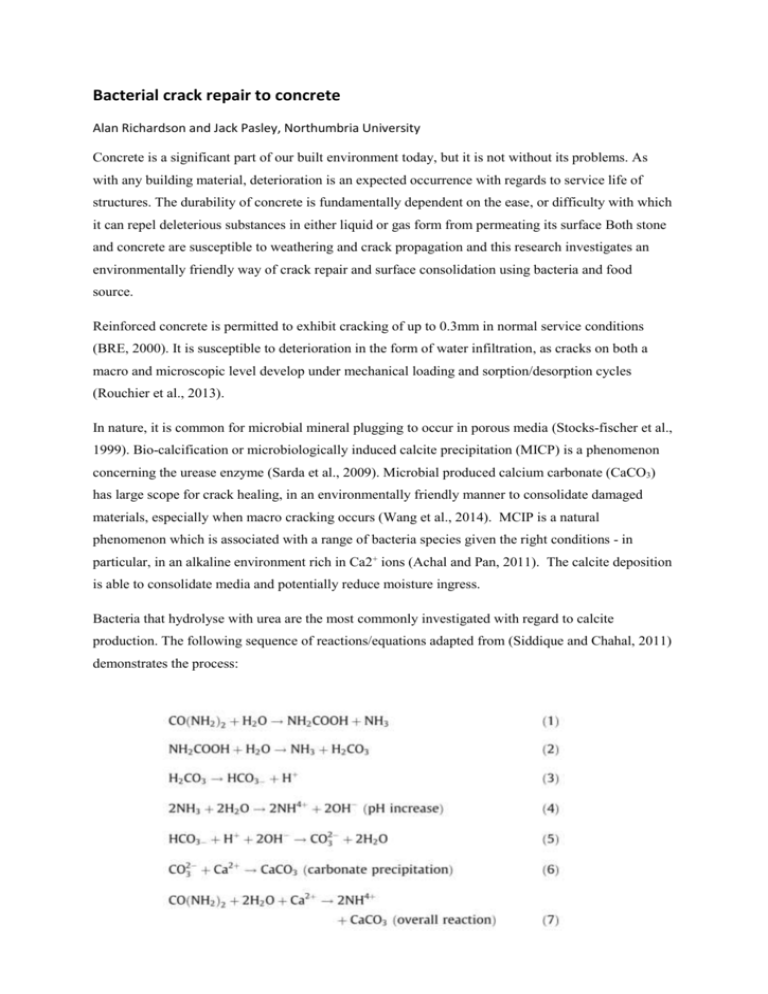
Bacterial crack repair to concrete Alan Richardson and Jack Pasley, Northumbria University Concrete is a significant part of our built environment today, but it is not without its problems. As with any building material, deterioration is an expected occurrence with regards to service life of structures. The durability of concrete is fundamentally dependent on the ease, or difficulty with which it can repel deleterious substances in either liquid or gas form from permeating its surface Both stone and concrete are susceptible to weathering and crack propagation and this research investigates an environmentally friendly way of crack repair and surface consolidation using bacteria and food source. Reinforced concrete is permitted to exhibit cracking of up to 0.3mm in normal service conditions (BRE, 2000). It is susceptible to deterioration in the form of water infiltration, as cracks on both a macro and microscopic level develop under mechanical loading and sorption/desorption cycles (Rouchier et al., 2013). In nature, it is common for microbial mineral plugging to occur in porous media (Stocks-fischer et al., 1999). Bio-calcification or microbiologically induced calcite precipitation (MICP) is a phenomenon concerning the urease enzyme (Sarda et al., 2009). Microbial produced calcium carbonate (CaCO3) has large scope for crack healing, in an environmentally friendly manner to consolidate damaged materials, especially when macro cracking occurs (Wang et al., 2014). MCIP is a natural phenomenon which is associated with a range of bacteria species given the right conditions - in particular, in an alkaline environment rich in Ca2+ ions (Achal and Pan, 2011). The calcite deposition is able to consolidate media and potentially reduce moisture ingress. Bacteria that hydrolyse with urea are the most commonly investigated with regard to calcite production. The following sequence of reactions/equations adapted from (Siddique and Chahal, 2011) demonstrates the process: Equation 1 - Hydrolysis of Urea (Siddique and Chahal, 2011) One molecule of urea is hydrolysed intracellularly to create 1 molecule of ammonia and 1 molecule of carbonate (Eq 1), which then spontaneously hydrolyses to form an additional molecule of ammonia and carbonic acid (Eq 2)). In water, the products then form bicarbonate, 2 molecules of ammonium, and 2 molecules of hydroxide ions Eqs. (3) and (4). The overall reaction is demonstrated in Eq 7 (Siddique and Chahal, 2011) which displays the formation of calcite. A bacterial cell surface can provide a nucleation site to non-specifically induce mineral deposition due to its variety of ions (Siddique and Chahal, 2011). Bacteria have the largest surface area to volume ratio of any life form (Fortin et al., 1996) thus being able to harbour calcium carbonate formation. It was concluded by Siddique and Chahal, (2011) that microbial mineral precipitation is a promising technique with regard to improvements in the compressive strength, permeability, lower water absorption and reduced chloride ingression. Wiktor and Jonkers, (2011) support this conclusion; their bio-chemical healing agent consisting of bacterial spores (Bacillus alkalinitrilicus, an alkali resistant soil bacterium) and a food source of calcium lactate, which was able to void cracks whilst being submersed in tap water for 100 days. Sporosarcina pasteurii (Previously Bacillus pasteurii), is able to aid in the urease production which, in turn, hydrolyses urea to ammonia and C02 (Bang et al., 2001). Such urease production from Sporosarcina pasteurii (S. pasteurii) was witnessed by (Sarda et al., 2009), stating among the other microbial cultures S. pasteurii NCIM 2477 was able to produce the most urease (Figure 1) . Figure 1- Urease Production by Different Bacteria (Sarda et al., 2009) Three fibre concrete beams were cast and once fully cured, they were cracked under a three point loading system, then a bead of silicone was applied around the cracked area to provide a reservoir to retain the liquid whilst the bacteria fed upon the food source, thus creating calcite as a repair agent. During this test, and prior to use, the S. pasteurii cultures were suitably incubated in an orbital incubator at 37° C at a rate of 200 rpm. The cultures were then measured at OD600 to see if the cell density is within the desired range (Approx. 0.9-3). A higher OD will provide more nucleation sites for calcite formation. Once the culture was prepared a100ml aliquot containing the cultures was added to a 900ml Duran container of nutrient broth with 50ml CaCl2 (calcium chloride). Immediately after mixing the solution, it was applied to the 3 beam sections. 15ml of solution per cube was applied followed by 1.5ml of urea to catalyse the reaction and help support the bacteria’s hydrolysis, leading to a higher calcite yield. The process outlined was repeated a total of three times, at 24 hour intervals and this layering effect gave the mixture time to be absorbed by the beams. Once the bacteria and the nutrient broth was mixed together, there was calcite formed almost immediately and this is a key finding in terms of an application process. This effect was noted in an earlier test. (Richardson et al 2014). The bacterial residue was examined at the surface (foreground) and at the intersection between the calcite and the concrete (background), using an Energy Dispersive Spectroscopy (EDS) image analysis technique and the chemical component parts are displayed in Table 1 and Figure 2. Oxygen (%) Carbon (%) Calcium (%) Background 58.0 17.5 24.5 Foreground 60.6 13.4 26.0 Table 1 – Chemical component parts of calcite deposit Figure 2 - Energy Dispersive Spectroscopy (EDS) image The Energy Dispersive Spectroscopy (EDS) results were tested alongside a MOH’s hardness test and hydrochloric acid test which showed the calcite formation was within normal hardness associated with the formation of calcite and the acid test showed the material to be of an alkaline nature, The effectiveness of the MICP crack healing properties are displayed in Figures 3 and 4 . Outline of Consolidated Micro-Crack Figure 3 – Above cracked fibre beam and below MICP sealing the crack Figure 4 – Cracked concrete beam on left and healed concrete beams to the right. This study has demonstrated the potential S. pasteurii and consequently MICP holds in improving the integrity of finish to concrete, without the use of chemical based sealants. This is part of an ongoing range of experimental work designed to find an effective way of using bacteria as a cementitious repair agent. There is scope for a partnership between industry and Northumbria University to develop this process further through a KTP or sponsored PhD. Biography Alan Richardson is currently a member of the RILEM team compiling a STAR report on Microorganisms-Cementitious material Interactions. TC 253-MCI Referenced work Achal, V. and Pan, X. (2011) “Characterization of urease and carbonic anhydrase producing bacteria and their role in calcite precipitation.” Current microbiology, 62(3) pp. 894–902. Bang, S. S., Galinat, J. K. and Ramakrishnan, V. (2001) “Calcite precipitation induced by polyurethane-immobilized Bacillus pasteurii.” Enzyme and Microbial Technology, 28(4-5) pp. 404– 409. BRE (2000) “Corrosion of steel in concrete - durability of reinforced concrete structures,” Digest 444(Part 1) pp. 2–9. Chahal, N., Siddique, R. and Rajor, A. (2012) “Influence of bacteria on the compressive strength, water absorption and rapid chloride permeability of concrete incorporating silica fume.” Construction and Building Materials. Elsevier Ltd, 37, December, pp. 645–651. Fortin, D., Davis, B. S. and Beveridge, T. J. (1996) “CHEMICAL Mineralization of bacterial surfaces,” 1. Richardson A E, Coventry KA, Forster A, Jamison C , (2014) "Surface consolidation of natural stone materials using microbial induced calcite precipitation", Structural Survey, Vol. 32 Iss: 3, pp 265-278 Rouchier, S., Woloszyn, M., Foray, G. and Roux, J.-J. (2013) “Influence of concrete fracture on the rain infiltration and thermal performance of building facades.” International Journal of Heat and Mass Transfer. Elsevier Ltd, 61, June, pp. 340–352. Sarda, D., Choonia, H. S., Sarode, D. D. and Lele, S. S. (2009) “Biocalcification by Bacillus pasteurii urease: a novel application.” Journal of industrial microbiology & biotechnology, 36(8) pp. 1111–5. Siddique, R. and Chahal, N. K. (2011) “Effect of ureolytic bacteria on concrete properties.” Construction and Building Materials. Elsevier Ltd, 25(10) pp. 3791–3801. Stocks-fischer, S., Galinat, J. K. and Bang, S. S. (1999) “Microbiological precipitation of CaCO 3,” pp. 31. Wiktor, V. and Jonkers, H. M. (2011) “Quantification of crack-healing in novel bacteria-based selfhealing concrete.” Cement and Concrete Composites. Elsevier Ltd, 33(7) pp. 763–770.
