Neuroscience Notes
advertisement
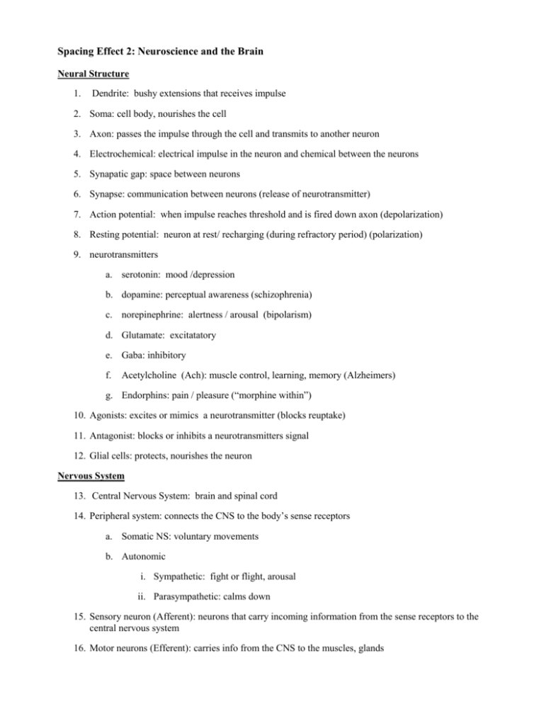
Spacing Effect 2: Neuroscience and the Brain Neural Structure 1. Dendrite: bushy extensions that receives impulse 2. Soma: cell body, nourishes the cell 3. Axon: passes the impulse through the cell and transmits to another neuron 4. Electrochemical: electrical impulse in the neuron and chemical between the neurons 5. Synapatic gap: space between neurons 6. Synapse: communication between neurons (release of neurotransmitter) 7. Action potential: when impulse reaches threshold and is fired down axon (depolarization) 8. Resting potential: neuron at rest/ recharging (during refractory period) (polarization) 9. neurotransmitters a. serotonin: mood /depression b. dopamine: perceptual awareness (schizophrenia) c. norepinephrine: alertness / arousal (bipolarism) d. Glutamate: excitatatory e. Gaba: inhibitory f. Acetylcholine (Ach): muscle control, learning, memory (Alzheimers) g. Endorphins: pain / pleasure (“morphine within”) 10. Agonists: excites or mimics a neurotransmitter (blocks reuptake) 11. Antagonist: blocks or inhibits a neurotransmitters signal 12. Glial cells: protects, nourishes the neuron Nervous System 13. Central Nervous System: brain and spinal cord 14. Peripheral system: connects the CNS to the body’s sense receptors a. Somatic NS: voluntary movements b. Autonomic i. Sympathetic: fight or flight, arousal ii. Parasympathetic: calms down 15. Sensory neuron (Afferent): neurons that carry incoming information from the sense receptors to the central nervous system 16. Motor neurons (Efferent): carries info from the CNS to the muscles, glands 17. Interneurons: transmits impulses between brain and CNS and connect to sensory and motor neurons 18. Spinal reflex: from sensory neuron to interneuron to motor neuron (without the brain!) Endocrine System 19. Glands: secrete hormones in to bloodstream (“snail mail”) 20. Pituitary gland: master gland / growth hormone 21. Adrenal glands: arouses sympathetic nervous system (epinephrine, norepinephrine, adrenaline, noradrenaline) The Brain 22. Lesion: tissue damage to the brain 23. Neuroimaging techniques: a. CAT: structure only (tumors etc.) b. MRI: Soft tissue c. PET: Traces glucose levels in brain (blood flow) d. EEG: Detects brainwaves (electrical activity) e. FMRI: Soft tissue plus glucose levels 24. Parts of Brain a. Medulla: heartbeat, breathing, blood pressure b. Pons: Coordinate facial expressions, active in dream state c. Cerebellum: balance, fine motor skills d. Reticular Formation: arousal, sleep e. Limbic System (emotions, drives, memory) i. Amygdala: emotions of fear, anger ii. Hypothalamus: drives (sexual, thirst, hunger) (regulates Pituitary gland!) iii. Hippocampus: processes new memories (stores memories in Cerebral Cortex) f. Thalamus: Sensory switchboard (all but smell!) Routes stimuli to different parts of brain. g. Brain hemispheres i. Left hemisphere: language, logic, literal , sequential, analysis ii. Right hemisphere: Spatial, Intuitive, creative, synthesis h. Lobes of Brain i. Frontal Lobe: Logic, judgement, speaking. Planning (Prefrontal Cortex: decisions, emotional control) ii. Occipital Lobe: vision iii. Temporal Lobe: hearing iv. Parietal Lobe: Sensory center i. Cerebral Cortex: consists of lobes and Sensory and Motor Cortex i. Motor Cortex: voluntary movements (located in back of frontal lobe) ii. Sensory Cortex: Sensory center (touch!) located in front of Parietal lobe iii. Association areas: complex, abstract thought processes (75% of Cortex) j. Language and the Brain i. Broca’s Area: Motor speech ii. Werniche’s area: Language comprehension k. Aphasia: Language impairment l. Corpus Callosum: bridge between the two hemispheres (neural impulses shared)
