Supplementary Table S1. Test characteristics.a Study ID Timing of
advertisement
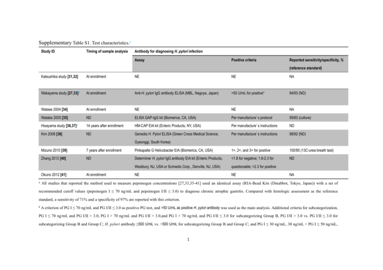
Supplementary Table S1. Test characteristics.a Study ID Timing of sample analysis Antibody for diagnosing H. pylori infection Assay Positive criteria Reported sensitivity/specificity, % (reference standard) Katsushika study [31,32] At enrollment NE NE NA Wakayama study [27,33]b At enrollment Anti-H. pylori IgG antibody ELISA (MBL, Nagoya, Japan) >50 U/mL for positive* 94/93 (ND) Watase 2004 [34] At enrollment NE NE NA Watabe 2005 [35] ND ELISA GAP-IgG kit (Biomerica, CA, USA) Per manufacture’ s protocol 95/83 (culture) Hisayama study [36,37]c 14 years after enrollment HM-CAP EIA kit (Enteric Products, NY, USA) Per manufacture’ s instructions ND Kim 2008 [38] ND Genedia H. Pylori ELISA (Green Cross Medical Science, Per manufacture’ s instructions 98/92 (ND) Gyeonggi, South Korea) Mizuno 2010 [39] 7 years after enrollment Pirikapalte G Helicobacter EIA (Biomerica, CA, USA) 1+, 2+, and 3+ for positive 100/95 (13C-urea breath test) Zhang 2012 [40] ND Determiner H. pylori IgG antibody EIA kit (Enteric Products, <1.8 for negative; 1.8-2.3 for ND Westbury, NJ, USA or Scimedix Corp., Denville, NJ, USA) questionable; >2.3 for positive NE NE Okuno 2012 [41] a At enrollment NA All studies that reported the method used to measure pepsinogen concentrations [27,33,35-41] used an identical assay (RIA-Bead Kits (Dinabbot, Tokyo, Japan)) with a set of recommended cutoff values (pepsinogen I ≤ 70 ng/mL and pepsinogen I/II ≤ 3.0) to diagnose chronic atrophic gastritis. Compared with histologic assessment as the reference standard, a sensitivity of 71% and a specificity of 97% are reported with this criterion. b A criterion of PG I ≤ 70 ng/mL and PG I/II ≤ 3.0 as positive PG test, and >50 U/mL as positive H. pylori antibody was used as the main analysis. Additional criteria for subcategorization, PG I ≤ 70 ng/mL and PG I/II > 3.0, PG I > 70 ng/mL and PG I/II > 3.0,and PG I > 70 ng/mL and PG I/II ≤ 3.0 for subcategorizing Group B, PG I/II > 3.0 vs. PG I/II ≤ 3.0 for subcategorizing Group B and Group C; H. pylori antibody ≤500 U/mL vs. >500 U/mL for subcategorizing Group B and Group C; and PG I ≤ 30 ng/mL, 30 ng/mL < PG I ≤ 50 ng/mL, 1 and PG I > 50 ng/mL for subcategorizing Group C were also used. c PG I ≤ 30 ng/mL and PG I/II ≤ 2.0 was also used as a “strong-positive” subgroup. Another cut-off values, PG I ≤ 59 ng/mL and PG I/II ≤ 3.9, estimated as the values for the maximum Youden’s index, were also estimated through an exploratory ROC analysis (sensitivity of 70% and specificity of 69% for predicting gastric cancer development, not for chronic atrophic gastritis) and used for the analysis of a 4-group risk model based on both PG test and H. pylori infection status. EIA = enzyme immunoassay; ELIZA= enzyme-linked immunosorbent assay; Ig = Immunoglobulin; NA = not applicable; ND = no data; NE = not evaluated; PG = pepsinogen; RIA = radioimmunoassay; 2
