Lecture No. 24 PARASITOLOGY DR.Raad HH. Protozology
advertisement
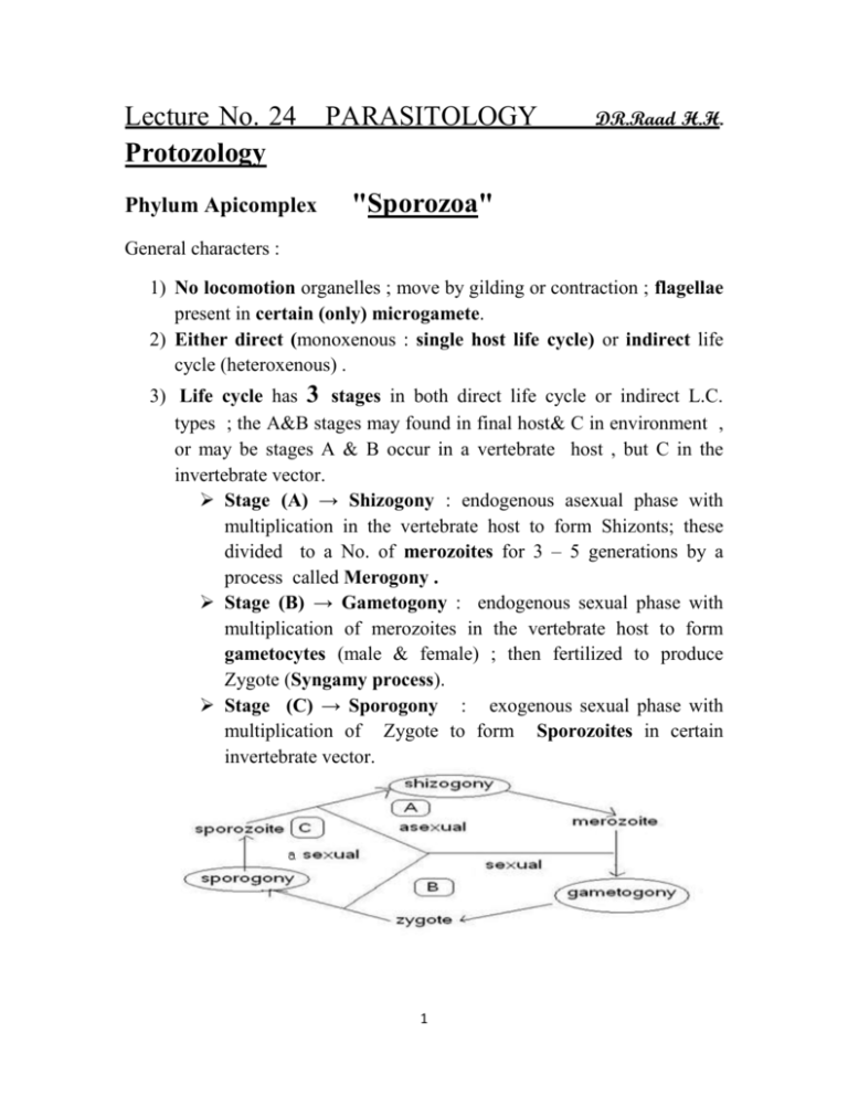
Lecture No. 24 Protozology Phylum Apicomplex PARASITOLOGY DR.Raad H.H. "Sporozoa" General characters : 1) No locomotion organelles ; move by gilding or contraction ; flagellae present in certain (only) microgamete. 2) Either direct (monoxenous : single host life cycle) or indirect life cycle (heteroxenous) . 3) Life cycle has 3 stages in both direct life cycle or indirect L.C. types ; the A&B stages may found in final host& C in environment , or may be stages A & B occur in a vertebrate host , but C in the invertebrate vector. Stage (A) → Shizogony : endogenous asexual phase with multiplication in the vertebrate host to form Shizonts; these divided to a No. of merozoites for 3 – 5 generations by a process called Merogony . Stage (B) → Gametogony : endogenous sexual phase with multiplication of merozoites in the vertebrate host to form gametocytes (male & female) ; then fertilized to produce Zygote (Syngamy process). Stage (C) → Sporogony : exogenous sexual phase with multiplication of Zygote to form Sporozoites in certain invertebrate vector. 1 Fig.: Diagram of Apicomplexan life cycle : 1-zygote, 2-sporozoites, 3-merozoites, 4-gametes. 4) The infective stage is sporozoite in plasmodium is capable to penetrate the host cell by their APICAL END which it is a compound of complex structure contains CONOID , MICRONEME ,and RHOPTRY ; OR sporulated cysts as in COCCIDIAL PARASITES 5) Most members are intracellular parasites in vertebrates (animals , birds ). 2 6) 7) 8) o - sexual reproduction by syngamy . all parasitic; no free-living members . Phylum Apicomplex classification: Class SOPROZOEA (merogony, gamogony, and sporogony normally present) Sub class COCCIDIA Order EUCOCCIDIIDA Sub order EIMERIINA ( genera : ISOSPORA , SARCOSYSTIS, , CRYPTOSORIDIUM ,CYCLOSPORA , ,TOXOPLASMA) - Sub order HAEMOSPORINA ( genus PLASMODIUM ) Sub class PIROPLASMIA - Order PIROPLASMIDA ( genus BABESIA) Terms defention: Sporozoans - parasitic spore-forming protozoan , nonmotile protozoans (that have no organelles for transportation ), many of which reproduce by the production of spores e.g. Plasmodium . Sporozoite - one of the minute active bodies into which sporozoans divide in one stage of their life cycle ; The sporozoite is an infective stage (e.g. Eimeria, Cryptosporidium). Trophozoite - a sporozoan in the active feeding stage of its life cycle Merozoite - a cell that arises from the asexual division of a parent sporozoan during its life cycle Coccidium, Eimeria - parasitic on the digestive epithelium of vertebrates and higher invertebrates Haemosporidian - minute protozoans parasitic at some stage of the life cycle in blood cells of vertebrates including many pathogens malaria parasite, - parasitic protozoan of the genus Plasmodium that causes malaria in humans e.g. Plasmodium vivax , & birds Leucocytozoan, leucocytozoon - parasitic in birds 3 Piroplasm - minute parasite of red blood cells of mammals transmitted by a tick and causing diseases of domestic animals Sarcocystidean, sarcocystieian, sarcosporidian - parasite of the muscles of vertebrates Microsporidian - parasite of arthropods and fishes that invade and destroy host cells Coccidian Parasites Found in animal Feces *** Species Cryptosporidium Cyclospora Size (µm) 4-5 8-10 Oocyst Structure 4 sporozoites, no sporocysts 2 sporocysts with 2 sporozoites each 30 x 12 2 sporocysts with 4 sporozoites each Sarcocystis Excreted Form sporulated oocysts unsporulated oocysts unsporulated oocysts sporulated oocysts 13 x 10 2 sporocysts with 4 sporozoites each Eimeria sporulated oocysts 10-30 x11- 4 sporocysts with 2 sporozoites each 40 Isospora Family Eimeriidae A. most species homoxenous . B. some form monozoic cysts in paratenic host C. merogony, gamogony, and oocyst wall formation in same host . D. in vertebrates or invertebrates. E. most genera distinguished by numbers of sporocysts and sporozoites within oocyst . F. 15-25 genera, depending upon author. "Most important genera "as follows: A. Eimeria (4 sporocysts in oocyst, each sporocyst with 2 sporozoites) 4 B. Isospora (2 sporocysts in oocyst, each sporocyst with 4 sporozoites) Genus Isospora 1. Classified as Sub order EIMERIINA Family Eimeriidae Genus Isospora The parasite infect epithelial tissue of Dogs and cats intestine are infected by the species I. bigemina, I. rivolta, I. felis and I. canis . Human intestine by Isospora belli (intestinal protozoon) ; where both Merogony (sexual) & gamogony (asexual) occurred in host while Sporogony Phase is the exogenous phase of the Isospora suis life cycle, during which the oocysts develop from the unsporulated, non-infectious stage to the infective stage. The oocysts are excreted in faeces; sporulation to the infectious stage takes place outside the host animal. The sporulated oocysts contain two sporocysts with four sporozoites each. Sporulation is dependent upon humidity, temperature and oxygen. Isospora suis oocysts sporulate rapidly at temperatures between 20° C and 40° C . In current swine production practices, where supplemental heat between 32° and 35° C is provided to newborn pigs, oocysts sporulation may occur between 12 and 16 hours. Once the oocysts are sporulated they are resistant to most disinfectants. Excystation Phase The excystation phase occurs immediately after the infectious oocysts have been ingested. During this phase, the sporozoites become active and are released into the intestinal lumen. Passage through the stomach alters the oocyst wall, allowing bile salts and digestive enzymes to activate the sporozoites. The active sporozoites then penetrate the villous epithelium, which starts the endogenous phase of parasite multiplication. 5 The greatest concentration is seen in the middle part of the jejenum, but the ileum is also infected. In severe infections the cecum and colon can also be involved. Endogenous Development The endogenous phase of the Isospora suis life cycle occurs mainly in cells of the small intestine, although in severe infections, the colon and cecum may also be involved. These stages usually take place in the distal portions of the villi, although in severe cases, they may also be located in the crypt region of the intestinal cells. During this phase, the sporozoites enter the intestinal cells and become binucleated type-1 meronts. The meronts divide within 24 hours to produce two type-2 daughter merozoites. Several divisional cycles may occur to produce many type-1 merozoites that in turn produce type-2 meronts and type-2 merozoites. Type-2 merozoites are multinucleated and smaller than type-1 merozoites, and can be seen as early as 4 days post-inoculation. The cells of the intestinal epithelium are destroyed with the release of the merozoites from the intestinal cells. According to Tubbs (1987), asexual reproduction repeats three times. Other authors stipulate that the number of asexual reproductions varies. Following the final asexual stage, the sexual stages of the I. suis life cycle begin to form: macrogamete (female) and microgamete (male). The microgamete is a biflagellate structure that fertilises the microgamete, producing a zygote that develops cell walls and becomes an oocyst. The oocyst is released into the lumen of the intestine and shed with faeces into the environment. These sexual stages may also be seen 4 days postinoculation, whereas oocysts are first seen in faeces 5 days post-inoculation (rarely 4 days). The prepatent period for Isospora suis is 5 to 7 days, and the patent time generally ranges from 5 to 16 days 6 2. Direct life cycle 3. The infective stage is Sporulated oocyst which called the (Di sporocyst tetrazoic oocyst type) i.e. it has 2 sporocyst each with 4 Sporozoites. 4. Acid-fast staining is the preferred method for coccidia which stain bright red. Cryptosporidium, Cyclospora, and Isospora are distinguished by size and oocyst structure (Table *** ). 5. Symptoms The disease is marked by a severe diarrhea, in which the feces are mucoid and frequently consist largely of blood. Usually marked 7 depression, loss of appetite, general weakness, anemia, and dehydration also are noted. Heavy infections in young animals commonly end in death. 6. Pathogenesis : Pathology associated with Isospora infections are primarily Jejunal villous atrophy, or blunting, and crypt hyperplasia as commonly seen in other intestinal infections. Villous Atrophy commonly associated with mal - absorption syndrome . 7. Diagnosis a) Direct stool exam. b) Indirect = = by i. concentration method with zinc Sulphate mixed with Iodine. ii. modified acid fast procedure. iii. Sheather’s sugar flotation = . 8. Differential diagnosis: The Sporulated oocyst is Differentiated from that of genus Eimeria (coccidia) which has 4 sporocysts each with 2 Sporozoites (Tetra sporocystic dizoic oocyst type). Eimeria 9. Treatment - Trimethoprim 160 mg. – sulpha methaxazole 800 mg. combination/ 4 times / 10days - Pyrimethamine 75 mg. & sulphadiazole 4 gm. /4times /21 days . 8 Genus Eimeria A. Host specificity - these parasities have a narrow host range (stenoxenous). B. Organ specificity - these parasities are usually found in a specific organ. C. Site specificity within organ or cell - these parasities inhabit specific sites within an organ or cell. D. Monoxenous - coccidia usually parasitize one host, the definitive host. E. Infects all animals except dogs and cats. F. Enteric parasites . G. Hepatic in rabbits e.g. E. stidae H. Younger age more susceptible , morbidity increases during wet season as spring & winter . I. Disease called coccidiosis J. Reproduction 1. Schizogony - (Merogony) -multiple fission. 2. Gamogony (syngamy) - union of similar gametes . 3. Structure of Eimeria sporulated oocyst - 4 sporocysts, each with 2 sporozoites (Tetra sporocystic dizoic oocyst type). K. Etiology: 9 L. Life cycle : M. Pathogenesis : Pathogenicity is influenced by host genetics, nutritional factors, concurrent diseases, and species of the coccidium. Eimeria necatrix and E tenella are the most pathogenic in chickens because schizogony occurs in the lamina propria and crypts of Lieberkühn of the small intestine and ceca, respectively, and causes extensive hemorrhage. Most species develop in epithelial cells lining the villi. Protective immunity usually develops in response to moderate and continuing infection. True age-immunity does not occur, but older birds are usually more resistant than young birds because of earlier exposure to infection. Hepatic infection , caused by Eimeria stiedae : Severity of disease depends on the number of oocysts ingested. Young rabbits are most susceptible. Affected rabbits may be anorectic and have a rough coat. Hepatic coccidiosis is most often subclinical, but growing rabbits may fail to make normal gains. Infrequently, death may 10 follow a short course. Rabbits usually succumb within 1 mo after a severe experimental exposure. At necropsy, small, yellowish white nodules are found throughout the hepatic parenchyma. In the early stages, they may be sharply demarcated, while in the later stages they coalesce. The early lesions have a milky content; older lesions may have a more cheese-like consistency. Microscopically, the nodules are composed of hypertrophied bile ducts or gallbladder. Diagnosis of this form of coccidiosis is based on the gross and microscopic changes, along with demonstration of the oocysts in the bile ducts. An impression smear of a lesion in the liver examined under light microscopy often reveals oocysts. The oocysts may also be demonstrated by fecal flotation. The following diagrams show the intestinal sites parasitised by various species of Eimeria in chickens : - E. tenella infections are found only in the ceca and can be recognized by accumulation of blood in the ceca and by bloody droppings. Cecal cores, which are accumulations of clotted blood, tissue debris, and oocysts, may be found in birds surviving the acute stage. 11 - E. necatrix - E. acervulina produces major lesions in the anterior and middle portions of the small intestine. Small white spots, usually intermingled with rounded, bright- or dull-red spots of various sizes, can be seen on the serosal surface. The white spots are diagnostic for E necatrix if clumps of large schizonts can be demonstrated microscopically. In severe cases, the intestinal wall is thickened, and the infected area dilated to 2-2.5 times the normal diameter. The lumen may be filled with blood, mucus, and fluid. Fluid loss may result in marked dehydration. Although the damage is in the small intestine, the sexual phase of the life cycle is completed in the ceca. Oocysts of E necatrix are found only in the ceca. Due to concurrent infections, oocysts of other species may be found in the area of major lesions, misleading the diagnostician. , the most common infection, is characterized by numerous, whitish, oval or transverse patches in the upper half of the small intestine and may be easily distinguished on gross examination. The clinical 12 course in a flock is usually protracted and results in poor growth, an increase in culls, and slightly increased mortality. - E. brunetti is found in the lower small intestine, rectum, ceca, and cloaca. In moderate infections, the mucosa is pale and disrupted but lacking in discrete foci, and may be thickened. In severe infections, extensive coagulative necrosis and sloughing of the mucosa occurs throughout most of the small intestine. 13 - E. maxima develops in the small intestine, where it causes dilatation and thickening of the wall; petechial hemorrhage; and a reddish, orange, or pink viscous mucous exudate and fluid. The oocysts and gametocytes (particularly macrogametocytes), which are present in the lesions, are distinctly large. - E. mitis is recognized as pathogenic in the lower small intestine. Lesions resemble moderate infections of E brunetti but can be distinguished by finding small, round oocysts associated with the lesion. 14 - E. praecox , which infects the upper small intestine, does not cause distinct lesions but may decrease rate of growth. It is considered to be of less economic importance than the other species. N. Symptoms: 1. Acute-chronic in extremely acute cases, the first thing noticed will be dead birds. 2. Unusually droopiness and depression are the first symptoms 3. The chicks stand huddles, with wings dropped, feathers ruffled and eyes closed 4. Diarrhea is commonly present and the loose droppings are frequently mixed with blood. 5. In the later stages of the disease there is paleness of the comb and wattles. 6. The greatest losses occurs within 6-10 days following the onset of symptoms. 7. In older fowl the chronic intestinal type of the disease is most common, Leg weakness is commonly manifested and in some cases is so severe that the affected fowl may be practically paralyzed. O. Diagnosis 1. Based on symptom and autopsy lesions. 2. A positive diagnosis of coccidiosis is dependent upon finding one or more of the various stages of the organisms by microscopic examination of a thin film of fecal material and intestinal mucosa. 3. The oocysts may also be demonstrated by fecal flotation. 15 P. Prevention, Treatment, and Control: 1. Sound management, dry litter, clean house, keep the house dry. Don't crowd the chicken. Proper feeding of Vitamin A and K. 2. Treatment with drugs. Following discovery of anticoccidial activity of the sulfa drugs in 1939, attention was directed toward treatment of coccidiosis. However, anticoccidial agents (coccidiostats) show activity so early in the life cycle that few benefits are derived from treatment after symptoms appear. For this reason the preventive approach for use of drugs has largely supersede- the treatment approach. S Q (Sulfaquinoxaline) is the drug of choice for treating this disease. Other commonly used drugs are Sulfamethazine, Sulfaquanidine, Sulfamerazine, Nitrofura-zone, ureomycine, Terramycin, Amprolium etc. There a various kinds of coccidiostats in the market for preventing against coccidia. Long term continuation administration of cocidiostats in water of feed will induce resistance to a specific drug thus, changing of coccidiostats after using it for certain period of time is important, among the commonly used cocidiostate, Monensin is the only drug that has not been running into the drug resistant problem, so far. Special of developing resistant of coccidia to various coccidiostats. 16
