Ch 8, Phylum Apicomplexa: Gregarines and Coccidia
advertisement
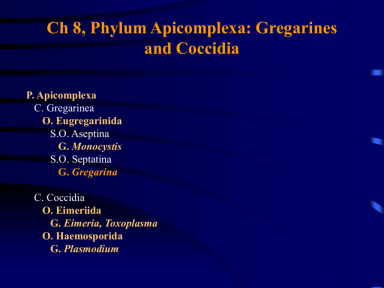
Ch 8, Phylum Apicomplexa: Gregarines and Coccidia P. Apicomplexa C. Gregarinea O. Eugregarinida S.O. Aseptina G. Monocystis S.O. Septatina G. Gregarina C. Coccidia O. Eimeriida G. Eimeria, Toxoplasma O. Haemosporida G. Plasmodium General Characteristics • All are parasitic and include a variety of parasites of medical importance • e.g., coccidians of the genus Eimeria that cause intestinal diseases of farm animals; Plasmodium that cause malaria • Possess a single type of nucleus; no cilia or flagella • This phylum is characterized by complex apical organelles the apical complex, found in the sporozoite and merozoite stages of the life cycles of these organisms Merozoites and Sporozoites • Asexual reproduction is via multiple fission or shizogony • Rrapid organelle and nuclear divisions, followed by multiple cytokinesis • The multinucleated cell is the shizont or meront • During merogony, daughter nuclei of the shizont arrange themselves peripherally and a membrane forms around each nucleus • After cytoplasmic divisions, each nucleus with its attendant cytoplasm forms a merozoite; breaks away from the aggregate to infect a new host cell • Merozoite enters another schizogonic cycle or undergoes gamatogony, yielding become a macro- or microgametocytes • Syngamy, the union of gametes derived from the gametocytes, initiates the sexual cycle • The resulting zygote undergoes sporogony (multiple fission of zygote) which results in the production of sporozoites (usually the infective stage) The Apical Complex • At the anterior end, beneath the cell membrane, are 1 or 2 electron dense structures call polar rings • In certain members of the order Eimeriida, a truncated cone of spirally arranged fibrillar structures - the conoid - lies within the polar rings • In most members of the phylum, subpellicular microtubules radiate from the polar rings parallel to the long axis of the cell; serve as support elements and possibly have a locomotive function • Within the polar rings are electron dense bodies called rhoptries The Apical Complex cont. • Parallel to the rhoptries are micronemes • Rhoptries and micronemes appear to be secretory organelles that may facilitate the parasite’s penetration into the host cell • At the lateral edges are one or more micropores; sites for endocytosis of nutrients during the intracellular life of the organism • At the edges of the micropore are 2 concentric, electron-dense rings • Host cytoplasm is drawn through the micropore rings into the parasite, where a food vacuole forms O. Eugregarinida • Gregarines are only parasitic of invertebrates, mainly annelids and arthropods • Parasitize the body cavity, intestine, or reproductive system of their hosts • Most members produce a resistant spore or oocyst (sporocyst containing sporozoites) • Hosts typically become infected by ingesting spores • Locomotion is by body flexion, gliding, or undulations of longitudinal ridges Monocystis lumbrica • Lives in the seminal vesicles of the earthworm Lumbricus terrestris • Worms become infected by ingesting mature spores containing sporozoites • Spores hatch in the gizzard and the released sporozoites penetrate the gut wall and enter the circulatory system via the dorsal vessel • They move forward to the hearts, leave the circulatory system and enter the seminal and • Sporozoites enter into tissue cells and become trophozoites (= sporadins or gamonts Monocystis lumbrica cont. • Gamonts attach to cells in the region of the sperm tunnel and undergo syzygy • Gamonts then secrete a wall around themselves, becoming a cyst (=gametocyst) • Gamont undergoes numerous nuclear divisions with the nuclei moving to the periphery of cytoplasm • Nuclei then bud off to become gametes (anisogametes); fusion of the gametes yield zygotes • After a spore or oocyst membrane forms around the zygote it undergoes sporogony to yield 8 sporozoites • A new host may become infected from ingesting a gametocyst or an oocyst Gregarina cuneata • Common parasite of the meal worm (Tenebrio spp.) • After ingestion, spores hatch and sporozoites penetrate the epithelial cells of the host’s gut • Undergo considerable growth as trophozoites; growth of the anterior end eventually slows relative to posterior region, and the organism becomes divided into the protomerite and deutomerite Gregarina cuneata cont. • Trophozoites become separated from the host cell, pair, and undergo syzygy • The trophozoites then encyst and form gametocysts • Protomerite and deuteromerite fuse to form a gametocyte which in turn form anisogametes • Anisogametes conjugate forming zygotes • Zygotes form resistant spore coats and their contents divide (sporogony) to form sporozoites within a sporocyst which are shed with the host’s feces Class Coccidia • Primarily parasites of vertebrates • Have a prevalent intracellular reproductive phase • Occur in the epithelium of the digestive tract, liver, kidneys, blood cells • Similar to other apicomplexans, the life cycle has 3 major phases: merogony (produces merozoites), gametogony (produces gametocystes), and sporognony (produces sprozoites) • (All three are kinds of schizogony - asexual reproduction by multiple or binary fission of a cell to form daughter cells) Overview of the Life Cycle • Sporozoites enter host cells becoming trophozoites • These eventually multiply by merogony to yield merozoites • Merozoites can enter other cells, undergo further merogony, and produce more merozoites • Some of the merozoites undergo gametogony and transform into gamonts The gamonts produce "female" macrogametocytes and "male" microgametocytes • The macrogametocyte yields a macrogamete; the microgametocyte undergoes multiple fission to yield microgametes • Upon fertilization, the zygote forms a protective wall around itself and sporogony yields a sporozoite-filled oocyst (oocysts are sporocysts, and within these are sporozoites which contain spores) • Sporozoites are released when the sporulated oocyst is eaten by another host Order Eimeriida • Macrogamete and microgamete develop independently without syzygy • Contains both heteroxenous and monoxenous species F. Eimeriidae • Many species within this family are of veterinary importance • The wall of the oocysts consist of 2 layers of resistant material Sporulated oocysts • Many species of Eimria form sporocysts, which contain the sporozoites within the oocyst • When the sporocysts reach the intestinal tract of their hosts, the sporocyst wall breaks down and the sporozoites are released • Eimeria species are highly host specific and species are highly site specific Life Cycle of Eimeria tenella • Common parasite in the epithelial cells of the cecae of chickens • Chickens become infected when they consume food or water that is contaminated with sporulated oocysts • Oocysts rupture in the gizzard and the sporozoites escape from the sporocyst, making their way to the cecum; penetrate epithelial cells • Sporozoite becomes a trophozoite and undergoes considerable growth • Merogony occurs giving rise to 1st generation merozoites which eventually break out into the lumen of the cecum Life Cycle of Eimeria tenella cont. • Some of the 1st generation merozoites enter cecal epithelial cells producing a 2nd merozoite generation; these can give rise to a third generation of merozoites • Some 2nd and 3rd generation merozoites enter epithelial cells of cecum and undergo gametogony • Both macro- and microgametocytes are produced; microgametes enter cells containing macrogametes, allowing for fertilization and zygote formation • The oocyst wall forms and oocysts are released from host cells; pass from the ceca to the large intestine and then out with the feces • Oocysts contain a singlecelled sporont; undergoes sporogony to produce sporocysts and sporozoites Pathogenesis • Infections with Eimeria are self limiting • If the chicken can survive through the oocyst release stage it has a good chance of surviving • Infection with Eimeria cause bloody diarrhea; here can be extensive damage to blood capillaries and as a result hemorrhaging • Cecum often becomes filled with clotted blood and plugs causing necrosis Family Sarcocystidae • Unlike the Eimriidae, members of this family are heteroxenous • Vertebrates serve as intermediate hosts; definitive hosts are mainly carnivorous birds and mammals General Life Cycle Information • Oocysts, with their sporozoites, are passed with the feces of the definitive host and are ingested by a intermediate host • The merozoites (tachyzoites or bradyzoites) multiply asexually in various tissues and eventually form cysts • These tissue cysts contain infective merozoites • Merozoite multiplication in the intermediate host is by merogony • When the carnivorous host has ingested its infected prey, some of the tissue merozoites develop into gametes • This sexual development leads to oocyst formation and sporogony leads to sporozoites Merozoites: Tachyzoites vs. Bradyzoites •The term tachyzoites has been coined for the first, actively multiplying merozoites that develop within the intermediate host, irrespective of whether infection is from oocysts or tissue cysts Tachyzoites in peritoneal macrophage • Metrocytes (noninfectious) and bradyzoites (infectious) are merozoites that develop within tissue cysts Pseudocyst: filled with bradyzoites Toxoplasma gondii • Causes human toxoplasmosis; originally discovered in the desert rodent • The primary means of acquiring the parasite is either ingestion of inadequately cooked meat (e.g. beef, pork, and lamb) containing tissue cysts or contact with feral or domestic cats with oocysts • Congenital toxoplasmosis is a very serious disease, and for this reason pregnant women should avoid contact with litter boxes used by cats • Flies and roaches have been implicated as carriers of the infective stages from cat feces to food Life Cycle • The parasite can attack a wide variety if tissue cells, but seems to favor muscle, lymph nodes and intestinal epithelium • Infection of intestinal epithelium cells occurs only in felines and this developmental pathway is termed the enteric or enteroepithelial phase • It is during this stage that sporozoite-containing oocysts are formed that serve as the primary source for human infection • In other hosts, including many species of carnivores, insectivores and primates, only the tissue or extraintestinal phase occurs Life Cycle cont. • The bradyzoites are released from the oocyst in the lumen of the host’s small intestine • In cats, some bradyzoites penetrate intestinal epithelial cells to begin the enteric phase, while others penetrate the mucosa and develop in cells of underlying tissues, including lymph nodes and leukocytes • In the enteric phase, the bradyzoites enter the host cell, become trophozoites and undergo merogony, producing merozoites Life Cycle cont. • Some of the merozoites invade host cells and develop into micro- and macrogametocytes; these undergo gametogony to yield gametes • The microgametes then invade cells with macrogametes to allow for fertilization and zygote formation • The zygote develops in an oocyst that breaks out of the cell into the lumen of the cat’s intestine to be passed out with the feces • The oocyst undergoes sporogony, forming 2 sporocysts each containing 4 sporozoites Life Cycle cont. • In extraintestinal development, the sporozoites invade cells other than those of the intestinal epithelium, reproduce, and form tachyzoites • In acute infections, an increase in the number of tachyzoites causes the cell to disintegrate, releasing the parasites to invade new cells •As the disease becomes chronic, parasites infecting the cells of the brain, heart, and skeletal muscle reproduce more slowly than during the acute phase • At this time they are bradyzoites and they accumulate in large numbers within a cell • Gradually thick walls develop around the masses to form pseudocysts, which may persist for years Epidemiology • T. gondii is cosmopolitan in distribution • Sporulated oocysts, tachyzoites and bradyzoites all serve as infective agents • Sources of infection vary, ranging from direct contamination (e.g. handling cat litter) to ingestion of inadequately cooked food or raw milk • Immunological surveys reveal that humans throughout the world carry antibodies to Toxoplasma • However, clinical toxoplasmosis is rare, and infections are generally asymptomatic • The level of pathology can be affected by a number of factors: • age of the host, with older hosts being more resistant to disease • virulence of the strain of T. gondii involved • natural susceptibility of the host • degree of acquired immunity of the host Symptomology • Acute toxoplasmosis in humans is characterized by parasitic invasion of the mesenteric lymph nodes and liver parenchyma • The most common symptom is painful swollen lymph glands, especially in the cervical region; accompanied by fever, headache, anemia, muscle pain and sometimes lung complications • Subacute toxoplasmosis is merely a prolongation of the acute stage • The duration of the chronic stage is limited by the host’s immunological system • However, if immunity develops slowly, the course of clinical toxplasmosis can be protracted • During this period, there is extensive lesions to the lungs, heart, liver, brain and eyes (caused by the tachyzoites) • The onset of the chronic phase occurs when immunity in the host becomes sufficient to suppress tachyzoite proliferation, which coincides with the formation of cysts Symptomology cont. • Cysts may remain intact for years, producing no clinical symptoms • A cyst wall may occasionally rupture , releasing bradyzoites, most of these are killed by host responses • Death of bradyzoites elicits a hypersensitive reaction • In the brain, nodules of glial cells gradually form at the sites of such reactions •And as a consequence, the victim may develop symptoms of chronic encephalitis, sometimes accompanied by spastic paralysis • Congential toxoplasmosis results from fetal transplacental infection; such infection may result in still birth
