A New Method Of Measurement Of Tibial Tubercle Trochlear Groove
advertisement
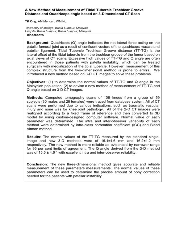
A New Method of Measurement of Tibial Tubercle Trochlear Groove Distance and Quadriceps angle based on 3-Dimensional CT Scan TK Ong, AM Merican, WM Ng. University of Malaya, Kuala Lumpur, Malaysia Hospital Kuala Lumpur, Kuala Lumpur, Malaysia Abstracts Background: Quadriceps (Q) angle indicates the net lateral force acting on the patella-femoral joint as a result of confluent vectors of the quadriceps muscle and patellar ligament. Tibial Tubercle Trochlear Groove distance (TT-TG) is the lateral offset of the tibial tubercle from the trochlear groove of the femur based on axial views of CT scans. Excessive high values of TT-TG and Q angle are often encountered in those patients with patella instability, which can be treated surgically with medialization of the tibial tubercle. However, measurement of this complex structure from the two-dimensional method is prone to errors. We introduced a new method based on 3-D CT images to solve these problems. Objectives: (1) to determine the normal values of TT-TG and Q angle in the Malaysian population; (2) to devise a new method of measurement of TT-TG and Q angle based on 3-D CT images. Methods: Computed tomography scans of 106 knees from a group of 59 subjects (30 males and 29 females) were traced from database system. All of CT scans were performed due to various indications, such as traumatic vascular injury and none was for knee joint pathology. All of the 2-D CT images were realigned according to a fixed frame of reference and then converted to 3D model by using custom-designed computer software. Normal value of each parameter was determined. The intra and inter-observer variability of each method were determined by intra-class correlation coefficient (ICC) and Bland Altman method. Results: The normal values of the TT-TG measured by the standard singleimage and new 3-D methods were of 16.1±4.6 mm and 16.2±4.2 mm respectively. The new method is more reliable as evidenced by narrower range for 95 per cent limits of agreement. The Q angle derived from the 3-D method was of 15.5 ± 4.6 ° with excellent intra and inter-observer reliability. Conclusion: The new three-dimensional method gives accurate and reliable measurement of these parameters measurements. The normal values of these parameters can be used to determine the precise amount of bony correction needed for the patients with patellar instability.
