Q1. (a) Cells of multicellular organisms may undergo differentiation
advertisement
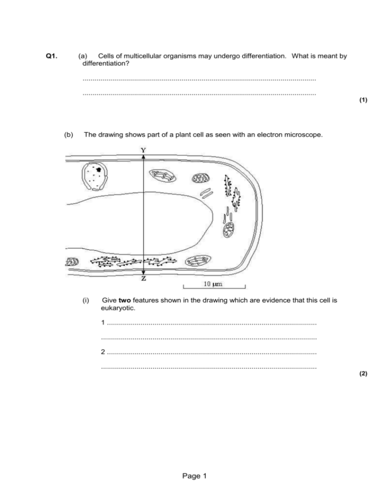
Q1. (a) Cells of multicellular organisms may undergo differentiation. What is meant by differentiation? ...................................................................................................................... ...................................................................................................................... (1) (b) The drawing shows part of a plant cell as seen with an electron microscope. (i) Give two features shown in the drawing which are evidence that this cell is eukaryotic. 1 .......................................................................................................... ............................................................................................................. 2 .......................................................................................................... ............................................................................................................. (2) Page 1 (ii) Calculate the actual width_ of the cell from Y to Z. Give your answer in micrometres (µm) and show your working. Answer ..................................... µm (2) (iii) Give one way in which a typical animal cell differs from the cell shown in the drawing. ............................................................................................................. ............................................................................................................. (1) (Total 6 marks) Page 2 Q2. The drawing shows an electron micrograph of parts of epithelial cells from the small intestine. (a) (i) Name the structures labelled A. ............................................................................................................. (1) (ii) Explain how these structures help in the absorption of substances from the small intestine. ............................................................................................................. ............................................................................................................. (1) (b) (i) The scale bar on this drawing represents a length of 0.1μm. Calculate the magnification of the drawing. Show your working. Magnification ............................................. (2) Page 3 (ii) Explain why an electron microscope shows more detail of cell structure than a light microscope. ............................................................................................................. ............................................................................................................. ............................................................................................................. (2) (c) The length of mitochondria can vary from 1.5 μm to 10 μm but their width_ never exceeds 1μm. Explain the advantage of the width_ of mitochondria being no more than 1μm. ...................................................................................................................... ...................................................................................................................... (1) (Total 7 marks) 3. Scientists use optical microscopes and transmission electron microscopes (TEMs) to investigate cell structure. Explain the advantages and the limitations of using a TEM to investigate cell structure. ...................................................................................................................... ...................................................................................................................... ...................................................................................................................... ...................................................................................................................... ...................................................................................................................... ...................................................................................................................... ...................................................................................................................... ...................................................................................................................... ...................................................................................................................... ......................................................................................................................(5) Page 4 M1. (a) cells become specialised/change to carry out a particular function; 1 (b) (i) named organelle e.g. nucleus/nuclear envelope; vacuole; chloroplast; RER; mitochondrion; no membrane bound organelles; (only award if no organelles named) (reject ribosomes, cell membrane, cell wall) ref to large(r) size 2 max (ii) 20.4 – 21.8 (correct answer 2 marks) 2 (iii) no cell wall (permanent) / (large) vacuole / chloroplasts / smaller; (accept microvilli) 1 max [6] M2. (a) (i) microvilli; (reject brush border) 1 (ii) increased surface area (for diffusion); 1 (b) (i) principle of (15 –17 tolerance) ; 160000; (correct answer award 2 marks) 2 (ii) electron microscope has a greater resolving Page 5 power / objects closer together can be distinguished; electron (beams) have a shorter wavelength; 2 (c) short diffusion pathway /short pathway to the centre / large SA:V ratio for faster, more diffusion; 1 [7] 3. Advantages: 1 Small objects can be seen; 2 TEM has high resolution; Accept better 3 Wavelength of electrons shorter; Advantages: allow maximum of 3 marks. Limitations: 4 Cannot look at living cells; 5 Must be in a vacuum; 6 Must cut section / thin specimen; 7 Preparation may create artefact 8 Does not produce colour image; Limitations: allow maximum of 3 marks. 5 max Page 6
