CT chest template RA.. - Radiology Associates of the Fox Valley, SC
advertisement
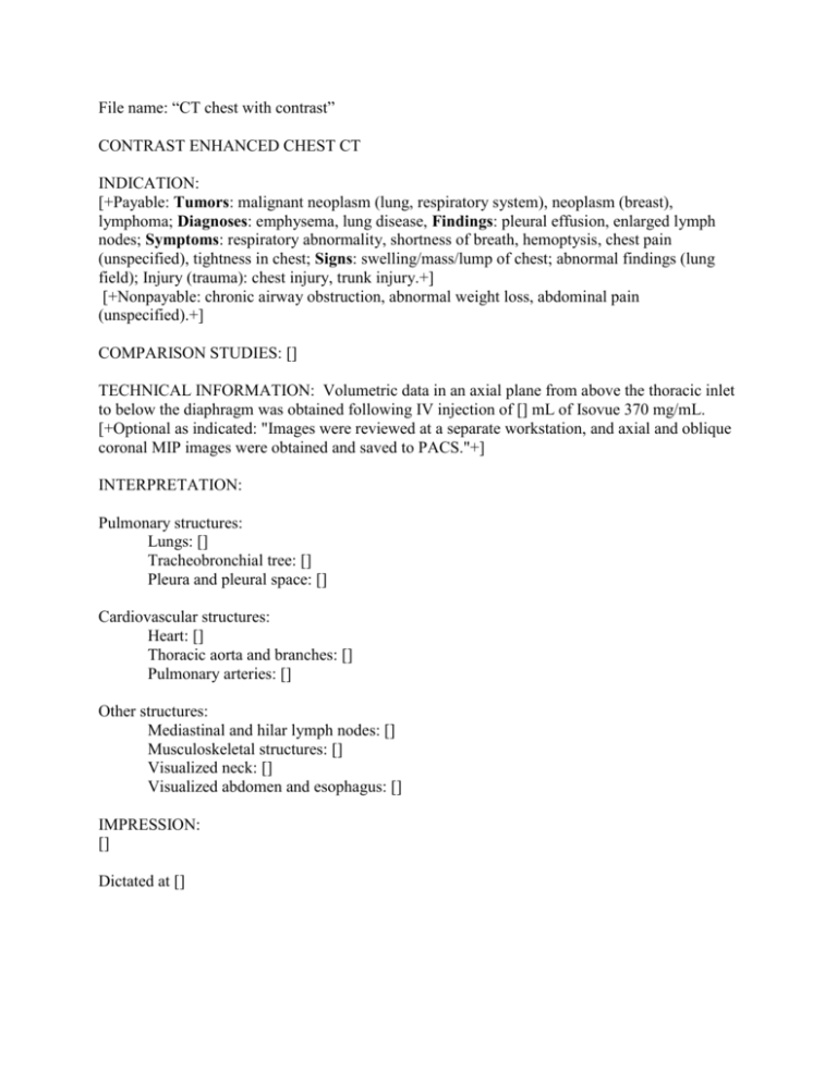
File name: “CT chest with contrast” CONTRAST ENHANCED CHEST CT INDICATION: [+Payable: Tumors: malignant neoplasm (lung, respiratory system), neoplasm (breast), lymphoma; Diagnoses: emphysema, lung disease, Findings: pleural effusion, enlarged lymph nodes; Symptoms: respiratory abnormality, shortness of breath, hemoptysis, chest pain (unspecified), tightness in chest; Signs: swelling/mass/lump of chest; abnormal findings (lung field); Injury (trauma): chest injury, trunk injury.+] [+Nonpayable: chronic airway obstruction, abnormal weight loss, abdominal pain (unspecified).+] COMPARISON STUDIES: [] TECHNICAL INFORMATION: Volumetric data in an axial plane from above the thoracic inlet to below the diaphragm was obtained following IV injection of [] mL of Isovue 370 mg/mL. [+Optional as indicated: "Images were reviewed at a separate workstation, and axial and oblique coronal MIP images were obtained and saved to PACS."+] INTERPRETATION: Pulmonary structures: Lungs: [] Tracheobronchial tree: [] Pleura and pleural space: [] Cardiovascular structures: Heart: [] Thoracic aorta and branches: [] Pulmonary arteries: [] Other structures: Mediastinal and hilar lymph nodes: [] Musculoskeletal structures: [] Visualized neck: [] Visualized abdomen and esophagus: [] IMPRESSION: [] Dictated at [] ALTERNATIVE FOR CHEST CT File name: “CT chest with contrast” CONTRAST ENHANCED CHEST CT INDICATION: [+Payable: Tumors: malignant neoplasm (lung, respiratory system), neoplasm (breast), lymphoma; Diagnoses: emphysema, lung disease, Findings: pleural effusion, enlarged lymph nodes; Symptoms: respiratory abnormality, shortness of breath, hemoptysis, chest pain (unspecified), tightness in chest; Signs: swelling/mass/lump of chest; abnormal findings (lung field); Injury (trauma): chest injury, trunk injury.+] [+Nonpayable: chronic airway obstruction, abnormal weight loss, abdominal pain (unspecified).+] COMPARISON STUDIES: [] TECHNICAL INFORMATION: Volumetric data in an axial plane from above the thoracic inlet to below the diaphragm was obtained following IV injection of [] mL of Isovue 370 mg/mL. [+Optional as indicated: "Images were reviewed at a separate workstation, and axial and oblique coronal MIP images were obtained and saved to PACS."+] INTERPRETATION: Visualized neck: [] Musculoskeletal structures: [] Tracheobronchial tree: [] Lungs: [] Pleura and pleural space: [] Heart: [] Thoracic aorta and branches: [] Pulmonary arteries: [] Mediastinal and hilar lymph nodes: [] Visualized abdomen and esophagus: [] IMPRESSION: [] Dictated at [] Report from radreport.org Exam Type: CT of the CHEST Date and Time: <<Order Observation End Time>> Clinical information: <<Clinical Information>> Comparison: [ report / film / date ] IMPRESSION: [ ] [<RECOMMENDATIONS:>] [ ] Procedure: [<Oral contrast was administered.>] [<100>] ml of [<IV Contrast Name>] was injected intravenously. Chest :[<with intravenous contrast>][<without intravenous contrast>][<without and with intravenous contrast>] FINDINGS: LUNG AND LARGE AIRWAYS: [<within normal limits.>] PLEURA: [<within normal limits.>] VESSELS: [<within normal limits.>][<Atherosclerotic changes in the aorta>] [<and coronary arteries>][<.>] HEART: [<normal size.>] [<No pericardial effusion.>] MEDIASTINUM AND HILA: [<within normal limits.>] CHEST WALL AND LOWER NECK: [<within normal limits.>] UPPER ABDOMEN:[<within normal limits.>] BONES: [<within normal limits.>] [Other]


