Table S1: Calculated and experimentally obtained pKa`s of different
advertisement
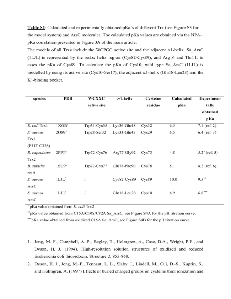
Table S1: Calculated and experimentally obtained pKa’s of different Trx (see Figure S3 for the model system) and ArsC molecules. The calculated pKa values are obtained via the NPApKa correlation presented in Figure 3A of the main article. The models of all Trxs include the WCPGC active site and the adjacent -helix. Sa_ArsC (1LJL) is represented by the redox helix region (Cys82-Cys89), and Arg16 and Thr11, to asses the pKa of Cys89. To calculate the pKa of Cys10, wild type Sa_ArsC (1LJL) is modelled by using its active site (Cys10-Ser17), the adjacent 1-helix (Gln18-Leu28) and the K+-binding pocket. species PDB WCXXC -helix active site Cysteine Calculated Experimen- residue pKa tally obtained pKa E. coli Trx1 1XOB1 Trp31-Cys35 Lys36-Glu48 Cys32 6.5 7.1 (ref. 2) S. aureus 2O893 Trp28-Ser32 Lys33-Glu45 Cys29 6.5 6.4 (ref. 3) 2PPT4 Trp72-Cys76 Arg77-Gly92 Cys73 4.8 5.2* (ref. 5) 1SU96 Trp72-Cys77 Glu78-Phe90 Cys76 8.1 8.2 (ref. 6) 1LJL7 / Cys82-Cys89 Cys89 10.0 9.5** 1LJL7 / Gln18-Leu28 Cys10 6.9 6.8*** Trx1 (P31T C32S) R. capsulatus Trx2 B. subtilis resA S. aureus ArsC S. aureus ArsC * pKa value obtained from E. coli Trx2 ** pKa value obtained from C15A/C10S/C82A Sa_ArsC, see Figure S4A for the pH titration curve. *** pKa value obtained from oxidized C15A Sa_ArsC, see Figure S4B for the pH titration curve. 1. Jeng, M. F., Campbell, A. P., Begley, T., Holmgren, A., Case, D.A., Wright, P.E., and Dyson, H. J. (1994). High-resolution solution structures of oxidized and reduced Escherichia coli thioredoxin. Structure 2, 853-868. 2. Dyson, H. J., Jeng, M.-F., Tennant, L. L., Slaby, I., Lindell, M., Cui, D.-S., Kuprin, S., and Holmgren, A. (1997) Effects of buried charged groups on cysteine thiol ionization and reactivity in Escherichia coli thioredoxin: structural and functional characterization of mutants of Asp26 and Lys57. Biochemistry 36, 2622-2636. 3. Roos, G., Garcia-Pino, A., Van Belle, K., Brosens, E., Wahni, K., Vandenbussche, G., Wyns, L., Loris, R., and Messens, J. (2007). The conserved active site proline determines the reducing power of Staphylococcus aureus thioredoxin. J. Mol. Biol. 368, 800-811. 4. Ye, J., Cho, S., Fuselier, J., Li, J., Beckwith, J., and Rapoport, T. (2007). Crystal structure of an unusual thioredoxin protein with a zinc finger domain. J. Biol. Chem. 282, 3494534957. 5. El Hajjaji, H., Dumoulin, M., Matagne, A., Colau, D., Roos, G., Messens, J. and Collet, J.-F. (2008). The zinc centre influences the redox and thermodynamic properties of Escherichia coli thioredoxin 2. J. Mol. Biol., in revision. 6. a) Crow, A., Acheson, R. M., Le Brun, N. E.,and Oubrie, A. (2004). Structural basis of redox-coupled protein substrate selection by the cytochrome c biosynthesis protein ResA. J. Biol. Chem. 279, 23654-23660. b) Lewin, A., Crow, A., Hodson, C. T. C., Hederstedt, L., and Le Brun, N. E. (2008). Effects of substitutions in the CXXC active-site motif of the extracytoplasmic thioredoxin ResA, Biochem J. 414, 81-91. 7. Zegers, I., Martins, J. C., Willem, R.,Wyns, L., and Messens, J. (2001). Arsenate reductase from S. aureus plasmid pI258 is a phosphatase drafted for redox duty. Nature Struct. Biol. 8, 843-847.
