Lung Donor Management - Organ Donation Alliance
advertisement
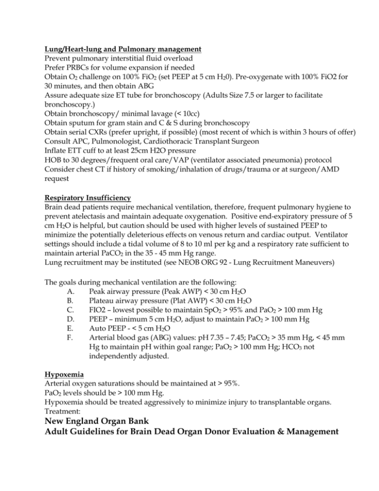
Lung/Heart-lung and Pulmonary management Prevent pulmonary interstitial fluid overload Prefer PRBCs for volume expansion if needed Obtain O2 challenge on 100% FiO2 (set PEEP at 5 cm H20). Pre-oxygenate with 100% FiO2 for 30 minutes, and then obtain ABG Assure adequate size ET tube for bronchoscopy (Adults Size 7.5 or larger to facilitate bronchoscopy.) Obtain bronchoscopy/ minimal lavage (< 10cc) Obtain sputum for gram stain and C & S during bronchoscopy Obtain serial CXRs (prefer upright, if possible) (most recent of which is within 3 hours of offer) Consult APC, Pulmonologist, Cardiothoracic Transplant Surgeon Inflate ETT cuff to at least 25cm H2O pressure HOB to 30 degrees/frequent oral care/VAP (ventilator associated pneumonia) protocol Consider chest CT if history of smoking/inhalation of drugs/trauma or at surgeon/AMD request Respiratory Insufficiency Brain dead patients require mechanical ventilation, therefore, frequent pulmonary hygiene to prevent atelectasis and maintain adequate oxygenation. Positive end-expiratory pressure of 5 cm H2O is helpful, but caution should be used with higher levels of sustained PEEP to minimize the potentially deleterious effects on venous return and cardiac output. Ventilator settings should include a tidal volume of 8 to 10 ml per kg and a respiratory rate sufficient to maintain arterial PaCO2 in the 35 - 45 mm Hg range. Lung recruitment may be instituted (see NEOB ORG 92 - Lung Recruitment Maneuvers) The goals during mechanical ventilation are the following: A. Peak airway pressure (Peak AWP) < 30 cm H2O B. Plateau airway pressure (Plat AWP) < 30 cm H2O C. FIO2 – lowest possible to maintain SpO2 > 95% and PaO2 > 100 mm Hg D. PEEP – minimum 5 cm H2O, adjust to maintain PaO2 > 100 mm Hg E. Auto PEEP - < 5 cm H2O F. Arterial blood gas (ABG) values: pH 7.35 – 7.45; PaCO2 > 35 mm Hg, < 45 mm Hg to maintain pH within goal range; PaO2 > 100 mm Hg; HCO3 not independently adjusted. Hypoxemia Arterial oxygen saturations should be maintained at > 95%. PaO2 levels should be > 100 mm Hg. Hypoxemia should be treated aggressively to minimize injury to transplantable organs. Treatment: New England Organ Bank Adult Guidelines for Brain Dead Organ Donor Evaluation & Management Respiratory Insufficiency (cont.) Hypoxemia Treatment (cont.) Maintain hematocrit > 27%; transfuse PRBCs as necessary Turn and suction patient frequently Increase FiO2 by increments of 10%, and then recheck ABG Diuretic therapy may be indicated if CXR is c/w pulmonary edema Consider pressure control ventilation Hypercapnia Apnea testing, to confirm the diagnosis of brain death, can result in significant hypercapnia leading to respiratory acidosis. Careful attention should be paid to the patient's ventilatory status and ABGs, especially following apnea tests. The ventilator rate should be increased as needed to normalize PaCO2 levels in the 35 to 45 mm Hg range.
