Pediatrics-Pediatric Respiratory Diseases
advertisement
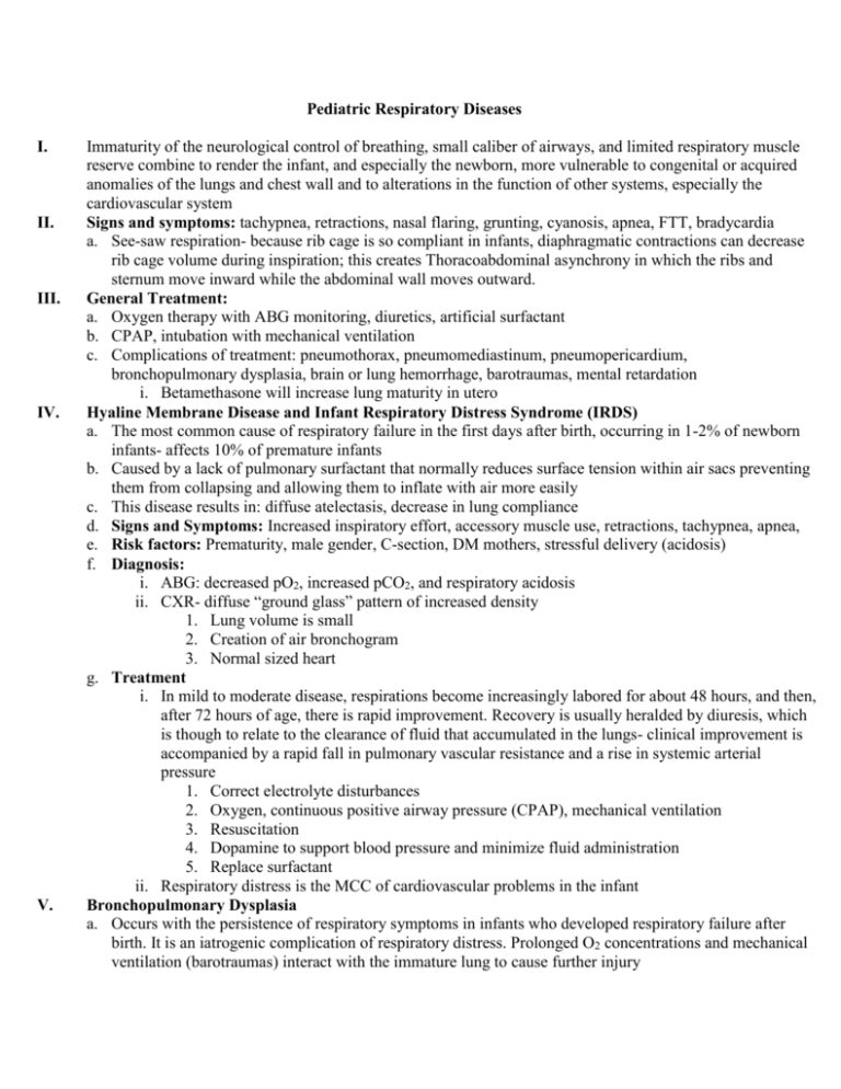
Pediatric Respiratory Diseases I. II. III. IV. V. Immaturity of the neurological control of breathing, small caliber of airways, and limited respiratory muscle reserve combine to render the infant, and especially the newborn, more vulnerable to congenital or acquired anomalies of the lungs and chest wall and to alterations in the function of other systems, especially the cardiovascular system Signs and symptoms: tachypnea, retractions, nasal flaring, grunting, cyanosis, apnea, FTT, bradycardia a. See-saw respiration- because rib cage is so compliant in infants, diaphragmatic contractions can decrease rib cage volume during inspiration; this creates Thoracoabdominal asynchrony in which the ribs and sternum move inward while the abdominal wall moves outward. General Treatment: a. Oxygen therapy with ABG monitoring, diuretics, artificial surfactant b. CPAP, intubation with mechanical ventilation c. Complications of treatment: pneumothorax, pneumomediastinum, pneumopericardium, bronchopulmonary dysplasia, brain or lung hemorrhage, barotraumas, mental retardation i. Betamethasone will increase lung maturity in utero Hyaline Membrane Disease and Infant Respiratory Distress Syndrome (IRDS) a. The most common cause of respiratory failure in the first days after birth, occurring in 1-2% of newborn infants- affects 10% of premature infants b. Caused by a lack of pulmonary surfactant that normally reduces surface tension within air sacs preventing them from collapsing and allowing them to inflate with air more easily c. This disease results in: diffuse atelectasis, decrease in lung compliance d. Signs and Symptoms: Increased inspiratory effort, accessory muscle use, retractions, tachypnea, apnea, e. Risk factors: Prematurity, male gender, C-section, DM mothers, stressful delivery (acidosis) f. Diagnosis: i. ABG: decreased pO2, increased pCO2, and respiratory acidosis ii. CXR- diffuse “ground glass” pattern of increased density 1. Lung volume is small 2. Creation of air bronchogram 3. Normal sized heart g. Treatment i. In mild to moderate disease, respirations become increasingly labored for about 48 hours, and then, after 72 hours of age, there is rapid improvement. Recovery is usually heralded by diuresis, which is though to relate to the clearance of fluid that accumulated in the lungs- clinical improvement is accompanied by a rapid fall in pulmonary vascular resistance and a rise in systemic arterial pressure 1. Correct electrolyte disturbances 2. Oxygen, continuous positive airway pressure (CPAP), mechanical ventilation 3. Resuscitation 4. Dopamine to support blood pressure and minimize fluid administration 5. Replace surfactant ii. Respiratory distress is the MCC of cardiovascular problems in the infant Bronchopulmonary Dysplasia a. Occurs with the persistence of respiratory symptoms in infants who developed respiratory failure after birth. It is an iatrogenic complication of respiratory distress. Prolonged O2 concentrations and mechanical ventilation (barotraumas) interact with the immature lung to cause further injury b. Persistence of respiratory symptoms in infants who developed respiratory failure after birth c. Risk factors: Prematurity, respiratory infection and failure, poor immune status, congenital heart disease, severe illness, dysregulated lung fluid balance d. Diagnosis: i. 50% of infants weighing <1500g at birth (premature) will experience this problem- eventually causes growth problems. Since the patient has difficulty eating, the infant needs nutritional, fluid, and electrolyte supplement ii. Clinical- continued respiratory distress in the case where oxygen was needed for >28 days and an abnormal CXR iii. After extubation, infant needs 60-80 mmHg during sleeping and eating- prevent pulmonary HTN iv. Labs: ABG- increase CO, decrease O2, acidosis 1. PFTs- increase airway resistance, decrease lung compliance e. Treatment: i. Fluid restriction iv. Bronchodilators ii. Oxygen and intubation v. Steroids iii. Diuretics vi. Vitamin A VI. Transient Tachypnea of the Newborn (persistent postnatal pulmonary edema) a. Liquid that fills the lungs during normal fetal development must be absorbed into the vascular system soon after birth to permit successful pulmonary gas exchange. Passage through the birth canal and the first few breaths resulting in lung expansion pushes the majority of fluid out of the lungs b. Significant amount of fluid remains in the lungs and is called “wet lungs” and is called IRDS-2 c. Typically recovers in 72 hours d. Risk factors: Prematurity, C-section delivery, excessive IV fluids to mother e. CXR findings: prominent pulmonary vascular (hilar) markings, hyperaeration, diaphragm flattening, fluid in interlobar fissures and pleural spaces f. Treatment: Diuretics and oxygen supportively, fluid restriction VII. Neonatal Pneumonia a. Occurs more commonly in at term infants who are born by C-section (not group B strep.) b. Major cause of infectious complications of newborn: Group B streptococcus (in mother’s vagina) c. Signs and symptoms: respiratory distress, cyanosis, apnea, poor perfusion, hypotension, lethargy d. CXR: fine, diffuse uniform opacification, patchy lung infiltrate; pleural effusion e. Diagnosis: chest x-ray, and cultures f. Treatment: antibiotics- for GBS: penicillin or Ampicillin +aminoglycoside VIII. Meconium Aspiration Syndrome a. Tachypnea, grunting and varying degrees of cyanosis in a meconium stained infant with meconium noted below the vocal cords during intubation b. Diagnosis: Meconium is visible below the level of the vocal cords when performing intubation c. Aspirated amniotic-meconium fluid causes mechanical obstruction of airways (if complete death) and peripheral chemical pneumonitis, resulting in alveolar collapse and necrosis d. Passage of meconium into amniotic fluid is associated with fetal distress e. CXR: coarse, fluffy infiltrate with alternating areas of lucency, hyperinflation with flattened diaphragm f. ABG: increase CO2, decrease O2, acidosis g. Complications: air-leak pneumonia, pneumothorax, pleural effusion h. Treatment: clear amniotic fluid and meconium by immediate postnatal suctioning before it disseminates peripherally; oxygen therapy and ventilator support IX. X. Persistent Pulmonary Hypertension of the Newborn (persistent fetal circulation) a. Disease results when the pulmonary blood pressure does not drop as it should, and blood continues to be shunted away from the lungs and back to the body without receiving oxygen since the pulmonary vascular resistance remains higher than the systemic resistance (RL shunt) b. The newborn remains in cyanosis even after the administration of 100% oxygen c. Causes: sepsis, diaphragmatic hernia, congenital heart disease d. CXR: cardiomegaly, diminished pulmonary vascularity e. Echo: dilation of the right atrium and ventricle, bowing of the interventricular septum f. Doppler: R L shunt through foramen ovale and ductus arteriosus g. Treatment: i. Correct the hypoxemia and acidosis to decrease vasospasm ii. 100% oxygen v. Artificial surfactant iii. Nitrous oxide vi. Positive ionotropes iv. Volume expanders vii. Heart/lung bypass machine Apnea- cessation of breathing for greater than 10 seconds and this is usually caused by immaturity of the central respiratory centers a. Periodic breathing- recurrent (3 or more) respiratory pauses of ≥3 seconds with less than 20 seconds of respiration between pauses; may be normal in premature infants b. Pathological apnea- prolonged respiratory pause (≥20 seconds) associated with cyanosis, pallor, hypotonia, and/or bradycardia c. Three types of apnea- distinguished by the presence of absence of obstruction i. Central apnea (10-15%)- no obstruction, no respiratory effort ii. Obstructive apnea (10-20%)- airway obstruction iii. Mixed apnea (50-70%)- obstructive and central apnea symptoms d. If the apnea occurs in infants >34 weeks of gestation is considered abnormal, must R/O other causes: hypoxia (sleep associated), e. GERD, infection l. CNS pathology f. Feeding intolerance m. Metabolic disorders g. Vomiting n. Perinatal drugs h. Lethargy o. Cardiac arrhythmias i. Seizures p. Temperature j. Fever q. Suctioning k. Sepsis r. Anatomic malformation s. Physical exam: neurological- lethargy, hypotonia, jitteriness t. Labs: CBC with diff, electrolytes, head CT, ABG, barium swallow u. Treatment: oxygen, antibiotics (infection), CPAP, caffeine, theophylline, mechanical ventilation
