Table 1 : Main clinical characteristics of fore-, mid-, and
advertisement

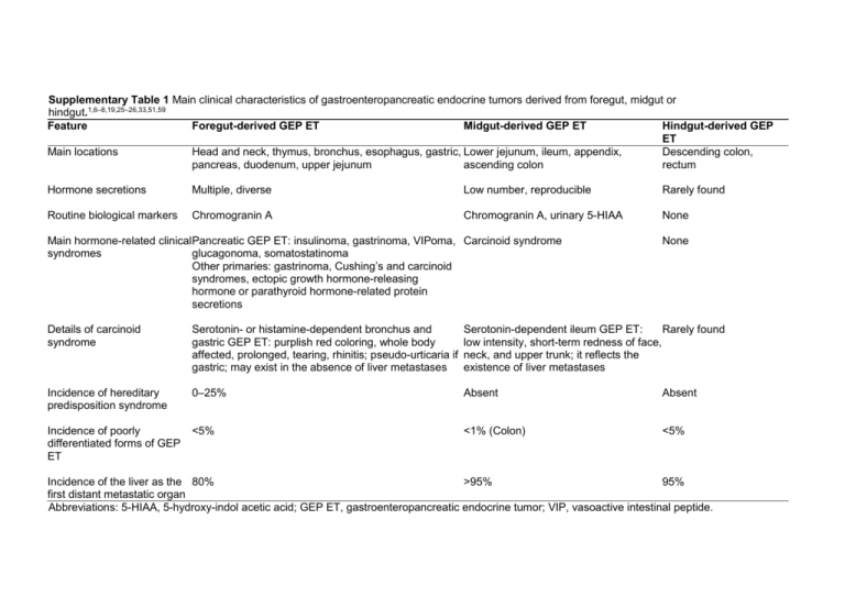
Supplementary Table 1 Main clinical characteristics of gastroenteropancreatic endocrine tumors derived from foregut, midgut or hindgut.1,6–8,19,25–26,33,51,59 Feature Foregut-derived GEP ET Midgut-derived GEP ET Hindgut-derived GEP ET Main locations Head and neck, thymus, bronchus, esophagus, gastric, Lower jejunum, ileum, appendix, Descending colon, pancreas, duodenum, upper jejunum ascending colon rectum Hormone secretions Multiple, diverse Low number, reproducible Rarely found Routine biological markers Chromogranin A Chromogranin A, urinary 5-HIAA None Main hormone-related clinicalPancreatic GEP ET: insulinoma, gastrinoma, VIPoma, Carcinoid syndrome syndromes glucagonoma, somatostatinoma Other primaries: gastrinoma, Cushing’s and carcinoid syndromes, ectopic growth hormone-releasing hormone or parathyroid hormone-related protein secretions None Details of carcinoid syndrome Serotonin- or histamine-dependent bronchus and gastric GEP ET: purplish red coloring, whole body affected, prolonged, tearing, rhinitis; pseudo-urticaria if gastric; may exist in the absence of liver metastases Serotonin-dependent ileum GEP ET: Rarely found low intensity, short-term redness of face, neck, and upper trunk; it reflects the existence of liver metastases Incidence of hereditary predisposition syndrome 0–25% Absent Absent Incidence of poorly differentiated forms of GEP ET <5% <1% (Colon) <5% Incidence of the liver as the 80% >95% 95% first distant metastatic organ Abbreviations: 5-HIAA, 5-hydroxy-indol acetic acid; GEP ET, gastroenteropancreatic endocrine tumor; VIP, vasoactive intestinal peptide. Supplementary Table 2 Histological classification of gastroenteropancreatic endocrine tumors according to the WHOa.6–8 Feature WHO 1999 classification WHO 2000 classification WHO 2004 classification Thymus and lung Well or poorly differentiated Well Well Digestive tract Poorly Typical Atypical Large-cell Phenotype carcinoid carcinoid carcinoma Features on histological Solid, illexamination Organoid Organoid defined Atypical cellularity No or mild Moderate Frequent CgA or synaptophysin Absent or staining Strong Strong weak >0.5 >0.5 Size (cm) NA Poorly Well Poorly Large- or Small-cell Benign Uncertain small-cell carcinoma behavior behavior Carcinoma carcinoma Solid, illSolid, illdefined Organoid Organoid Organoid defined Frequent No or mild No or mild No or mild Frequent Absent or Absent or weak Strong Strong Strong weak b b 1–2 >1–2 NA NA NA 2 2 >2 >10 (Usually) (Usually) (Usually) >10 2 2 >2 NA (Usually) (Usually) (Usually) >15 Pancreas Well Benign behavior Poorly Uncertain behavior LargeCarcinoma cell ca Organoid Organoid No or mild No or mild Organoid No or mild Solid, Frequ Strong <2 Strong NA Absen NA Strong 2 <2 2–10 >10 <2 2–10 Mitoses per 10 HPF NA Cells staining for Ki67 <2 2 (%) NA NA NA NA T stage (in-depth T1c T1c T2c invasion) NA NA NA NA NA NA NA NA Vascular and/or Prominent Absent Present Absent Present perineural invasion NA NA NA NA NA (often) NA Absent Rare Frequent Frequent NA Frequent Necrosis NA NA NA NA NA Absent Absent Present Absent Absent Present Local spread NA NA NA NA NA Absent Absent Present Absent Absent Present Metastases NA NA NA NA NA a Among the features, the major criteria for each classification are shown in bold. bOne centimeter for gastric, duodenum, jejunum, and ileum endocrine tumors but 2|cm for appendix and colon–rectum. cT2 for all digestive endocrine tumors except the appendix, for which invasion of mesoappendix or beyond is required: T1, confined to the mucosa–submucosa; T2, invasion of the muscularis propria or beyond except for appendix for which invasion of mesoappendix or beyond (so-called T4) is required. Abbreviations: CgA, chromogranin A; HPF, high-powered fields; NA, not taken into account for classification. >10 NA NA NA Frequ NA NA Supplementary Table 3 Main clinical and prognostic characteristics of well-differentiated endocrine carcinomas as compared with poorly differentiated large-cell carcinomas.1,2,4,5,12–14,19,22,27,29,39 Feature Prevalence among non-small-cell ET Tobacco habit Gender Positive diagnosis Main differential diagnosis Time elapsing before diagnosis Mixed tumor General condition Primary Well-differentiated GEP ET 95% Not all Same in women and men Light microscopy, intense chromogranin A staining Medullary thyroid carcinoma, pheochromocytoma, melanoma, kidney or other GEP ET metastasis, mixed tumors, large cell endocrine carcinoma Long (years) Rare Good Entire body Revealing functional tumors 5–10% General biological markers Chromogranin A > neuron-specific enolase Hereditary syndrome of predisposition 0–25% Metastases at diagnosis <50% First-line scintigraphic imaging Somatostatin receptor scintigraphy Cisplatin-based chemotherapy objective <10% response Five-year survival >50% Abbreviation: GEP ET, gastroenteropancreatic endocrine tumor. Poorly differentiated (large-cell) GEP ET 5% Virtually all Preponderance in men Immunostaining positive for >1 neuroendocrine marker Poorly differentiated carcinoma, sarcoma, thymoma, lymphoma, mixed tumors, welldifferentiated endocrine tumor, melanoma Short (months) Frequent Poor Mainly foregut; not reported in the appendix and ileum <1% Neuron-specific enolase > chromogranin A Absent >50% PET with fluorodeoxyglucose 50% <20%


