Surgical Incisions – Guidelines and Recommendations
advertisement
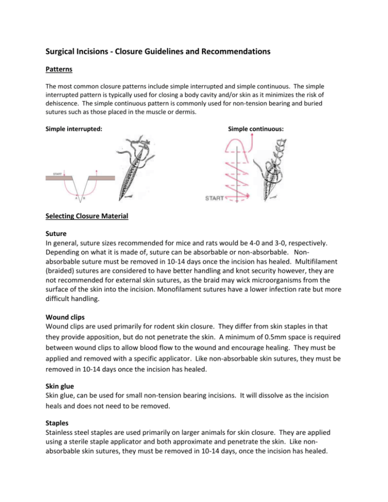
Surgical Incisions - Closure Guidelines and Recommendations Patterns The most common closure patterns include simple interrupted and simple continuous. The simple interrupted pattern is typically used for closing a body cavity and/or skin as it minimizes the risk of dehiscence. The simple continuous pattern is commonly used for non-tension bearing and buried sutures such as those placed in the muscle or dermis. Simple interrupted: Simple continuous: Selecting Closure Material Suture In general, suture sizes recommended for mice and rats would be 4-0 and 3-0, respectively. Depending on what it is made of, suture can be absorbable or non-absorbable. Nonabsorbable suture must be removed in 10-14 days once the incision has healed. Multifilament (braided) sutures are considered to have better handling and knot security however, they are not recommended for external skin sutures, as the braid may wick microorganisms from the surface of the skin into the incision. Monofilament sutures have a lower infection rate but more difficult handling. Wound clips Wound clips are used primarily for rodent skin closure. They differ from skin staples in that they provide apposition, but do not penetrate the skin. A minimum of 0.5mm space is required between wound clips to allow blood flow to the wound and encourage healing. They must be applied and removed with a specific applicator. Like non-absorbable skin sutures, they must be removed in 10-14 days once the incision has healed. Skin glue Skin glue, can be used for small non-tension bearing incisions. It will dissolve as the incision heals and does not need to be removed. Staples Stainless steel staples are used primarily on larger animals for skin closure. They are applied using a sterile staple applicator and both approximate and penetrate the skin. Like nonabsorbable skin sutures, they must be removed in 10-14 days, once the incision has healed. Table 4, below, summarizes the characteristics of some common wound closure materials. If you are unfamiliar with wound closure materials and/or how to utilize these materials, please consult a ULAR veterinarian before choosing a material or attempting wound closure. ULAR provides training, free of charge. Please contact us at ulartraining@osu.edu. Material Characteristics/Common Uses Polyglactin 910 (e.g. Vicryl®) Absorbable. Completely absorbed after 60-90 days. Braided. Used most often for soft tissue approximation and ligation. Low reactivity. Polyglycolic acid (e.g. Dexon®) Absorbable. Completely absorbed after 60-90 days. Braided. Excellent handling. Used most often for soft tissue approximation and ligation. Low reactivity. Polydiaxanone (e.g. PDS®) Absorbable. Excellent retention of tensile strength (maintains strength longer than all other absorbable suture materials). Completely absorbed after 6 months. Monofilament. Used most often for soft tissue approximation and ligation. Low reactivity. Optimal when extended wound support but ultimate absorption is desired. Chromic Gut Absorbable. Absorption is variable but is generally complete after 70 days. Used most often for soft tissue approximation and ligation, but not recommended for cardiovascular or neurological surgery. Tissue reactive. Not suitable for skin suturing. Nylon (Ethilon®) Non-absorbable. Inert. Used most often for general closures. Should be removed after wound has healed (10-14 days). Available as a monofilament and braid (braided is coated with silicon). Silk Considered non-absorbable, but is undetectable in the site after 2 years. Tissue reactive. High coefficient of friction Braided. Excellent handling. Preferred for cardiac procedures. Not for use in skin closure. Monofilament Stainless steel Non-absorbable. Inert. Used most often in orthopedics, abdominal wall closures, and sternum closures. Long-term wound support. Stainless steel wound clips or staples Cyanoacrylate (e.g. Vetbond®, Nexband®) Non-absorbable. Inert. Requires removal from skin after wound has healed (10-14 days). Require special instrument for insertion and removal Skin glue. Used for non-tension bearing skin incisions. Reference: http://vpr.utsa.edu/files/larc/PrinciplesVeterinarySuturing.pdf
