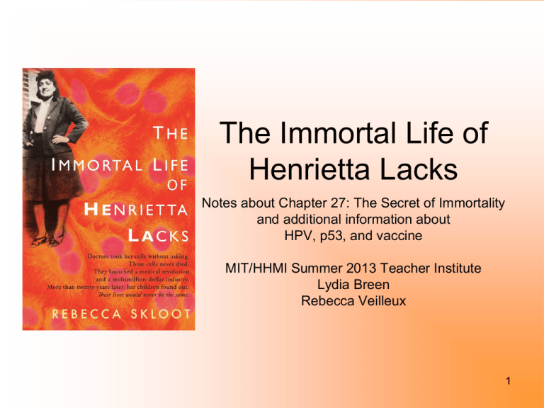
The Immortal Life of
Henrietta Lacks
Notes about Chapter 27: The Secret of Immortality
and additional information about
HPV, p53, and vaccine
MIT/HHMI Summer 2013 Teacher Institute
Lydia Breen
Rebecca Veilleux
1
HeLa Cell Immortality
• 1984- Dr. Harald zur
Hausen discovered Human
Papilloma Virus 18 (HPV 18)
along with HPV-16 (1983)
caused cervical cancer
• Tissue from Henrietta Lack’s
original biopsy tested
positive for multiple copies
of HPV18
A papsmear with healthy cells (blue)
and HPV-infected cells (pink).
Photomicrograph by Ed Uthman,
MD. Creative Commons.
2
HPV
• 100+ strains
• 13 strains of HPV cause
cervical, anal, oral, labia, and
penal cancers
• 90% of sexually active adults
have been infected with at
least on of the strains
• 1980’s HeLa used to study
how HPV causes cancer and
how if HPV DNA is blocked,
cells stop being cancerous
• Leads to HPV vaccine
• Nobel prize for zur Hausen
3
More about HPV and Cancer
Slides 5-8 from NCI: Understanding Cancer
http://www.cancer.gov/cancertopics/understandingcancer
4
Many Types of HPVs
Different HPVs–Different Infections
Harmless
No warts or cancer
Warts-Linked
Genital warts
Cancer-Linked
Most clear up
Some persist, but no abnormalities in cervix
Some persist, some abnormalities in cervix
A few persist and progress to cervical cancer
There are three groups of genital HPV strains: many no-risk types cause neither warts nor cancer; a few types cause genital warts; and 15 or so
high-risk types can increase one’s risk of cancer. If left untreated, genital warts do not turn into cancer. High-risk HPV, on the other hand, may
trigger an infection that leads to cervical cancer. The majority of infections with high-risk HPVs clear up on their own. Some infections persist
without causing any additional abnormal cell changes. However, a few infections caused by high-risk HPVs end up triggering cervical cancer
over many years.
5
Virus Penetrates Cervix
Papillomavirus
Uterus
Layers of
epithelial cells
Cervix
HPV
infection
Vagina
Both harmless and cancer-linked human papillomaviruses pass by skin-to-skin contact. The high-risk types of HPVs need to penetrate deeply into the lining of
the cervix to establish a chronic infection. A vaginal sore or sex, which can abrade the lining, may provide a point of entry for the papillomavirus. Once inside the
cervical lining, the virus attaches to epithelial cells. As these cells take in nutrients and other molecules that are normally present in their environment, they also
take in the virus. Over 99 percent of cervical cancer cases are linked to long-term infections with high-risk human papillomaviruses.
6
Virus Uncoats
Nucleus
Viral DNA
enters
nucleus
mRNAs
for viral
proteins
E6 and E7
Virus
“uncoats”
Epithelial cell
interior
The HPV sits inside the epithelial cells housed in a protective shell made of a viral protein called L1. After the virus enters the cell, the viral
coat is degraded, leading to the release of the virus’ genetic material into the cell and its nucleus. From the nucleus, the genes of the virus are
expressed, including two genes called E6 and E7, which instruct the cell to build viral proteins called E6 and E7.
7
Virus Disables Suppressors
Mucus
Healthy cells
E6 viral
protein
Suppressor
protein 1
Degraded
suppressors
Cancerous
epithelial cells
E7 viral
protein
Suppressor
protein 2
Viral proteins E6 and E7 then disable the normal activities of the woman’s own suppressor genes, which make suppressor
proteins that do “damage surveillance” in normal cells. These proteins usually stop cell growth when a serious level of
8
unrepaired genetic damage exists. Even after suppressors are disabled in a woman’s cervical cells, it usually takes more
than 10 years before the affected tissue becomes cancerous.
9
Henrietta’s cancer
• HPV DNA inserted into long arm of chromosome 11
(*chromosome 17)
• HPV DNA turned off p53 gene (tumor suppressor gene)
• Still unknown why Henrietta’s cancer cells were so virulent
•
*research states p53 gene in on the 17th chromosome in the human)
Super website:
http://p53.free.fr/index.html
10
What is the p53 Gene?
The p53 gene is
responsible for proteins
that can either repair
damaged cells, or cause
damaged cells to die, a
process called
apoptosis.
When the gene is not
working due to a
mutation, these proteins
that repair cells or
eliminate damaged cells
are not produced, and
abnormal cells are
allowed to divide and
grow.
By Lynne Eldridge MD, About.com Guide
Updated October 10, 2012
http://www.aschoonerofscience.com
11
Cell Cycle Checkpoints
• DNA in chromosomes can
be damaged by a number of
agents including radiation,
toxic chemicals, and free
radicals.
• At this checkpoint, a protein
known as p53 will inspect
the chromosomes’ DNA for
damage.
•
* super website
http://lifesciences.envmed.rochester.edu/lessonsCancer.
html
12
HeLa cells and HIV 1980’s
• 2004 Nobel Prize winner Richard
Axel infected HeLa cells with HIV
DNA sequence that made the
cells susceptible to HIV infection
and thus these cells could be
used to study the virus in the lab.
• Led to law suit by activist Jeremy
Rifkin to stop HIV HeLa
research……later dismissed
• Brought about the discussion
about the “evolution” of the HeLa
cells and are HeLa cells a “new
species”
Dr. Richard Axel
13
HeLa Term Project
•
•
Dr. Axel also used recombinant DNA
technology to discover the molecule CD4,
that is a surface protein on a T-cell responsible for the transmission of
HIV (Axel, 2004). A T-cell is a white blood
cell which protects the body from infection,
called lymphocytes (Medicine Net, 2010).
He and Ellen Robey hypothesized that HIV’s
glycoprotein, called gp120, is what that
reacts to T-cell’s CD4. They worked on nonT-cells, including HeLa cells, by injecting
them with CD4 to see if that made them
susceptible to HIV. It did. They also
isolated the gp120 and CD4 proteins, mixed
them in solution and found that there was an
affinity for the two to bind together (Robey &
Axel, 1990). The hope is through this
research there may be a treatment or
vaccine found for HIV.
–
http://helatermproject.blogspot.com/2012/03/richardaxel.html
14
Scientific Use of HeLa cells
•
Well done interactive website for students to use when researching the use of HeLa cells in
scientific research
http://whenintime.com/ShowEvents.aspx?tlurl=/tl/kelseypitts/HeLa_3a_Scientific_and_Medical_Breakt
hroughs_Through_the_Years/
HeLa: Scientific and Medical Breakthroughs Through the Years
–
Created by
Kelsey Pitts
Henrietta Lacks' immortal cells, known as HeLa, made many scientific and medical
advances possible ever since they were harvested. This timeline details these advances
and how HeLa was involved in each.
15
HeLa and Immortality
• Hayflick Limit- cells can only
divide a finite number of times1961 Leonard Hayflick cells
reach their limit when they’ve
doubled 50 times
• HeLa cells studied for their
immortality
• Cancer cells have the enzyme
telomerase that maintains the
telomeres on each chromosome
making them immortal
•
“the day after the 58th anniversary of Henrietta Lacks’s
death — the Nobel Prize in medicine has been awarded
to Elizabeth Blackburn, Carol Greider, and Jack Szostak
for the discovery of how telomeres and the enzyme
telomerase protect chromosomes from degrading over
time.” Rebecca Skloot’s blog 2009
16
Telomerase
Telomere illustration. (Credit: Copyright The Nobel Committee for
Physiology or Medicine 2009 / Illustration: Annika Röhl)
17
HPV Vaccine
http://www.cdc.gov/hpv/
From: The Journal of Infectious Diseases
June 2013
New study shows HPV vaccine helping lower HPV infection rates in teen girls
A new study looking at the prevalence of human papillomavirus (HPV) infections in girls and women
before and after the introduction of the HPV vaccine shows a significant reduction in vaccine-type HPV
in U.S. teens. The study, published in [the June issue of] The Journal of Infectious Diseases reveals
that since the vaccine was introduced in 2006, vaccine-type HPV prevalence decreased 56 percent
among female teenagers 14-19 years of age.
About 79 million Americans, most in their late teens and early 20s, are infected with HPV. Each year,
about 14 million people become newly infected.
“This report shows that HPV vaccine works well, and the report should be a wake-up call to our nation
to protect the next generation by increasing HPV vaccination rates,” said CDC Director Tom Frieden,
M.D., M.P.H. “Unfortunately only one third of girls aged 13-17 have been fully vaccinated with HPV
vaccine. Countries such as Rwanda have vaccinated more than 80 percent of their teen girls. Our low
vaccination rates represent 50,000 preventable tragedies – 50,000 girls alive today will develop
cervical cancer over their lifetime that would have been prevented if we reach 80 percent vaccination
rates. For every year we delay in doing so, another 4,400 girls will develop cervical cancer in their
lifetimes.”
18

