PowerPoint bemutató
advertisement
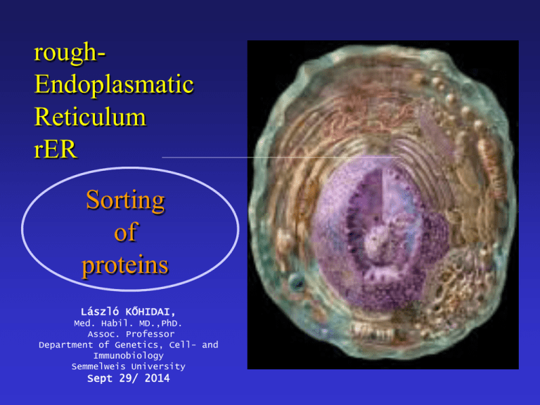
roughEndoplasmatic Reticulum rER Sorting of proteins László KŐHIDAI, Med. Habil. MD.,PhD. Assoc. Professor Department of Genetics, Cell- and Immunobiology Semmelweis University Sept 29/ 2014 Endoplasmic = inside the cell; reticulum = network • Extensive membrane system • Includes up to half of membrane of cell • Tubules and sacs = cisternae • Continous with the nuclear envelope • Two types: rough ER (ribosomes) smooth ER rER s-ER (smooth ER) Structure: tubular Function: • synthesis of phospholipids, cholesterol, ceramide • synthesis of steroids • storage and regulation of Ca2+ • detoxification – cyt P450 TEM of ribosomes attached to the rER in a pancreatic exocrine cell mRNA peptide polyribosome Ribosomes – mRNA – Polyribosome Molecular composition of ribosome 60S rRNA + peptides rRNA Ribosome subunits Comparison of prokaryotic and eukaryotic ribosomes Structure of ribosome ? t-RNA activator enzyme of AA ribosome anticodon codon Initiation Elongation Peptide bond formation peptidyl transferase peptide bond Termination Internalization of peptides into the rER Synthesis of secretory proteins on the rER Structure of SRP • Universal • 300 base RNA • Six proteins • P54 - signal peptide • P9, P14 - ribosome • P68, P72 move the peptide Synthesis of secretory proteins on the rER Electron microscopic view of a translocon channel The ribosome-translocon-ER membrane complex Translocon complex • TRAM – (= translocating chain-associated membrane protein) binds the signal sequence • Sec61p – major constituent of the translocon channel; assembles into a donut-like structure •The Sec 61 complex binds the ribosome, participates the transmembrane transfer Cycles of GDP/GTDP exchange and GTP hydrolysis that drive insertion of nascent secretory protein into the translocon Topologies of some integral membrane proteins synthesized on the rER Synthesis and insertion into the ER membrane of the insulin receptor and similar proteins N-terminus faces to ER lumen C-terminus faces to cytosol A signal sequence is cleaved Stop-transfer membrane-anchor signal Synthesis and insertion into the ER membrane of the asialoglycoprotein receptor and similar proteins C-terminus faces to ER lumen N-terminus faces to cytosol No N-terminal ER signal sequence an uncleaved integral signal membrane-anchor sequence Synthesis and insertion into the ER membrane of proteins with multiple transmembrane a-helical segments - An uncleaved internal signal membrane-anchor sequence - A stop-transfer membrane-anchor sequence - An uncleaved internal signal membrane-anchor sequence Etc. Post-translational modification • Proteolytic cleavage of proteins • Glycosilation • Acylation • Methylation • Phosphorylation • Sulfation • Prenylation • Vitamin C-dependent modifications • Vitamin K-dependent modifications • Selenoproteins Proteolytic cleavage • • Removal of signal peptide from preproproteins preproteins Signal peptidase Properties of uptake-targeting signal sequences Target organelle Usual signal location within protein Signal removal Nature of signal rER N-terminal + „core” of 6-12 mostly hydrophobic amino acids, often proceeded by one or more basic amino acids Mitochondrium N-terminal + 3-5 nonconsecutive Arg or Lys residues often with Ser and Thr; no Glu or Asp Chloroplast N-terminal + No common motives, generally rich in Ser,Thr, poor in Glu and Asp Perixisome C-terminal - Ser-Lys-Leu Nucleus Internal - Cluster of 5 basic amino acids or two samller clusters separated by 10 amino acids Glycoproteins Predominant sugars are: glucose, galactose, mannose, fucose, GalNAc, GlcNAc, NANA O-glycosidic linkage – hydroxyl group of Ser, Thr, hydrLys N-glycosidic linkage – consensus sequence N-X-S(T) (BUT No P) Major N-linked families: high mannose type, hybride type, complex type (sialic acids) Glycosilation rER N-linkage to GlcNAc rER O-linkage to GalNAc O-linked sugars: sugars coupled to UDP, GDP (mannose), CMP (NANA) glycosprotein glycosylttransferase N-linked sugars: Requires a lipid intermediate dolichol phosphate N-Glycosilation Glycosylphosphatodyl inositol (GPI) -anchored peptides GPI-anchored peptides become the outer surface of the surface membrane Protein folding: Protein Disulfide Isomerase (PDI) • Provides mechanism for breaking incorrectly paired disulfide bonds. • The most stable folded sate is reached Protein folding: • Peptidyl-prolyl isomerase: accelerates rotation about peptidyl-prolyl bonds • Oligosaccharide protein transferase: transfers carbohydrate chains to the nascent polypeptide as they enter the lumen of ER • Calnexin, calreticulin: interact with CHO groups of glycoproteins Protein signals: • Integral, soluble proteins of ER, Golgi retrieved by the KDEL-receptors. They recognize the KDEL signal (Lys-Asp-Glu-Leu at C-terminus). • ER membrane proteins have a KKXX (dilysine motif) on the C-terminus. • Other ER membrane proteins possess di-arginine motif on the N-terminus. Chase-pulse technique Antibiotics They inhibit different steps of protein synthesis • Actinomycin D • Rifamycin • Amanitin • Streptomycin • Tetracycline • Erythromycin • Cycloheximide • Chloramphenicol • Puromycin - transcription (complex with DNA) - transcription (RNA polymerase) - transcription (RNA polymerase II) - iniciation - aminoacyl-tRNA - A locus interaction - translocation of tRNA from A to P locus “ (only in eukaryotes) - peptide bond formation - termination Penicillins and Cephalosporins - synthesis of bacterial cell wall (proteoglycans) actinomycin rifamycin amanitin streptomycin chloramphenicol tetracycline A puromycin P A erythromycin, cycloheximide A


