What is chest pain?
advertisement
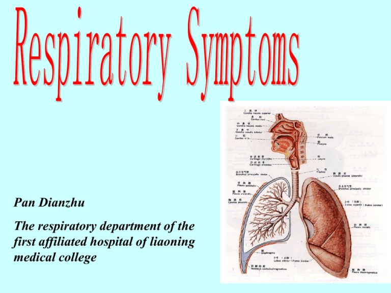
Pan Dianzhu The respiratory department of the first affiliated hospital of liaoning medical college 1 Common respiratory symptoms • Cough • Expectoration • Hemoptysis • Chest pain • Dyspnea • Cyanosis 2 What is chest pain? Chest pain is one of the common symptoms and often likely to be accompanied by fear of heart disease. we shall pay more attention to the history taking because there may be no physical signs associated with chest pain. 3 Attributes of chest pain Provocative-palliative (Influential) factors Quality /feature Exertional, respiration, food intake, administration (pricking /bursting /pressing /blunt/colic/distention/burning/stabbing/ crushing /throbbing) Region /location Severity Timing/ Duration – angina pectoris myocardial infarction Referred pain 4 Diagnostic approach First : assess the risk for major vascular disease and acute coronary events Second :develop the differential diagnosis of noncardiovascular explanations Acute coronary events may be missed in younger patients, women, and people with normal ECGs, so careful risk factor assessment and a high index of suspicion are necessary. Chest pain Chest pain with tenderness Deep retrosternal or precordial pain 5 Chest pain with tenderness The distinction between respiratory pain with tenderness and chest pain with tenderness is somewhat artificial; the conditions may present in either manner, or with both pain at rest and with respirations. The patient may recognize the pain as superficial ,sharp, and well localized. Almost always, this type of pain is accompanied by localized tenderness. The structures involved are the skin and subcutaneous tissues, the fat, skeleton, or the breasts. 6 Skin and Subcutaneous Structures Inflammation, trauma, and neoplasm in these tissues offer no special diagnostic problems, provided that they are considered and searched for. The presence of bruises, lacerations, ulcers, hematomas, masses, or tenderness is usually diagnostic. 7 Chest Wall Syndrome Ask your patient to point to the region of pain.Next, perform four maneuvers: (1) Palpate the chest wall for tenderness by applying firm, steady pressure to the sternum, the costosternal junctions the intercostal spaces, the ribs, and the pectoralis major muscles and their insertions. (2) Flex the arms horizontally by lifting one arm after the other by the elbow and pulling it across the chest toward the contralateral side, with the head rotated toward the ipsilateral side . (3) Extend the neck by having the patient look toward the ceiling as the arms are pulled backward and slightly upward. (4) Exert vertical pressure on the head. If any of these tests reproduces the patient's pain, review the history to ascertain whether your patient forgot recent minor trauma or strain of the chest muscles. 8 Costochondritis of Rib and Tietze Syndrome This is a common cause of chest pain. The onset may be sudden or gradual. The pain is usually dull. It may be intensified by respiratory motion and movements of the shoulder girdle . The sole physical sign is tenderness at the costochondral junction of bone and cartilage . There is no swelling and there are no X-ray findings. In Tietze syndrome the pain is accompanied by tender, fusiform swelling of one or more costicartilages, often that attached to the second rib. The overlying skin is reddened. Pain may radiate to the shoulder, neck, or arm. There is no lymphadenopathy. It may subside in a few weeks or persist for months. The swelling may persist for months after the pain and tenderness subside. The cause is unknown and the condition must be distinguished from osteitis , periostitis , rheumatic chondritis , and neoplasm of the ribs. 9 Fractured Rib The history may suggest pleurisy. The patient complains of pain in the chest with breathing. Movement of rib fragments causes well-localized, sharp,lancinating pain. Inspiration is limited and palpation discloses point tenderness on a rib. There may be a history of direct chest trauma; if not, ask about recent severe coughing which the patient may not recognize as a cause of fracture. The edges of the fracture may be felt, but bone crepitus is absent when the fragments are well opposed. With one hand supporting the back, compression of the sternum with the other elicits pain at the untouched fracture site (Figure 1-1). The diagnosis is made readily when the patient gives a history of trauma to the thorax.Fractures of several contiguous ribs are usually caused by external violence; the chest wall may be so weakened as to produce the flail chest. 10 11 Cough Fracture Any rib from the fifth to the tenth is likely to break. The fracture is usually anterior to the attachments of the serratus anterior that pulls the rib upward , and posterior to the fixations of the abdominal external oblique muscle that pulls the rib downward creating a shearing force on the rib. A single cough is not enough to produce fracture; breaking is attributed to the fatigue ( stress fracture ) from repeated coughing. Cough fracture was described by Robert Graves sometime before 1833. The patient has been coughing for some time and pain begins to occur with respiratory movements and coughing. The typical signs of fracture of a rib are present. When palpation of the ribs is not .performed, the condition is usually diagnosed as pleurisy. 12 Tender Sternum Many normal persons have slight tenderness in the lower third of the sternum, elicited when the finger is drawn over it. Fractured Sternum The profile of the sternum usually has abnormal angulation, the site of which is tender. Pain prompts the patient to bend the head and thorax forward with the shoulders rotated inward. 13 Thrombophlebites of the Thoracoepigastric Vein (Mondor Disease) The pain is felt along the anterolateral chest wall with radiation to the axilla or inguinal region. A tender cord, 3—4mm in diameter, is usually palpable and often visible when the skin is stretched. The disease is selflimited and lasts 2—4weeks. Tender Muscle in the Thorax Frequently, a tender muscle is mistaken by the patient and the physician for intrathoracic disease. 14 Fat In a rare form of obesity, symmetrical fat lobuleson the trunk and limbs are painful and tender. The condition is known as adiposis dolorosa ( Dercum disease) . If inflammation is present, consider one of the forms of panniculitis. Breasts Painful lesions are fissures of the nipples, cystic mastitis, fibroadenosis, acute breast abscess, and,occasionally, breast carcinoma. Xiphisternal Arthritis The pain may be ascribed to myocardial ischemia unless the xiphoid cartilage is palpated and the pain reproduced. 15 Deep retrosternal or precordial pain Deep visceral pain behind the sternum or in the precordial region is not specific for cardiac disorders. Rather, it is the primary symptom for the entire region supplied by dermatomes Tl to T6. The neuroanatomy of the region furnishes the structural basis for this concept, which clinical experience confirms. 16 Pathophysiology 17 Clinical Occurrence Congenital hypertrophic cardiomyopathy. Endocrine retrostemal thyroid. Idiopathic esophageal spasm , gastroesophageal reflux. Inflammatory/Immune esophagitis, pericarditis, pleuritis, myocarditis , postcardiotomy syndrome , pancreatitis, cholecystitis , gastritis . Infectious infectious pericarditis and pleuritis, myocarditis, subphrenic abscess. Metabolic/Toxic acid or alkali ingestion . Mechanical/Trauma pneumothorax, esophageal rupture, esophageal obstruction ( extrinsic, foreign body, neoplasm, web, or ring), esophageal diverticulum , gastric perforation . Neoplastic carcinoma ( primary or metastatic ) of the esophagus, pericardium, lung, mediastinum, pleura, lymphoma . Thymoma . Teratoma ; testicular cancer . Neurologic postherpetic neuralgia , diabetic radiculopathy , intercostal neuritis . Psychosocial somatization disorder , panic attack , hypochondriasis , Malingering , Munchausen syndrome. Vascular myocardial ischemia ( coronary atherosclerosis , spasm, embolism, thrombosis, vasculitis), pulmonary embolism and infarction, aortic dissection. . 18 But the pain may extend to the upper band of Tl to T4 through posterior, connections in the sympathetics, so that the pattern may be indistinguishable from that arising above the diaphragm. To determine the cause of deep chestpain: ( 1 ) Ask the location of the pain, accepting the location of the pain as indicating only that the source is somewhere in the six-dermatome band (the myocardium, pericardium, aorta, pulmonary artery, mediastinum, esophagus, gallbladder, pancreas, duodenum, stomach, or subphrenic region). (2) Ask the patient to state the intensity of the pain on a scale of 1 to 10. (3) Shorten the list of possibilities by carefully searching for provocative-palliative factors and timing. (4) Make appropriate tests to distinguish between the disorders on the shortened list. 19 Cardiovascular disease • Cardiac ischemia pain is a dull,crushing retrosternal pain, often radiating to the jaw or arms, building up over a few minutes and may brought on by exercise, emotion, or cold weather, resolving on resting or with glyceryl trinitrate(GTN), • Dissecting aortic aneurysm cause a tearing interscapular pain with sudden onset. 20 The radiating pain of angina pectoris 21 Respiratory disease • Pleuritic chest pain is a sharp pain that is worse on deep inspiration, coughing, or movement. • Spontaneous pneumothorax and pulmonary embolism usually cause sudden onset of pleuritic pain (the patient often remembers exactly what they were doing at the time). • Pulmonary disease causes unilateral pain which the patient can often localize specifically. 22 Chest pain: accompany symptoms • Cough, sputum and/or fever – Respiratory diseases • Dyspnea – Severe pneumonia, pneumothorax, pleurisy, pulmo embolism • Hemoptysis – Carcinoma, pulmo embolism • Shock – myocardial infarction, dissecting aneurysm (rupture ), large area pulmo embolism • Dysphagia – Esophageal disease 23 Common respiratory symptoms • • • • Cough Expectoration Chest pain Hemoptysis • Dyspnea • Cyanosis 24 25 Dyspnea results from abnormalities of gas exchange ( decreased oxygenation, hypoventilation, hyperventilation) , and increased work of breathing because of changes in respiratory mechanics and/or anxiety. 26 Definition subject feeling: insufficiency of air、breathing Exertion objective manifestation: respiratory movement Exertion buccal respiration、flaring of alae nasi orthopnea cyanosis accessory respiratory muscles partake action Changes in breathing frequency, vertical extent and rhythm 27 The process of gas exchange arteriole alveoli capillary network venule alveoli capillary pulmonary alveoli O2 O CO2 28 reduced hemoglobin turn into oxyhemoglobin causes air(oxygen) all over the body lung blood heart 29 30 31 Dyspnea: causes • Respiratory system – – – – – Obstruction: asthma, COPD, tumor Pulmo Diseases: pneumonia, interstitial lung disease, Chest wall or pleural: pleurisy, pneumothorax, trauma neuro-muscles: poliomyelitis , myasthenia gravis) Diaphragma movement disorder: obviously elevated pressure in abdominal cavity • Cardiovascular system – Heart failure – Pulmo embolism 32 Dyspnea: causes • Poisoning – ketoacidosis • Central nervous system – Cerebral tumor , trauma, abscess, hemorrhage, encephalitis, meningitis • hematological system – Severe anemia 33 Mechanism and manifestation respiratory dyspnea Respiratory diseases → ventilation, ventilatory dysfunction → O ↓and/or CO ↑ 2 2 34 CLINICAL OCCURRENCE • Decreased Fraction of Inspired Oxygen High altitudes. Qinghai-Tibet Plateau • Airway Obstruction Larynx and Trachea infections (laryngeal diphtheria , acute laryngitis, epiglottitis , Ludwig angina ) , angioedema , trauma ( hematoma or laryngeal edema) , neuropathic ( abductor paralysis of vocal cords ) , foreign body, tumors of the neck ( goiter ,carcinoma, lymphoma , aortic aneurysm) , ankylosis of the cricoarytenoid joints ; Bronchi and Bronchioles (acute and chronic bronchitis, asthma, retrosternal goiter , aspirated foreign bodies, extensive bronchiectasis, bronchial stenosis). 35 CLINICAL OCCURRENCE • Abnormal Alveoli Alveolar Filling pulmonary edema from left ventricular failure or acute lung injury, pulmonary infiltrations (infectious and aspiration pneumonia, carcinoma, sarcoidosis, pneumoconioses) , pulmonary hemorrhage, pulmonary alveolar proteinosis. Alveolar Destruction pulmonary emphysema, pulmonary fibrosis, cystic disease of the lungs. Compression of the Alveoli atelectasis, pneumothorax, hydrothorax,abdominal distention. 36 pneumonia Alveolar Filling with Inflammatory exudate 37 CLINICAL OCCURRENCE • Restrictive Chest and Lung Disease Paralysis of the respiratory muscles ( especially the intercostals and the diaphragm) , thoracic deformities ( kyphoscoliosis , thoracoplasty ) , scleroderma of the thoracic wall, pulmonary fibrosis. • Abnormal Pulmonary Circulation Pericardial tamponade , pulmonary artery stenosis , arteriovenous shunts in heart and lungs, pulmonary thromboemboli and infarction, other emboli( fat, air, amniotic fluid ) , arteriolar stenosis ( primary pulmonary hypertension-Ayerza disease, irradiation) . 38 PE triad Dyspnea Chest pain Hemoptysis pulmonary thromboembolism 39 CLINICAL OCCURRENCE • Oxyhemoglobin Deficiency Anemia, carbon monoxide poisoning ( carboxyhemoglobinemia ) , methemoglobinemia and sulfhemoglobinemia, cyanideand cobaltpoisoning.Abnormal Respiratory Stimuli • Pain from respiratory movements exaggerated consciousness of respiration ( effort syndrome) , hyperventilation syndrome, secondary respiratory alkalosis (increased intracranial pressure, metabolic acidosis ). 40 CLINICAL OCCURRENCE • Paroxysmal Dyspnea There is a transient increase in pulmonary capillary pressure associated with redistribution of fluid from edematous extremities to the lungs with recumbency, or ischemia-induced transient decreases in left ventricular performance. This is characterized by sudden paroxysms of breathlessness. When sleep is interrupted, it is termed paroxysmal nocturnal dyspnea. These attacks are attended by orthopnea and coughing. The patient often finds that walking a few minutes relieves the dyspnea, permitting the patient to resume sleep. This can be distinguished ''from true asthma by finding that the lungs do not clear when the patient inhales a bronchodilator. 41 CLINICAL OCCURRENCE • Orthopnea Redistribution of extracellular fluid from the periphery to the lungs, elevation of the diaphragm from obesity or ascites, and muscular weakness all contribute to dyspnea when lying flat. Orthopnea is dyspnea associated with recumbency leading the patient to assume a resting position that elevates the head and chest. Many patients experience awakening severely short of breath in the supine position (paroxysmal nocturnal dyspnea). Severity may be judged by the number of pillows the patient requires to achieve a comfortable position. Orthopnea is often overlooked if the patient does not mention it and is only examined in the seated position; the physician must specifically ask about it or observe the patient supine. 42 Respiratory Dyspnea • Inspiratory dyspnea ____obstruction in large airway – Three depression sign • depression in suprasternal fossa, supraclavicular fossa, intercostal space • Expiratory dyspnea —obstruction in small airway or alveolar elasticity decreased – Prolonged expiratory time – Expiratory rhonchi • Mixed dyspnea ____deficient gas exchange – Respiratory rate increased – Shallow breathing 43 supraclavicular fossa suprasternal fossa intercostal space Three depression sign 44 Cardiac dyspnea ____ Heart failure Mechanism of left heart failure Pulmo edema Alveolar tension increased receptor diffusion capacity decreased (blood retention) stimulating stretch excitation of vagus nerve excitation of respiratory center Alveolar elasticity decreased vital capacity decreased Increased pressure of pulmo circulation stimulating respiratory center 45 Features of dyspnea by left heart failure • Underlying diseases • Mixed dyspnea exercise and Position related dyspnea • crackles or rhonchi in both lungs • Relief of symptoms after digitalis, diuretic, vasodilator agent used 46 Nocturnal paroxysmal dyspnea Characteristics • Awoken due to chest tightness or dyspnea • Forced sitting position or orthopnea • Severe sweat • Pink frothy sputum Cardiac asthma • crackles or rhonchi in both lungs • Tachycardia • gallop rhythm 47 Nocturnal paroxysmal dyspnea Mechanism Elevated excitation of vagus nerve Contraction of coronary artery myocardial ischemia Contraction of bronchiole Vital capacity decreased in supine position Returned blood volume increased Sensitivity of respiratory center decreased decreased alveolar ventilation pulmo edema reaction after obvious hypoxia 48 Poisoning dyspnea • Underlying diseases of metabolic acidosis (uremia, diabetic ketoacidosis ) • deep breathing (Kussmaul breathing) 49 Other dyspnea Nervous and psychogenic dyspnea Cerebral tumor , trauma, abscess, hemorrhage, encephalitis, meningitis Hematologic dyspnea Severe anemia methemoglobinemia large blood loss 50 Dyspnea: accompany signs (1) • Rhonchi – – – – Asthma Acute left heart failure (cardiac asthma) Foreign body in large airway Acute laryngeal edema • Chest pain – – – – – Infection Pneumothorax Pulmo embolism Lung cancer Acute myocardial infarction 51 Dyspnea: accompany signs (2) • Fever – Infection • Cough and sputum – COPD – Infection – Left heart failure • Unconsciousness – CNS disorder – Uremia – diabetic ketoacidosis 52 Questions: 1.the common causes of hemoptysis? 2.the common causes of dyspnea? 53 54
