Macrocytic Anemia: Vitamin B12, Folic Acid, and Bone Marrow Analysis
advertisement
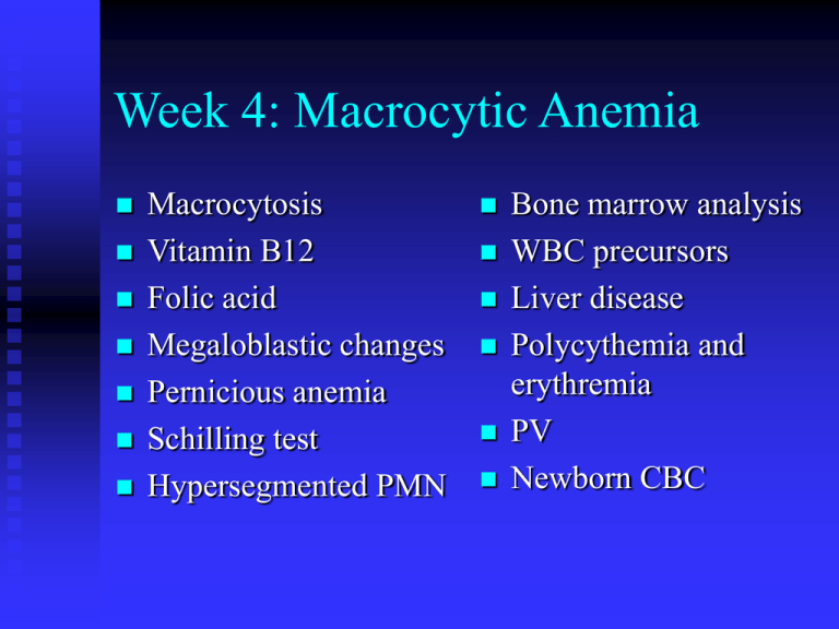
Week 4: Macrocytic Anemia Macrocytosis Vitamin B12 Folic acid Megaloblastic changes Pernicious anemia Schilling test Hypersegmented PMN Bone marrow analysis WBC precursors Liver disease Polycythemia and erythremia PV Newborn CBC Bone Marrow Analysis Aspirate Particles in fluid part of marrow Differential count Biopsy Solid part of the marrow Cellularity Clot section Aspiration Biopsy WBC Precursors Myeloblast Promyelocyte Primary azurophilic granules Meylocyte Specific granules Metamyelocyte Band, stab Segmented neutrophil, PMN Myeloblast Promyelocyte Neutrophilic Myelocyte Segmented neutrophil and lymphocyte Mast cell and histiocyte Mast cell Megakaryocyte Plasma cell Histiocyte Fat cell Macrophage Osteoclasts Osteoblasts Giant cell Non-Megaloblastic Macrocytosis Alcoholism Poor nutrition Cirrhosis Liver disease Reticulocytosis Hemolytic anemia Hemorrhage Megaloblastic Anemia Hemolytic changes Waxy pallor Neurologic changes with B12 deficiency Glossitis, beefy red or smooth pale tongue Causes of Megaloblastic Anemia Folate deficiency Dietary (green leafy vegetables) Malabsorption B12 deficiency Dietary Fish tapeworm (D latum) Pernicious anemia Lack of gastric intrinsic factor (IF) for absorption B12 Metabolism Bind to cobalophilin (cobalamine binding protein) in saliva and gastric secretion Bind to IF (from parietal cell) in the duodenum Cbl-IF complex bind to receptor (cubilin) at distal ileum for mucosal absorption 1% B12 can be absorbed without IF (megadose of B12 for IF deficiency, no need for IV) Autoimmune mechanism for PA Megaloblastic Lab Changes MCV up to 140 fL Howell-Jolly bodies and Cabot rings Hypersegmented PMN Pancytopenia BM with low M:E ratio Megaloblastic morphology (giant bands and metas) B12 and folate assays RBC folate (long term indicator) IF antibody (PA) Schilling test (obsolete) Hypersegmented PMN Megaloblastic anemia Megaloblastic BM Other Apparent Macrocytoses Cold agglutinin Prewarm samples Hyperglycemia Osmotic changes Clot Rouleaux Polycythemia Vera A type of myeloproliferative disorder (MPD) Independent of EPO High total RBC mass (by radio tagging) Secondary Polycythemia Dehydration (decreased plasma volume) Increased O2 demand (e.g., emphysema, high elevation) High EPO (e.g., kidney tumor)
