Digestive System II Lecture 22 24-1
advertisement
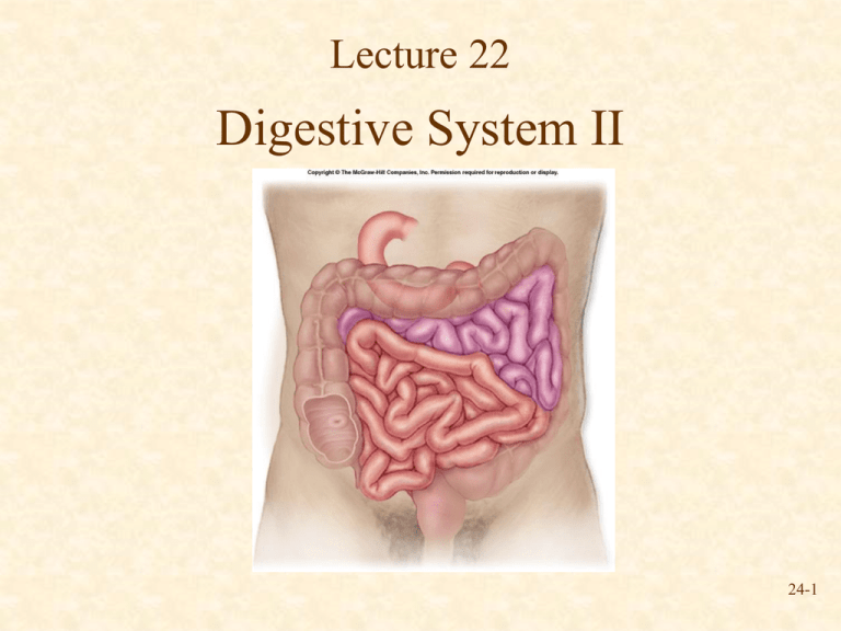
Lecture 22 Digestive System II 24-1 Small Intestine Duodenum Jejunum Ileocecal valve Ileum Fig. 26.14 • Site of greatest amount of digestion and absorption • Divisions – Duodenum (“twelve finger widths”) – Jejunum (“empty”) – Ileum (“twisted”) 24-2 Small Intestine Secretions • Mucus – Protects against digestive enzymes and stomach acids • Digestive enzymes – Disaccharidases: Break down disaccharides to monosaccharides – Peptidases: Hydrolyze peptide bonds – Nucleases: Break down nucleic acids 24-3 Duodenum and Pancreas Duodenum • 25cm in adult • Accessory glands empty secretions into duodenum Fig. 26.20 24-4 Histology of Small Intestine Fig. 26.15 • Circular folds, villi and microvilli increase surface area • Epithelial cells produced by intestinal glands 24-5 Liver Inferior vena cava Posterior Left lobe Right lobe Caudate lobe Left lobe Inferior vena cava Hepatic portal vein Gallbladder (a) Anterior view Right lobe Common hepatic duct Quadrate lobe Cystic duct Gallbladder Anterior (b) Posteroinferior view Fig. 26.18 • 4 Lobes • Ducts – Cystic duct joins with common hepatic duct to form common bile duct – Common bile duct joins pancreatic duct 24-6 Functions of the Liver • Bile production – Salts emulsify fats, neutralizes stomach acid – Stored in gall bladder – Gallstones can form as precipitate of cholesterol • Storage – Glycogen, fat, vitamins, copper and iron • Nutrient interconversion • Detoxification – Removal of ammonia and conversion to urea (eliminated by kidneys in urine) • Phagocytosis – Removal of worn-out and dying red and white blood cells, some bacteria • Synthesis – Blood proteins 24-7 Pancreas • Anatomy – Endocrine • Insulin – Exocrine • Pancreatic juice – – – – Digestive enzymes Released in inactive form Become active in duodenum Digest protein, fats, carbohydrates, and nucleic acids 24-8 Review Question Digestion of proteins is started in the ______________ and completed in the ________________. (a) Oral cavity, stomach (b) Stomach, small intestine (c) Esophagus, small intestine (d) Oral cavity, small intestine (e) Esophagus, stomach 24-9 Large Intestine Transverse colon • Extends from ileocecal valve to anus • Consists of cecum, colon, rectum, anal canal • Movements sluggish (1824 hours) Descending colon Ascending colon Cecum Vermiform appendix Sigmoid colon Ileocecal valve Cecum Vermiform appendix Ileum Rectum Anal canal Fig. 26.16 24-10 Movement in Large Intestine • Mass movements Rectal valve – Common after meals • Defecation reflex Rectum Anal canal – Distension of the rectal wall by feces • Defecation – Usually accompanied by voluntary movements to expel feces through abdominal cavity pressure caused by inspiration (breathing in) and contraction of abdominal wall muscles Veins Internal anal sphincter Anus External anal sphincter Fig. 26.16 24-11 Points to Remember • Stomach – Mixes food – Protein digestion – Limited absorption (aspirin) • Small intestine – Receives secretions of liver and pancreas – Chemical and mechanical digestion – Transports undigested material • Large intestine – Absorb water – Form, store and expel feces 24-12 Questions? 24-13
