Cardiovascular - Heart
advertisement
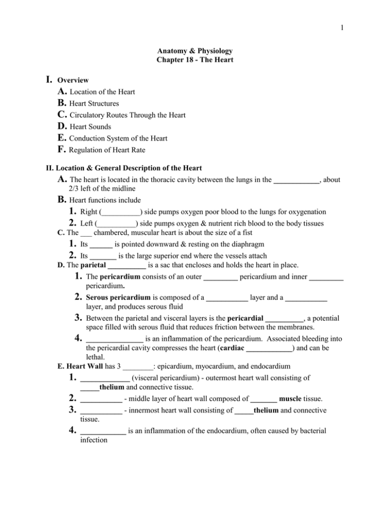
1 Anatomy & Physiology Chapter 18 - The Heart I. Overview A. Location of the Heart B. Heart Structures C. Circulatory Routes Through the Heart D. Heart Sounds E. Conduction System of the Heart F. Regulation of Heart Rate II. Location & General Description of the Heart A. The heart is located in the thoracic cavity between the lungs in the ____________, about 2/3 left of the midline B. Heart functions include 1. Right (__________) side pumps oxygen poor blood to the lungs for oxygenation 2. Left (__________) side pumps oxygen & nutrient rich blood to the body tissues C. The ___ chambered, muscular heart is about the size of a fist 1. 2. Its ______ is pointed downward & resting on the diaphragm Its _______ is the large superior end where the vessels attach D. The parietal __________ is a sac that encloses and holds the heart in place. 1. The pericardium consists of an outer _________ pericardium and inner _________ pericardium. 2. Serous pericardium is composed of a ___________ layer and a ___________ layer, and produces serous fluid 3. Between the parietal and visceral layers is the pericardial __________, a potential space filled with serous fluid that reduces friction between the membranes. 4. _______________ is an inflammation of the pericardium. Associated bleeding into the pericardial cavity compresses the heart (cardiac ____________) and can be lethal. E. Heart Wall has 3 ________: epicardium, myocardium, and endocardium 1. _____________ (visceral pericardium) - outermost heart wall consisting of _____thelium and connective tissue. 2. 3. ___________ - middle layer of heart wall composed of _______ muscle tissue. 4. ____________ is an inflammation of the endocardium, often caused by bacterial infection ___________ - innermost heart wall consisting of _____thelium and connective tissue. 2 III. Heart Chambers & Valves A. Four chambers include 2 upper ______ and 2 lower _________. 1. L. & R. atria have ______ muscle walls a. ___________ muscles make atria surfaces rough b. The __________ is an appendage on surface of the atrium that increases atrial volume. c. An interatrial _______ separates L. & R. atria; the ______ ________ (remnant of fetal foramen ovale) is found in this septum d. Both atria contract simultaneously and empty blood into the ventricles though atrioventricular (___) valves (R.= _________; L. = bicuspid or ________) e. These valves are secured via chordae __________ tendons attached to __________ muscle outgrowths of the myocardium in the ventricles 2. L. & R. ___________ have thicker walls a. Internal surface is wrinkled due to trabeculae ________ (myocardium covered by endocardium) b. c. _______ ventricle wall is thickest, to pump blood throughout body. d. An interventricular ________ separates the ventricles. Both ventricles contract simultaneously, pushing blood through _____________ (SL) valves (L. = _______; R. = ____________) into the aorta & pulmonary artery IV. Circulation of blood through the heart and body A. Cardiac ________ is one complete heart beat, which involves 1. Atrial ________ (atria contract) – blood flows into ventricles 2. Atrial _________ (atria relax) 3. Ventricular _________ (ventricles contract) – blood is pushed into the aorta and pulmonary artery; AV valves snap shut 4. Ventricular __________ (ventricles relax) – SL valves close B. ____________ Circulation progresses in the following sequence: 1. Oxygenated blood from lungs returns to heart via __________ veins. 2. Pulmonary veins feed blood into the left ________. 3. Contraction of L. atrium pushes blood through the bicuspid valve into the L. 4. ___________ Contraction of L. ventricle pushes blood through the aortic semilunar valve into the ________ (largest artery in the body), which then feeds blood to: 5. Arteries arterioles ____________, from which O2 is delivered to body tissues and CO2 is picked up. 6. Deoxygenated blood flows from capillaries venules veins and returns to heart via the superior & inferior ______ ________ (largest veins in body). 3 C. ______________ Circulation 1. Deoxygenated blood, high in CO2, is delivered to the R. ________ (via superior & inferior vena cava). 2. Contraction of R. atrium pushes blood through the tricuspid valve into R. ____________. 3. Contraction of R. ventricle pushes blood through the pulmonary semilunar valve into the ___________ artery, which has L. & R. branches that take blood to lungs. 4. In _______, blood flows through arteries arterioles capillaries where blood releases CO2 and picks up O2. ______________ blood then returns to heart via venules pulmonary veins. D. _________ Circulation - blood supply to & from the heart tissue 5. 1. Coronary (atrioventricular) _________ - external groove between atria and ventricles, contains coronary blood vessels 2. Coronary __________ emerge from the base of the _______ and deliver oxygenated blood to heart tissues. a. ______ coronary artery (in coronary sulcus) branches into 1) Left anterior _______________ (widow maker) artery passes down the anterior interventricular sulcus and delivers blood to left ____________ tissues b. ________ coronary artery (in rt. coronary sulcus) 3. Coronary ________ take deoxygenated blood away from heart tissues. a. 4. Coronary ________ - largest vein on posterior surface of heart; empties into R. _________ ____________ of coronary arteries can occur a. Cholesterol ________ can build up on the inside of the artery wall, narrowing the passageway b. ___________ – stationary blood clot in a blood vessel c. ___________ – blood clot that breaks off and moves d. Result of blockage is ___________, a decrease in blood to the tissues, which leads to e. __________ pectoris - pain or heaviness in the chest, often accompanied by radiating pain or numbness in the jaw &/or left arm f. Myocardial __________ – a heart attack in which an area of the heart tissue dies from lack of oxygen g. Coronary _________ surgery attaches blood vessels around the blocked area 4 V. Heart Sounds A. “Lub-dup” heart sounds are caused by the opening and closing of heart ________ 1. The “____” is caused by the closing of ____ valves, when the ventricles contract at 2. systole The “____” is caused by the closing of the ___________ valves, when the ventricles relax at the beginning of diastole B. Body areas at which the valves may be listened to are called valvular _____________ areas, and include: 1. 2. 3. __________ area - 2nd intercostal space right of sternum _____________ area - left, 2nd intercostal space left of sternum Tricuspid & __________ areas are both near the 5th intercostal space, with the bicuspid more lateral C. Heart __________ are caused by valve leakage or restriction and the turbulence of the blood as it passes through the heart. Four types of murmurs are: 1. Valvular _____________ - heart valves do not form a tight seal; causes a blowing sound after the valve closes 2. ___________ - walls surrounding a valve are roughened or constricted; stiffened valves don’t open properly; produces a “click” sound during ventricular systole 3. Mitral valve ___________ - inherited weakness of valve collagen & chordae tendinae that allows blood backflow through valve into atrium during systole; detected by a click followed by a swish 4. ________ murmur - caused in children by the blood turbulence during heavy exercise; not serious VI. ______________ System of the Heart - consists of interconnected strands of specialized cardiac muscle/nerve tissue that conducts electrical impulses through the heart, coordinating contractions. Components include: A. Sinoatrial (___) node - the “pacemaker;” sets HR at _______ bpm 1) Found in R. _____ posterior wall, inferior to superior vena cava 2) Initiates the cardiac cycle by producing an __________ impulse that spreads over 3) 4) both atria, causing contraction Atria _________ an instant after receiving the impulse from the SA node (ventricles are relaxed = diastole) The impulse then passes along the _____________ pathway to B. Atrioventricular (___) node - in the lower interatrial septum between the atrium and ventricle. (Can set heart rhythm at __-__ bpm if SA node is damaged). The AV node passes the impulse to C. Atrioventricular bundle septum; passes impulse to (bundle of ____) - extends from AV node into interventricular 5 D. L. & R. __________ branches - extensions of AV bundle on L. & R. sides of the septum; passes impulse to E. __________ fibers - extend from the branches into the ventricular walls; Impulses through C, D, & E cause ventricular contraction (systole) F. An electrocardiogram (_____) records the electrical impulses through the heart as waves: 1. 2. 3. 4. ___ = SA node depolarization atrial contraction 5. Abnormal waves indicate heart __________ ____ = ventricular depolarization ventricular contraction __ = ventricular repolarization ventricular relaxation Atrial repolarization occurs during ventricular depolarization, thus its small wave is obscured by the ____ complex G. ______________ are abnormal heart patterns, which include: 1. ______cardia – faster than normal heart rate (100 bpm or more) 2. ______cardia – slower than normal heart rate (60 bpm or less) 3. __________ – rapid heart rate (250-350 bpm), with controlled contractions 4. Heart ______ – blocked impulse transmission, usually from AV node to ventricles, interferes with ventricular contraction; requires an artificial pacemaker to bypass the blockage 5. Ventricular __________ - ventricles contract rapidly in a weak, uncoordinated manner; caused by a damaged conduction system; can result in death if not corrected quickly 6. _______________ – an electrical shock to the heart, which depolarizes it and allows the SA node to reestablish its rhythm VII. Regulation of Heart Rate A. ____ Node (pacemaker) normally controls heart rate, but when blood volume drops or the heart is weakened, other systems intervene B. Medulla oblongata _______ center can decrease or increase HR 1. _____sympathetic nerves (i.e., _______ nerves) slow heart rate a. Branches extend to ____ and ____ nodes b. Secretes acetylcholine (____) neurotransmitter c. Decreases SA and AV node activity, _________ the heart 2. _____________ nerves increase heart rate a. Branches extend to ____ and ____ nodes b. Secretes ____________ c. Increases SA and AV node activity, ___________ the heart C. ____________ Gland Medulla (endocrine) increases HR 1. ___________ NS stimulates the adrenal medulla to secrete epinephrine (______________) into the blood 2. Epinephrine increases ___ and ____ node activity 6 3. Heart rate ____________ D. ___________ – pressure/stretch receptors found in the carotid and aortic arteries 1. Sensitive to changes in blood ___________ (BP) 2. When BP increases, baroreceptors signal the Cardiac Center in the medulla, which sends out a parasympathetic response, __________ heart rate 3. When BP decreases, baroreceptors again signal the Cardiac Center, which sends out a sympathetic response, _________ HR E. Cerebrum and ___________ can also send out impulses to increase of decrease HR F. _____________ changes can alter heart rate 1. Increased body temp. __________ heart rate 2. Decreased body temp. (as in hypothermia) ___________ HR G. Potassium (___) and Calcium (____) levels influence HR 1. Excess K+ (___________) interferes with depolarization, which can lead to cardiac arrest 2. Low K+ (___________) causes the heart to beat weakly and arrhythmically 3. Reduced Ca2+ levels (___________) depress the heart 4. High Ca2+ levels (____________) can lead to spastic heart contractions VIII. Cardiac ______ - the volume of blood ejected from the ventricles each minute. A. Cardiac output (___) is the product of heart rate (___) and stroke volume (___): [CO = HR x SV] B. ________ volume is the volume of blood ejected from the ventricles with each contraction (usually about 70 ml) C. Heart ______ is the number of beats per minute (about 75/min) D. So an ___________ cardiac output is: 75/min x 70 ml = 5250 ml/min. = ______ L/min E. Total blood volume in the body is ______ L, so all of our blood circulates through the heart every minute! F. Cardiac output is increased or decreased by increasing or decreasing _______ volume and/or heart _______.
