File
advertisement
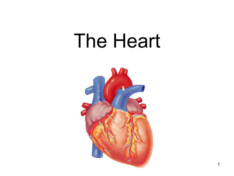
The Heart 1 The heart=a muscular double pump with 2 functions Overview The right side receives oxygen-poor blood from the body and tissues and then pumps it to the lungs to pick up oxygen and dispel carbon dioxide Its left side receives oxygenated blood returning from the lungs and pumps this blood throughout the body to supply oxygen and nutrients to the body tissues 2 Simplified… Four chambers Two atria, two ventricles Double pump – the ventricles Two circulations Systemic circuit: blood vessels that transport blood to and from all the body tissues Pulmonary circuit: blood vessels that carry blood to and from the lungs 3 Chest x rays Normal female Lateral (male) 4 Starting from the outside… Pericardium (see next slide) Without most of pericardial layers 5 Chambers of the heart divided by septae: Two atria-divided by interatrial septum Right atrium Left atrium Two ventriclesdivided by interventricular septum Right ventricle Left ventricle 6 Valves three tricuspid one bicuspid (cusp means flap) “Tricuspid” valve RA to RV Pulmonary or pulmonic valve RV to pulmonary trunk (branches R and L) Mitral valve (the bicuspid one) LA to LV Aortic valve LV to aorta 7 Function of AV valves 8 Function of semilunar valves (Aortic and pulmonic valves) 9 more on valves 10 11 Simplified flow: print and fill in details 12 Heartbeat Definition: a single sequence of atrial contraction followed by ventricular contraction See http://www.geocities.com/Athens/Forum/6100/1heart.html Systole: contraction Diastole: filling Normal rate: 60-100 Slow: bradycardia Fast: tachycardia ***Note: blood goes to RA, then RV, then lungs, then LA, then LV, then body; but the fact that a given drop of blood passes through the heart chambers sequentially does not mean that the four chambers contract in that order; the 2 atria always contract together, followed by the 13 simultaneous contraction of the 2 ventricles Conducting System of Heart Conduction System of the Heart SA node: sinoatrial node. The pacemaker. Specialized cardiac muscle cells. Generate spontaneous action potentials (autorhythmic tissue). Action potentials pass to atrial muscle cells and to the AV node AV node: atrioventricular node. Action potentials conducted more slowly here than in any other part of system. Ensures ventricles receive signal to contract after atria have contracted AV bundle: passes through hole in cardiac skeleton to reach interventricular septum Right and left bundle branches: extend beneath endocardium to apices of right and left ventricles Purkinje fibers: Large diameter cardiac muscle cells with few myofibrils. Many gap junctions. Conduct action potential to ventricular muscle cells (myocardium) Impulse Conduction through the Heart Artificial Pacemaker 17 Another flow chart 18 Use to study 19
