B New Developments in Display Quality Control Ehsan Samei, PhD
advertisement

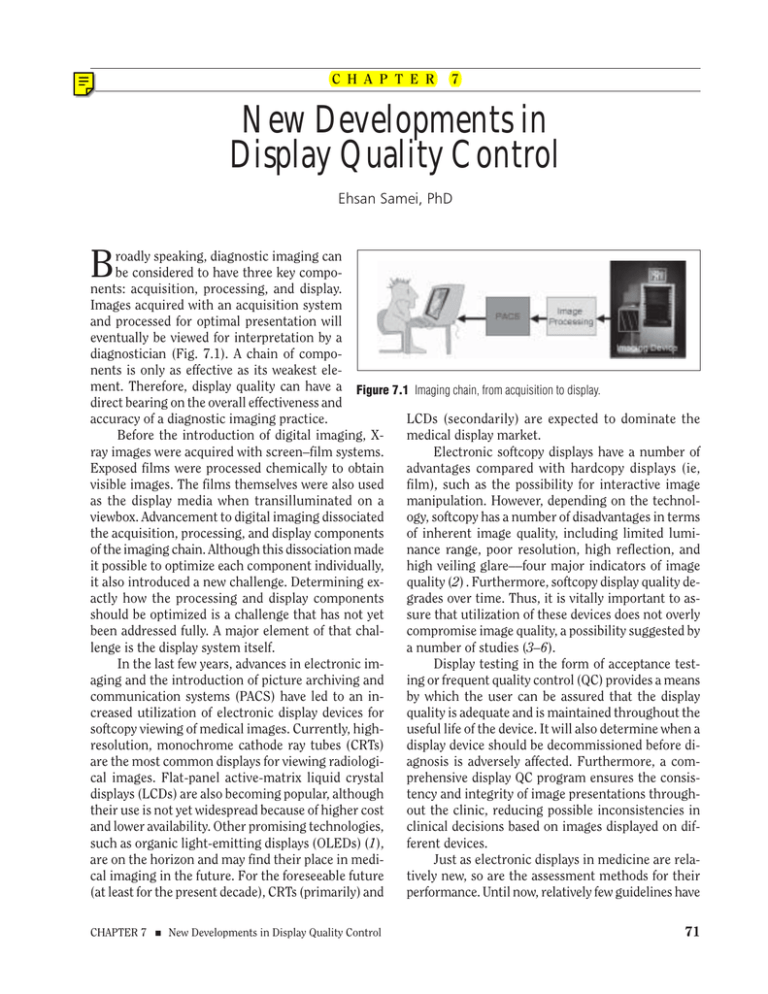
C H A P T E R 7 New Developments in Display Quality Control Ehsan Samei, PhD B roadly speaking, diagnostic imaging can be considered to have three key components: acquisition, processing, and display. Images acquired with an acquisition system and processed for optimal presentation will eventually be viewed for interpretation by a diagnostician (Fig. 7.1). A chain of components is only as effective as its weakest element. Therefore, display quality can have a Figure 7.1 Imaging chain, from acquisition to display. direct bearing on the overall effectiveness and LCDs (secondarily) are expected to dominate the accuracy of a diagnostic imaging practice. medical display market. Before the introduction of digital imaging, XElectronic softcopy displays have a number of ray images were acquired with screen–film systems. advantages compared with hardcopy displays (ie, Exposed films were processed chemically to obtain film), such as the possibility for interactive image visible images. The films themselves were also used manipulation. However, depending on the technolas the display media when transilluminated on a ogy, softcopy has a number of disadvantages in terms viewbox. Advancement to digital imaging dissociated of inherent image quality, including limited lumithe acquisition, processing, and display components nance range, poor resolution, high reflection, and of the imaging chain. Although this dissociation made high veiling glare––four major indicators of image it possible to optimize each component individually, quality (2) . Furthermore, softcopy display quality deit also introduced a new challenge. Determining exgrades over time. Thus, it is vitally important to asactly how the processing and display components sure that utilization of these devices does not overly should be optimized is a challenge that has not yet compromise image quality, a possibility suggested by been addressed fully. A major element of that chala number of studies (3–6). lenge is the display system itself. Display testing in the form of acceptance testIn the last few years, advances in electronic iming or frequent quality control (QC) provides a means aging and the introduction of picture archiving and by which the user can be assured that the display communication systems (PACS) have led to an inquality is adequate and is maintained throughout the creased utilization of electronic display devices for useful life of the device. It will also determine when a softcopy viewing of medical images. Currently, highdisplay device should be decommissioned before diresolution, monochrome cathode ray tubes (CRTs) agnosis is adversely affected. Furthermore, a comare the most common displays for viewing radiologiprehensive display QC program ensures the consiscal images. Flat-panel active-matrix liquid crystal tency and integrity of image presentations throughdisplays (LCDs) are also becoming popular, although out the clinic, reducing possible inconsistencies in their use is not yet widespread because of higher cost clinical decisions based on images displayed on difand lower availability. Other promising technologies, ferent devices. such as organic light-emitting displays (OLEDs) (1), Just as electronic displays in medicine are relaare on the horizon and may find their place in meditively new, so are the assessment methods for their cal imaging in the future. For the foreseeable future performance. Until now, relatively few guidelines have (at least for the present decade), CRTs (primarily) and CHAPTER 7 ■ New Developments in Display Quality Control 71 been available for performing display quality testing in a clinical setting. Previous studies have focused on fundamental design aspects of the display technology (7–9) or have attempted to address QC aspects of electronic displays (10–15). A number of efforts have been launched to standardize display measurements (16). Among the most recent developments are acceptance testing guidelines by the Deutsches Institut für Normung (17), the definition of advanced testing procedures for flat-panel displays by the Video Electronics Standards Association (18), and the recommendation of a standardized display luminance response by the Digital Imaging and Communications in Medicine (DICOM) Grayscale Display Standard Function (GDSF) working group (19). However, until now, the recommended methods have been either too complicated or too subjective, have not included assessment of some critical medical display characteristics, and have not been endorsed by the medical community. The American Association of Physicists in Medicine (AAPM) recently formed a Task Group (TG18) to address acceptance testing and QC of medical electronic display devices. The purpose of TG18 was to provide standard guidelines for performing display quality evaluations and to facilitate inter- and intrainstitutional comparisons. The TG18 effort has led to the generation of a detailed document for testing of display devices and a set of newly designed display test patterns (20). Based on the current standard of practice, recommendations have been made for acceptable ranges of display performance with limited impact on diagnostic efficacy. The document is expected to be finalized in 2002. Based on the tentative recommendations of TG18, this chapter outlines some methods and procedures that can be utilized to evaluate medical display devices. RESPONSIBILITY For current commercial medical devices, manufacturers often provide device-specific testing methods and tools. They also often perform acceptance testing of a device upon installation. Although these evaluations and utilities can be effective in assuring system performance and can ease the burden on the user, it is important that the user independently verify the acceptability of the system performance at installation and frequently throughout the life of the system. This task falls within the professional responsibilities of medical physicists (21). Monthly or quarterly routine QC procedures may be performed by a 72 trained QC technician under the direct supervision of a medical physicist. The more extensive tests involved in acceptance testing and annual QC, however, are better performed by the physicist. The daily QC procedure should be performed by the actual user of the system. LEVELS OF DISPLAY EVALUATION Display testing is undertaken at multiple levels, depending on whether the purpose is acceptance testing or QC. In acceptance testing, the goal is to evaluate the performance and functionality of the system before its clinical utilization to assure that the system meets previously delineated performance requirements. In QC, the main goal is to ensure that the performance of the system has not degraded beyond the acceptable ranges and is still closely comparable to that established at acceptance testing. For testing any given display quality characteristic, three types of tests may be used: visual, quantitative, and advanced. The visual and quantitative tests are used in acceptance testing and QC procedures, whereas the advanced tests are oriented toward researchers in laboratory settings. In evaluating the performance of a display device, it is also important to take into account the intended purpose of the device. In clinical practice today, display devices are often used in two capacities: primary (so-called “diagnostic”) and secondary (socalled “clinical”). Primary displays are used for the primary diagnostic interpretation of medical images. Secondary displays are used for clinical review of images for which diagnostic interpretations have already been rendered. The performance criteria are usually less stringent for secondary class display devices. Acceptance Testing New display devices should be evaluated using standard recommended acceptance testing procedures. These tests usually are more extensive and quantitative than QC evaluations and may take 1–2 hours to complete. Before starting the tests, a number of preparatory steps should be undertaken: 1. The display monitors should be placed in a way that prevents visible specular reflection of light sources (lights, other monitors, viewboxes, windows, etc.) on the monitor from a normal viewing direction. 2. The normal boot-up procedure for the system should be executed. The device should be warmed up for SAMEI ties for conformance with the DICOM GSDF, the calibration procedure should be executed. After the completion of the prerequisite steps, a number of display-specific characteristics should be evaluated in sequential order. Figure 7.2 The TG18-QC test pattern. The pattern contains: (1) 16 luminance patches at the corners of each of which are four lowcontrast targets for evaluating luminance and low-contrast response; (2) two continuous grayscale ramp bars for evaluating display bit-depth; (3) Cx and line–pair targets at the center and four corners and a scoring Cx reference for evaluating display resolution; (4) white–black bars for evaluating video artifacts; and (5) 5% and 95% embedded patches and low-contrast “QUALITY CONTROL” letters with variable contrast within different luminance backgrounds, for evaluating the grayscale rendition/luminance response of the display device. about 30 minutes before starting the measurements, and, if needed, the faceplate of the display device should be cleaned using nonabrasive material. The general functionality of the system should be verified by inspecting the appearance of TG18QC, a standard test pattern (Fig. 7.2). In particular, all 16 luminance steps of this pattern should be distinctly resolvable and the continuous grayscale ramp bars should be without any contouring artifacts that indicate inadequate bit-depth. (Bit-depth resolution can be more fully evaluated using the TG18-MP test pattern, not shown.) The alignment and centeredness of the active display area also should be verified and adjusted if necessary. 3. The level of ambient lighting within the room and the minimum luminance value of the display should be measured and adjusted based on the reflection characteristics of the monitor and the desired luminance ratio (Lmax/Lmin) as described later in this chapter. 4. If the device is equipped with self-calibration utiliCHAPTER 7 ■ New Developments in Display Quality Control Geometrical Distortion Potential geometrical distortions of images are of concern in CRT display devices. The distortions can be in concave, convex, skewed, or other nonlinear forms. Such distortions can be evaluated by judging the straightness of lines in the TG18-QC test pattern (Fig. 7.2). Directional measurements of the line lengths are made using a flexible ruler. The difference between expected and measured lengths in different directions, within each quadrant of the pattern, between the quadrants, and within the whole pattern should not exceed 2% for primary class displays or 5% for secondary class displays. If the device does not meet these criteria, the active display area should be adjusted via monitor controls before proceeding to the rest of the tests. Reflection Electronic display devices have specular and diffuse reflections that can reduce image contrast and affect image quality (Fig. 7.3). Ambient light reflections are more pronounced in display devices with thick faceplates (eg, CRTs) than in those with thinner faceplates (eg, LCDs). They usually are reduced by the application of antireflective (AR) coating on the faceplate and/or the addition of light absorbers within the faceplate of the display. But they cannot be completely eliminated. The magnitude of display reflection can be characterized by specular and diffuse reflection coefficients. For a given display device, these coefficients can be used to establish the maximum allowable level of ambient lighting at which the device can be operated without overly compromising the desired luminance ratio and contrast threshold. That level is devised so that in the darkest areas of an image the specular reflection is less than the contrast threshold and so that the contrast reduction resulting from diffuse reflection is <20%. Both reflection coefficients are measured with the monitor turned off (or with a black screensaver activated) and with no or minimal ambient lighting. For the specular coefficient, the faceplate is illuminated from a 75-cm distance with a small source of diffuse white light at 15º from the display normal (Fig. 7.4A). The luminance of the reflection of the light 73 these equations, for a typical CRT with AR coating (Rs = 0.004, Rd = 0.02 nit/lux) operated at minimum luminance values of 0.5, 1, 1.5, and 20 nit, the ambient lighting should be <~7, 12, 19, and 25 lux, respectively. Displays without AR coating have notably more restrictive ambient lighting criteria. Increasing the minimum luminance would allow a higher ambient lighting level but would necessitate increasing Lmax to maintain the desired luminance ratio (Lmax/ Lmin), an important display quality parameter. Figure 7.3 The effect of ambient lighting on image contrast. The overall reduction in contrast is the result of diffuse reflection, whereas the “structured” reflection of identifiable objects is an example of specular reflection. source is measured with a telescopic photometer from a distance of 75 cm. The luminance of the light source is also measured directly from a distance of 1.5 m. The ratio of the former over the latter is the dimensionless specular reflection coefficient, Rs. For the diffuse coefficient, the monitor is illuminated with a standard illuminator device (Fig. 7.4B), as described in the TG18 document (20), providing a uniform illumination of the display faceplate. The ratio of the measured display luminance to the illuminance falling on the faceplate is the diffuse reflection coefficient, Rd, in units of nit/lux (22). (The nit is the Standard International [SI] unit of luminance equal to 1 lumen/steradian-m2, or candela/m2, and 0.292 footLambert. Lux is the SI unit of illuminance equal to 1 lumen/m2. Lumen is the unit of luminous flux and is equal to 0.001464 W at 555 nm wavelength) (22). Depending on the specular or diffuse reflections the maximum allowable levels of ambient lighting in units of lux, Is and Id, are calculated as: Is = RCt-min Lmin/(0.9Rs), and Id = 0.25 Lmin/Rd, where Lmin is the minimum luminance and Ct-min is the contrast threshold of the human visual system at Lmin, a value that ranges between 0.032 and 0.018 for typical Lmin values between 0.5 and 1.5 nit. Based on 74 Luminance Response The human visual system perceives brightness in a nonlinear fashion (23). Ideally, the luminance response of a display device should match this nonlinear response, so that image values are displayed in equally perceptible luminance increments. The DICOM GDSF (19) offers a way to approach this goal, in a limited sense, by applying a specific look-up table to the display values, so that the display values present equally discriminable levels of brightness. The limitation of this approach comes from the fact that the perceptual linearization achieved by GDSF only applies to scenes without much luminance variation (23). The contrast sensitivity of the human eye can be different in scenes in which a wide range of luminance levels is present (eg, medical images) (2,20). Figure 7.4 Measuring the specular (a) and diffuse (b) reflection coefficients. The illuminator in the latter is made of continuous light-reflecting material, except for a small hole on the back, from which the luminance on the faceplate is measured using a telescopic luminance meter. SAMEI Figure 7.5 Three examples of the TG18-LN test patterns representing the patterns for the maximum, the minimum, and midvalue luminance measurements. In spite of this limitation, the GDSF approach can be effective in achieving reasonably consistent and comparable image presentations on different display devices if it is used with caution. The intrinsic luminance response (ie, luminance versus display value) of most display devices is markedly different from the GDSF. It usually follows a power-law relationship for CRTs and a linear relationship for LCDs (7,24). In addition, the response may vary over time. In CRTs, for example, the phosphor efficiency decreases as the device ages. High-quality display devices have circuitries to compensate for the loss of CRT phosphor efficiency and to maintain the luminance characteristics of the display devices over time. Modern display devices also have utilities that automatically calibrate the luminance response of the device to GDSF. However, the functionality and accuracy of these utilities should be independently verified by the user. In addition to adherence to a standard luminance response function, the absolute luminance values of a display device should be such that the luminance ratio of the device does not limit the contrast threshold of the human visual system. In an image with a wide range of luminance values, the desired ratio should be >250. To achieve this ratio, however, it is important to have an Lmin that is not so low that the dark regions of the image are affected by ambient light reflection (as discussed previously) and to have an Lmax, in the case of CRTs, that is not so high as to compromise display resolution. In order to characterize the luminance response of a display device, the luminance of the display is measured at 16–18 points ranging between the minimum and maximum display values using the TG18LN display patterns (Fig. 7.5). Measurements are made with no or minimal ambient lighting. If a telescopic luminance meter is used, it might be necessary to use a cone or baffle to shield the probe from CHAPTER 7 ■ New Developments in Display Quality Control the surround luminance of the pattern (Fig. 7.6), particularly for low-luminance measurements. After the measurements, the monitor is turned off (or a black screensaver is activated), the ambient lighting is turned on to the level determined from the reflection considerations discussed above, and the luminance on the faceplate is measured, Lamb. With Lamb added to the other measured luminance values, the DICOM’s just-noticeable-difference (JND) indices (19) of measured Lmin and Lmax values are identified, and the display values of the patterns are subsequently mapped to the JND indices. This allows a direct comparison of the measured luminance response with the GDSF. The response should be such that luminance contrast (ie, the slope of the response calculated as ∆L/Lavg at JNDavg) does not deviate more than 10% from the GDSF (Fig. 7.7). In addition, for primary class displays, the maximum luminance should be >170 nit and the luminance ratio should be >250. For secondary class displays, the luminance ratio should be >100. If a display workstation has more than one monitor, the maximum luminance of the monitors should not differ by more than 5%. Luminance Uniformity Luminance nonuniformity across the active display area of a monitor is common, particularly in CRTs, in which the luminance at the center is intrinsically higher than at the corners and edges, primarily as a result of variations in light transmission through the faceplate glass. Compensation strategies correct for such nonuniformities by using a variable gain amplifier, applying a digital look-up table, or Figure 7.6 The use of a cone for luminance or veiling glare measurements. The cone has a light-absorbing tip and a lightabsorbing interior surface to minimize internal reflections and the contribution of surround lights to the measured luminance at the center. A similar cone may also be used for blocking the surround brightness for visual evaluation of veiling glare. 75 Figure 7.9 The effect of display blur on image resolution. The images are captured from a display device (a) with acceptable resolution characteristics and from another device (b) with excessive blur. The displays had similar matrix sizes. Figure 7.7 The Digital Imaging and Communications in Medicine grayscale display standard function (GDSF) and the GDSF-based standard contrast function for evaluating the luminance response of display devices. increasing the beam current at the edges of the display area. However, such compensations are often applied with moderation to preserve display resolution characteristics at the edges. The level of luminance nonuniformity in a display can be ascertained by measuring the luminance at the center and four corners of the display area using two uniform test patterns at two luminance levels: 10% and 80% of maximum display value (TG18UNL patterns) (Fig. 7.8). If a telescopic luminance meter is used, it may be necessary to use a cone or baffle to minimize the influence of the probe’s flare on the measurements (Fig. 7.6). The difference between any two measurements should not exceed 30% of their average. Figure 7.8 The TG18-UNL80 test pattern for luminanceuniformity and color-uniformity evaluations. 76 Display Resolution In display terminology, resolution often refers to the nominal (or maximum) resolving power of a display device described by its number of display pixels (eg, 1k or 2k, 3 or 5 megapixel). However, from an image science standpoint, resolution is the actual ability of a display device to present the spatially detailed elements of an image (Fig. 7.9). This ability is related to both the number of pixels and the actual spatial extent of each pixel. Because of various optical and electronic processes, a display pixel can have a breadth that is larger than its nominal value, degrading the display resolution from its ideal level. In CRTs, the pixels have a near-Gaussian profile. Resolution is sometimes expressed in terms of the resolution-addressability ratio (RAR), which is the ratio of the full-width-at-half-maximum (FWHM) of a pixel over its nominal size at maximum luminance. An RAR of 0.9–1.1 is believed to be optimal for medical imaging (9). But even RARs within this range represent notable display blur, and the acceptability of this range has not yet been confirmed by objective observer experiments. Furthermore, in CRTs, the resolution is nonisotropic and varies as a function of luminance and position on the faceplate. CRT resolution also varies as a display monitor ages, because of the deterioration of the electron optics and the systematic increase in the beam current to compensate for the reduction in phosphor efficiency. The most objective method to measure display resolution is to capture a magnified image of a singlepixel-wide, low-contrast line displayed by the monitor using a high-quality digital camera. The image is processed to deduce the line–spread function (LSF) and the modulation transfer function (MTF) of the display in the direction perpendicular to the line (Fig. 7.10) (25). A simpler method to quantify resolution is to evaluate the appearance of Cx test targets using SAMEI a method proposed by Kohm et al. (26) . The TG18QC or TG18-CX test patterns (Figs. 7.11 and 7.12) are displayed with one pattern pixel represented by one display pixel. The Cx targets at various locations in the pattern are evaluated visually using a magnifier, and their appearance is scored in comparison to the digitally magnified reference scale that is also embedded in those two patterns. Based on the current tentative criteria of TG18, the scores should be <5 (judged sharper than the 5th sharpest reference) for primary class displays, and <7 for secondary class displays. These criteria correspond to RARs ≤1.15 and 1.47, respectively (26). Figure 7.10 The modulation transfer function of a CRT display device evaluated using a test pattern containing single-pixel-wide low-contrast lines and a digital camera. The measurements show the variation in resolution response as a function of direction and closely correlate with full-width-at-half-maximum (FWHM) measurements in the two directions (25). Figure 7.11 The TG18-QC test pattern with one of the Cx targets and one of the scoring references magnified. The magnified views of the Cx targets are scored against the reference as a visual evaluation of display resolution. Figure 7.12 The TG18-CX test pattern for the evaluation of display resolution and resolution uniformity. CHAPTER 7 ■ New Developments in Display Quality Control Display Noise Display noise refers to statistical fluctuations in the image that vary either spatially (so-called spatial noise) or in time (so-called temporal noise). Temporal noise, which is usually dominant in the dark regions of displayed images, is difficult to characterize outside of a laboratory setting and has less well-understood perceptual influence. Spatial noise is dominant in the brighter areas of displayed images. In CRTs, phosphor granularity is the main contributor to spatial noise. In current monochrome CRT devices, two types of phosphors are primarily used, singlecomponent phosphors (eg, P45) and blended phosphors (eg, P104 and P4). The blended phosphors have a notably higher level of spatial noise. They are more efficient in light production. However, their efficiency also decreases more rapidly as the monitor ages. The most objective method to measure display noise is to capture a magnified image of a displayed uniform image using a scientific-grade digital camera. The image is processed to deduce the noise power spectrum of the display (20). This method, however, is complex and might be difficult to implement clinically. Alternatively, spatial noise can be evaluated by assessing the visibility of low-contrast objects in a midluminance background using the TG18-AFC test pattern (Fig. 7.13), based on a general method previously established by Hangiandreou et al. (27). The pattern consists of four quadrants. Each quadrant consists of multiple squares at the corner of each of which is a small square target of a specific size and contrast. Four size–contrast combinations are used for these targets. For primary display devices, all targets except the smallest ones should be visible. For secondary class displays, the two largest target sizes should be visible. It should be pointed out that if a display fails this test, the failure might be the result of inadequate bit-depth. That is why the adequacy of 77 identical to the GQ and GQN patterns, except for the presence of five low-contrast targets at the center of the central dark circle. Using a cone or funnel that prevents the surround light from reaching the eye (Fig. 7.6), the observer discerns the visibility of these targets in the two patterns. In both patterns, at least three or four targets should be visible for primary or secondary class displays, respectively. Figure 7.13 The TG18-AFC test pattern. The contrast of the pattern is enhanced to illustrate the elements of the pattern. Based on a four-alternative forced-choice methodology, the visibility of small low-contrast targets is used for visual evaluation of display noise. display bit-depth should be verified beforehand as one of the preparatory steps mentioned previously. Veiling Glare Veiling glare in a display device is a light-spreading phenomenon that leads to degradation of image contrast. Veiling glare is caused by internal light-scattering processes in the device’s faceplate, light leakage, or electron backscattering (on CRTs). It is more pronounced in display devices with thick faceplates (eg, CRTs) and in CRTs with substantial electron backscattering (eg, color CRTs). Veiling glare in a CRT can be reduced by applying AR coatings on the faceplate, and/or by adding light absorbers in the glass of the faceplate. Veiling glare in a display device can be quantified in terms of glare ratio (GR) using a sequence of three test patterns (TG18-GQ, TG18-GQB, and TG18GQN) (Fig. 7.14) and a telescopic luminance meter. The luminance meter should be collimated with the aid of a funnel or cone to “view” only the central 5mm diameter of the patterns (Fig. 7.6). First, using the TG18-GQ test pattern, the luminance (L) at the center of a 1-cm central dark circle surrounded with a 20-cm diameter bright region is measured. Next, the dark circle is removed (TG18-GQB pattern) and the measurement is repeated (Lb). Finally, the luminance is similarly measured in a continuously black pattern (TG18-GQN) (Ln). GR is calculated as (Lb – Ln)/(L – Ln). GR should be >400 and >50 for primary and secondary class display devices, respectively. Veiling glare also may be evaluated visually by discerning the visibility of low-contrast objects in the TG18-GV and TG18-GVN test patterns, which are 78 Color Uniformity In display devices, color uniformity refers to the intrinsic magnitude and uniformity of color tint of the device when displaying a white monochrome image. In monochrome CRTs, color tint is dictated by the phosphor type and can vary slightly from monitor to monitor. Color tint is usually considered a preference issue. However, it can be a cause of distraction, especially in multiple-monitor workstations where the color tints mismatch. Quantitatively, the color coordinates of each display, defined by the Commission Internationale de l’Eclairage (u’,v’) (28), are measured at the center and corners of the display area using a colorimeter and a uniform test pattern at 80% maximum display value (TG18-UNL80, Fig. 7.8). Color uniformity is calculated as the maximum distance in the color space (ie, ∆[u’,v’]) within and between monitors. For primary class displays, that distance should be <0.004. No requirements are specified for secondary class displays. Other Tests In addition to the tests outlined above, a display device may be evaluated in terms of a number of other miscellaneous, technology-specific characteristics. For CRTs, those include display artifacts (such as ghosting, shadowing, moiré, and yoke rings), color purity in color CRTs, physical defects, DAC artifacts, and flicker. For flat-panel displays, these characteristics include angular dependencies, electronic crosstalk, and pixel defects. The details of these miscellaneous tests are described in the TG18 document (20). Figure 7.14 The TG18-GQ, GQB, and GQN test patterns for quantitative evaluation of veiling glare via glare ratio. SAMEI Quality Control Display devices that are in clinical use should be evaluated on a regular basis to assure that they maintain a minimum performance level. Depending on the frequency of QC inspections, different levels of testing in terms of depth and thoroughness are recommended. Daily QC It is recommended that a QC procedure be performed on a daily basis by the actual user of the system. This procedure is based on a visual evaluation of the TG18-QC test pattern (Fig. 7.2) and should take <1 minute to complete. The following is a list of characteristics that should be quickly inspected: 1. Geometrical distortion: Verify that the borders and lines of the pattern are visible and straight, and that the pattern is centered within the active area of the display device. 2. General image quality and artifacts: Evaluate the overall appearance of the pattern. Note any nonuniformities or artifacts, especially at blackto-white and white-to-black transitions. Verify that the ramp areas appear continuous without any contouring artifacts. 3. Luminance, reflection, and noise: Verify that all 16 luminance patches are distinctly visible. Verify that the 5% and 95% patches are visible. Evaluate the appearance of low-contrast letters and confirm the visibility of targets at the corners of all luminance patches. 4. Resolution: Evaluate the appearance of Cx targets at the center and the corners of the pattern and score them compared with the reference. The scores should be <5 for primary displays and <7 for secondary ones. Also, verify the visibility of the line–pair patterns at Nyquist frequency at the center and corners of the pattern. Any noncompliance should be brought to the attention of the medical physicist in charge of display QC. Such instances should be investigated by more in-depth evaluations and followed up by readjustment, service, repair, or replacement of the device. Monthly/Quarterly QC Monthly or quarterly inspections of a display system are more involved than the daily QC. But they still rely more on testing procedures that are less time consuming than acceptance testing. The procedures, to be performed by a medical physicist or a QC technician, should take about 20 minutes. It is recomCHAPTER 7 ■ New Developments in Display Quality Control Figure 7.15 The central portion of the TG18-AD test pattern for visual evaluation of diffuse reflection. One of the square targets is magnified and contrast enhanced to illustrate the low-contrast line–pair targets of the pattern. mended that this level of QC be performed on a monthly basis at first. Once the stability of the system is established, the tests can be performed less frequently, on a bimonthly or quarterly basis. Before starting the tests, a number of preparatory steps should be undertaken, including the measurement and possible re-adjustment of the ambient lighting level, as outlined under the acceptance testing methods above. In addition, the results of the acceptance testing and previous QC inspections should be available for reference. Geometrical distortion is evaluated visually by inspecting the straight lines in the TG18-QC pattern (Fig. 7.2). The lines should appear straight with equal lengths in the orthogonal directions. For reflection and ambient light effects, the evaluation also relies on visual appearance: the specular reflection should be low enough so that white objects (eg, lab coats) are not visible in the display field when the display is off (or a blank screensaver activated). The visual evaluation of diffuse reflection is based on the visibility of low-contrast targets in a dark background using the TG18-AD test pattern (Fig. 7.15). The pattern consists of low-contrast line–pair patterns at 1/2 Nyquist frequency embedded in an otherwise black background. When viewed with and without ambient lighting, the detection threshold of the line–pair patterns should not change. For luminance response, it is recommended that the quantitative method described previously under acceptance testing be undertaken. However, this test may also be performed visually by evaluating the visibility of low-contrast targets at the corners of the 79 Figure 7.16 The TG18-CT test pattern with one of the elements magnified and contrast enhanced to show the four low-contrast targets at the four corners of each luminance area. Similar lowcontrast targets are present in the TG18-QC test pattern (Fig. 7.2). luminance patches in the TG18-CX pattern (Fig. 7.16). All targets should be discernable. Luminance uniformity may also be ascertained either quantitatively, as described previously, or visually, by evaluating the brightness uniformity of the TG18-UNL test patterns (Fig. 7.8). Finally, the resolution evaluation is performed using the Cx test targets of the TG18QC or TG18-CX test patterns as described. Noise, veiling glare, and color uniformity are not evaluated routinely as a part of monthly/quarterly QC. The results of each of the these evaluations should be compared with the previous QC evaluations, acceptance testing results, and acceptance criteria. Noncompliance or a significant deviation from previous results should be more thoroughly investigated and should prompt corrective actions. Annual QC Annual inspection of display systems is necessary for in-depth monitoring of display performance via direct comparison with the acceptance testing results. The annual QC tests, thus, are nearly identical to the acceptance testing procedures described here. Again, noncompliance or significant deviations from previous results should prompt corrective actions in the form of re-adjustments, service, repairs, or replacement of the display device. REFERENCES 1. He Y, Gong S, Hattori R, Kanicki J. High performance organic polymer light-emitting heterostructures. Appl Phys Lett. 1999;16: 661–663. 2. Flynn MJ, Kanicki J, Badano A, Eyler WR. High-fidelity electronic display of digital radiographs. RadioGraphics. 1999;19:1653–1669. 80 3. Ackerman SJ, Gitlin JN, Gayler RW, Flagle CD, Bryan RN. Receiver operating characteristic analysis of fracture and pneumonia detection: comparison of laserdigitized workstation in1ges and conventional analog radiographs. Radiology. 1993;186:263–268. 4. Scott WW, Rosenbaum JE, Ackerman SJ, et al. Subtle orthopedic fractures: teleradiology workstation versus film interpretation. Radiology. 1993;187:811–815. 5. Scott WW, Bluemke DA, Mysko WK, et al. Interpretation of emergency department radiographs by radiologists and emergency medicine physicians: teleradiology workstation versus radiograph readings. Radiology. 1995;195:223–229. 6. Parr LF, Anderson AL, Glennon BK, Fetherson P. Quality control issues on high-resolution diagnostic monitors. J Digit Imaging. 2001:14:22–26. 7. Muka E, Blume H, Daly S. Display of medical images on CRT soft-copy displays: a tutorial. Proc SPIE. 1995;2431:341–359. 8. Senol E, Muka E. Spatial frequency characteristics of CRT soft-copy displays. Proc SPIE. 1995;2431:302– 315. 9. Muka E, Mertelmeier T, Slone RM, Senol E. Impact of phosphor luminescence noise on the specification of high-resolution CRT displays for medical imaging. Proc SPIE. 1997;3031:210–221. 10. Roehrig H, Blume H, Ji TL, Browne M. Performance tests and quality control of cathode ray tube displays. J Digit Imaging. 1990;3:134–145. 11. Gray JE. Use of SMPTE test pattern in picture archiving and communication systems. J Digit Imaging. 1992;5:54–58. 12. Nawfel RD, Chan KH, Wagenaar DJ, Judy PF. Evaluation of video gray-scale display. Med Phys. 1992;19:561–567. 13. Reimann DA, Flynn MJ, Ciarelli JJ. A system to maintain perceptually linear networked display devices. Proc SPIE. 1995; 2431:316–326. 14. Eckert MP, Chakraborty DP. Video display quality control measurements for PACS. Proc SPIE. 1995; 2431:328–340. 15. Kato H. Hard- and soft-copy image quality. Radiological Society of North America Categorical Course in Physics 1995. Chicago, IL: Radiological Society of North America; 243–258. 16. Kelley EF, Jones GR, Boynton PA, Grote MD, Bechis DJ. A survey of the components of display measurement standards. SID Digest. 1995:637–640. 17. Deutsches Institut für Normung. Image Quality Assurance in X-Ray Diagnostics.5 Acceptance Testing for Image Display Devices. DIN 6868-57-2000. Munich, Germany: Deutsches Institut für Normung e.V.; 2000. 18. Video Electronics Standards Association. Flat Panel Display Measurements Standard. Version 2.0, June 2001. Milpitas, CA: Video Electronics Standards AssoSAMEI ciation; 2001. Available at: www.vesa.org. 19. National Electrical Manufacturers Association. Digital Imaging and Communications in Medicine (DICOM) Part 14: Grayscale Standard Display Function. PS 3.14-2000. Rosslyn, VA: National Electrical Manufacturers Association; 2001. Draft available at: http://medical.nema.org/dicom/2000/draft/ 00_14DR.PDF 20. Samei E, Badano A, Chakraborty D, et al. Assessment of Display Performance for Medical Imaging Systems. Draft Report of the American Association of Physicists in Medicine (AAPM) Task Group 18. Version 7.1. College Park, MD: American Association of Physicists in Medicine; 2001. 21. American Association of Physicists in Medicine. The Role of the Clinical Medical Physicist in Diagnostic Radiology. Report of the AAPM Task Group No. 2. College Park, MD: American Institute of Physics; 1994. 22. Ryer AD. Light Measurement Handbook. Newburyport, MA: International Light; 1998. CHAPTER 7 ■ New Developments in Display Quality Control 23. Barten PGJ. Contrast Sensitivity of the Human Eye and Its Effects on Image Quality. Bellingham, WA: SPIE Press; 1999. 24. MacDonald LW, Lowe AC, eds. Display Systems Design and Applications. New York, NY: John Wiley and Sons; 1997. 25. Samei E, Flynn MJ. A method for in-field evaluation of the modulation transfer function of electronic display devices. Proc SPIE. 2001;4319:599–607. 26. Kohm KS, Cameron AW, Van Metter RL. Visual CRT sharpness estimation using a fiducial marker set. Proc SPIE. 2001;4319:286–297. 27. Hangiandreou NJ, Fetterly KA, Flemlee JP. Optimization of a contrast-detail-based method for electronic image display quality evaluation. J Digit Imaging. 1999;12:60–67. 28. International Commission on Illumination, Commission Internationale de l’Eclairage. Colorimetry. 2nd ed. Publication No. 15.2. Vienna, Austria: IEC/CIE; 1986. Also available at: http://www.cie.co.at/cie/. 81


