Assessment of display performance for medical imaging systems: Chairman: Ehsan Samei
advertisement
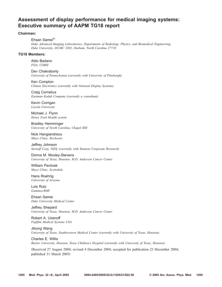
Assessment of display performance for medical imaging systems: Executive summary of AAPM TG18 report Chairman: Ehsan Sameia兲 Duke Advanced Imaging Laboratories, Departments of Radiology, Physics, and Biomedical Engineering, Duke University, DUMC 3302, Durham, North Carolina 27710 TG18 Members: Aldo Badano FDA, CDRH Dev Chakraborty University of Pennsylvania (currently with University of Pittsburgh) Ken Compton Clinton Electronics (currently with National Display Systems) Craig Cornelius Eastman Kodak Company (currently a consultant) Kevin Corrigan Loyola University Michael J. Flynn Henry Ford Health system Bradley Hemminger University of North Carolina, Chapel Hill Nick Hangiandreou Mayo Clinic, Rochester Jeffrey Johnson Sarnoff Corp, NIDL (currently with Siemens Corporate Research) Donna M. Moxley-Stevens University of Texas, Houston, M.D. Anderson Cancer Center William Pavlicek Mayo Clinic, Scottsdale Hans Roehrig University of Arizona Lois Rutz Gammex/RMI Ehsan Samei Duke University Medical Center Jeffrey Shepard University of Texas, Houston, M.D. Anderson Cancer Center Robert A. Uzenoff Fujifilm Medical Systems USA Jihong Wang University of Texas, Southwestern Medical Center (currently with University of Texas, Houston) Charles E. Willis Baylor University, Houston, Texas Children’s Hospital (currently with University of Texas, Houston) 共Received 27 August 2004; revised 4 December 2004; accepted for publication 23 December 2004; published 31 March 2005兲 1205 Med. Phys. 32 „4…, April 2005 0094-2405/2005/32„4…/1205/21/$22.50 © 2005 Am. Assoc. Phys. Med. 1205 1206 Ehsan Samei et al.: Performance assessment of medical displays 1206 Digital imaging provides an effective means to electronically acquire, archive, distribute, and view medical images. Medical imaging display stations are an integral part of these operations. Therefore, it is vitally important to assure that electronic display devices do not compromise image quality and ultimately patient care. The AAPM Task Group 18 共TG18兲 recently published guidelines and acceptance criteria for acceptance testing and quality control of medical display devices. This paper is an executive summary of the TG18 report. TG18 guidelines include visual, quantitative, and advanced testing methodologies for primary and secondary class display devices. The characteristics, tested in conjunction with specially designed test patterns 共i.e., TG18 patterns兲, include reflection, geometric distortion, luminance, the spatial and angular dependencies of luminance, resolution, noise, glare, chromaticity, and display artifacts. Geometric distortions are evaluated by linear measurements of the TG18-QC test pattern, which should render distortion coefficients less than 2%/5% for primary/secondary displays, respectively. Reflection measurements include specular and diffuse reflection coefficients from which the maximum allowable ambient lighting is determined such that contrast degradation due to display reflection remains below a 20% limit and the level of ambient luminance 共Lamb兲 does not unduly compromise luminance ratio 共LR兲 and contrast at low luminance levels. Luminance evaluation relies on visual assessment of low contrast features in the TG18-CT and TG18-MP test patterns, or quantitative measurements at 18 distinct luminance levels of the TG18-LN test patterns. The major acceptable criteria for primary/ secondary displays are maximum luminance of greater than 170/ 100 cd/ m2, LR of greater than 250/ 100, and contrast conformance to that of the grayscale standard display function 共GSDF兲 of better than 10% / 20%, respectively. The angular response is tested to ascertain the viewing cone within which contrast conformance to the GSDF is better than 30% / 60% and LR is greater than 175/ 70 for primary/secondary displays, or alternatively, within which the on-axis contrast thresholds of the TG18-CT test pattern remain discernible. The evaluation of luminance spatial uniformity at two distinct luminance levels across the display faceplate using TG18-UNL test patterns should yield nonuniformity coefficients smaller than 30%. The resolution evaluation includes the visual scoring of the CX test target in the TG18-QC or TG18-CX test patterns, which should yield scores greater than 4 / 6 for primary/secondary displays. Noise evaluation includes visual evaluation of the contrast threshold in the TG18-AFC test pattern, which should yield a minimum of 3 / 2 targets visible for primary/secondary displays. The guidelines also include methodologies for more quantitative resolution and noise measurements based on MTF and NPS analyses. The display glare test, based on the visibility of the low-contrast targets of the TG18-GV test pattern or the measurement of the glare ratio 共GR兲, is expected to yield scores greater than 3 / 1 and GRs greater than 400/ 150 for primary/secondary displays. Chromaticity, measured across a display faceplate or between two display devices, is expected to render a u⬘,v⬘ color separation of less than 0.01 for primary displays. The report offers further descriptions of prior standardization efforts, current display technologies, testing prerequisites, streamlined procedures and timelines, and TG18 test patterns. © 2005 American Association of Physicists in Medicine. 关DOI: 10.1118/1.1861159兴 Key words: medical display, liquid crystal display, cathode ray tube, image quality, quality assurance, quality control, acceptance testing, picture archiving and communication system 共PACS兲 I. INTRODUCTION The adoption of digital detectors and Picture Archiving and Communication Systems 共PACS兲 has provided healthcare institutions an effective means to electronically archive and retrieve radiological images. Medical display workstations, an integral part of PACS, are used to display these images for diagnostic and clinical purposes. Considering the fundamental importance of image quality to the overall effectiveness of a diagnostic imaging practice, it is vitally important to assure that electronic display devices 共also termed softcopy displays兲 do not compromise image quality as a number of studies have suggested.1–3 According to the American Association of Physicists in Medicine 共AAPM兲 professional guidelines,4 the performance Medical Physics, Vol. 32, No. 4, April 2005 assessment of electronic display devices falls within the professional responsibilities of medical physicists. While many prior publications have addressed some aspect of medical display performance,5–15 prior evaluation and standardization efforts have fallen short of providing an unified approach for testing the performance of display devices such that the tests would take into consideration all the important aspects of display performance, be specific to medical displays, and be relatively easy to implement in a clinical setting. AAPM Task Group 18 共TG18兲 recently completed a report which suggests standard guidelines and criteria for acceptance testing and quality control of medical display devices.16 The intended audience of the report is practicing medical physicists, engineers, researchers, radiology administrative staff, manufacturers of medical displays, radiolo- 1207 Ehsan Samei et al.: Performance assessment of medical displays gists, and students interested in display quality evaluation. The report is developed such that while addressing the current dominant medical display technologies, cathode-ray tubes 共CRTs兲 and liquid crystal displays 共LCDs兲, many of the tests and concepts could be adapted to future display technologies. The report is divided into six sections. Section one summarizes prior standardization efforts in the performance evaluation of medical display devices. Section two is a tutorial on the current and emerging medical display technologies. Section three sets forth prerequisites for the assessment of the display performance and includes a description of required instrumentation and TG18 test patterns. Section four is the main body of the report containing the description, quantification methods, and acceptance criteria for each key display characteristic. Sections five and six outline procedures for acceptance testing and quality control of display devices. The report further includes appendices providing guidelines for evaluating the performance of “closed” display systems, requirements for equivalent appearance of monochrome images, a full tabular description of TG18 test patterns, and a selected bibliography. Considering the significant extent of the TG18 report, this paper aims to provide an executive summary of the report in a more condensed format. This paper focuses mainly on the testing procedures and criteria of the most direct relevance to acceptance testing and quality control procedures. The educational, advanced, and detailed descriptive portions of the report are not included. Interested individuals are referred to the full report for a complete description of the eliminated, summarized, and referenced sections. II. GENERAL PREREQUISITES FOR DISPLAY ASSESSMENTS II.A. Classification of Display Devices In recognition of the currently accepted practice and in accordance with the guidelines set forth by the American College of Radiology17 and the Food and Drug Administration, display devices for medical imaging are characterized in the TG18 report as either primary or secondary. Primary display systems are those used for the interpretation of medical images. They are typically used in radiology and in certain medical specialties such as orthopedics. Secondary systems are those used for viewing medical images by medical staff or specialists other than radiologists after an interpretive report is rendered. The operator’s console monitors commonly used to “adjust” the images before they are sent for interpretation are treated as a primary display in terms of contrast response but secondary otherwise. II.B. Required Tools II.B.1. Instrumentation Although many display tests can be performed visually, a more objective and quantitative evaluation of display performance requires special test tools. The required instruments vary in their complexity and cost depending on the context Medical Physics, Vol. 32, No. 4, April 2005 1207 of the evaluation 共research, acceptance testing, or quality control兲 and how thorough the evaluation needs to be. Table I summarizes the required instruments for display quality evaluation. The readers are advised to consult Sections 3.1 and 4-6 of the TG18 report to determine the subset of the tools and their performance requirements for the particular tests being performed. II.B.2. Test Patterns The TG18 report recommends the use of specific test patterns for performance evaluation of display devices in order to facilitate comparisons of measurements. The recommended patterns are designated with a nomenclature of the form TG18-xyz, where x, y, and z describe the type and derived variants of a pattern. The patterns are listed in Table II and a few examples are illustrated in Fig. 1. The full description of the patterns are in Sec. 3.2 and Appendix III of the TG18 report. While the electronic copy of the TG18 report provides the patterns in multiple formats, they may also be generated with the aid of the information provided in the report. When displaying the patterns, no special processing functions should be applied. The 16-bit version of the patterns should be displayed with a window width and level set to cover the range from 0 to 4095 共window width, WW= 4096, window level, WL= 2048兲, except for the TG18-PQC, TG18-LN, and TG18-AFC patterns, where a WW of 4080 and WL of 2040 should be used. For 8-bit patterns, the displayed range should be from 0 to 255 共WW= 256, WL= 128兲. For some of the patterns, it is also essential to have a one-on-one relationship between the image pixels and the display pixels. II.B.3. Software Though not essential, software tools can facilitate the performance assessment of display devices. They include software for semiautomated generation of test patterns, processing software for assessment of resolution and noise, and spreadsheets for recording and manipulating the evaluation results. Some tools are provided along with the electronic copy of the TG18 report. Further information is available in Sec. 3.3 of the report. II.C. Initial Steps for Display Assessment II.C.1. Availability of Tools Before starting the tests, the availability of the applicable tools and test patterns should be verified. Lists of desired tools for acceptance testing and quality control purposes are provided in the following section of this paper. The TG18 test patterns should be stored on the display workstation during installation, or otherwise be accessible from a network archive. This approach ensures that the same pattern will be utilized for all future testing. 1208 Ehsan Samei et al.: Performance assessment of medical displays 1208 TABLE I. Instrumentation used for display quality evaluation. Instrument Near-range luminance meter Desired requirements • Calibration traceable to NIST • 0.05–1000 cd/ m2 luminance range Purpose Luminance and luminance uniformity measurements • Better than 5% accuracy • Better than 10−2 共ideally 10−3兲 precision • Aperture range 艋5 deg • Better than 3% compliance with the Commission Internationale de L’Eclairage 共CIE兲 standard photopic spectral response Telescopic luminance meter • Those listed above for near-range meter • Acceptance angle 艋1 deg • Ability to focus to an area 艋6 mm Illuminance meter • Calibration traceable to NIST • 1–1000 lux illuminance range Luminance, luminance uniformity, reflection, angular response, and glare measurements Reflection and ambient lighting measurements • Better than 5% accuracy • Better than 3% compliance with the CIE standard photopic spectral response • 180 deg cosine 共Lambertian兲 response to better than 5% out to 50° angulation Colorimeter • Calibration traceable to NIST • 1–1000 cd/ m2 luminance range Chromaticity measurements • Better than 0.004 共u⬘ , v⬘兲 accuracy Digital camera • Low noise and wide dynamic range • 1–500 cd/ m2 luminance range Quantitative resolution and noise measurements • ⬎512⫻ 512 matrix size • 10- to 12-bit depth • Equipped with a focusable macro lens • Variable frame rate/integration times up to 1 s • Digital interface to a computer • Calibrated for camera luminance, flat-field response, noise, and MTF • Equipped with a stable stand or tripod with directional adjustments Light source • Uniform luminance ⬎200 cd/ m2 Quantitative specular reflection measurement • Small enough to subtend 15° from center of display Illumination device • See TG18 report Sec. 3.1.3 Quantitative diffuse reflection Baffle • Light absorbing characteristics • 5–15 mm opening Glare and luminance measurements Cone • Light absorbing characteristics • 5 mm opening and 艋60 deg angular divergence Glare and luminance measurements Light absorbing cloth or hood • Light absorbing characteristics Display evaluation in the areas that have no control over the level of ambient lighting Medical Physics, Vol. 32, No. 4, April 2005 Ehsan Samei et al.: Performance assessment of medical displays 1209 1209 TABLE I. 共Continued.兲 Instrument Measuring microscope or magnifier Desired requirements • Magnification 艌25–50x Purpose Visual resolution measurements • Equipped with a metric reticle with 艋0.05 mm divisions • Focusing capabilities • Allow a working distance of 艌12.5 mm Flashlight • None Allow inspections in dark Lint-free cleaning tissue glass-cleaning solution Two rulers and angle measurement device • Recommended by the display manufacturer Used for cleaning the faceplate, if needed • 1 m in length Tape measure • Flexible and 20–30 cm in length Angular response and specular reflection coefficient measurements Geometric distortions measurements II.C.2. Display Placement Prior to testing, the proper placement of a display device should be verified and adjustments made as appropriate. In the placement of a display device, the following should be considered: 1. 2. 3. Display devices should always be positioned to minimize specular reflection from direct light sources such as ceiling lights, film illuminators, or surgical lamps. The reflection of such light sources should not be observed on the faceplate of the display in the commonly used viewing orientations. Many display devices, such as CRTs, are affected by magnetic fields; they should not be placed in an area with strong magnetic fields 共i.e., vicinity of MRI scanners兲, unless properly shielded. Displays should be placed ergonomically to avoid neck and back strain at reading level with the center of the display slightly below eye level. II.C.3. Start-up Procedures Prior to evaluation, the display device should be warmed up for approximately 30 min. In addition, the general system functionality should be verified by a quick review of the TG18-QC 关Fig. 1共a兲兴 test pattern. The pattern should be evaluated for distinct visibility of the 16 luminance steps, the continuity of the continuous luminance bars at the right and left of the pattern, the absence of gross artifacts, and the proper size and positioning of the active display area. Any adjustments to vertical and horizontal size must be made prior to performing the luminance measurements. Dust and smudges on the face of the display will absorb, reflect, or refract emitted light possibly resulting in erroneous test results. In addition, newly installed displays are sometimes covered with a protective plastic layer, which upon removal can leave residual marks on the faceplate. Before testing a display device, the cleanliness of the faceplate Medical Physics, Vol. 32, No. 4, April 2005 should be verified. If the faceplate is not clean, it should be cleaned following the manufacturer’s recommendations. II.C.4. Ambient Lighting Level The artifacts and loss of image quality associated with reflections from the display surface depend on the level of ambient lighting. As shown in Table III, illumination of display device surfaces in various locations of a medical facility may vary by over two orders of magnitude. The reflection measurement described in a later section of this document delineates a method to determine the maximum ambient light level appropriate for any given display device based on its reflection and luminance characteristics. It is important to verify that the ambient lighting in the room is below this maximum. The condition for the tests should be similar to those under normal use of the equipment. By recording ambient light levels at a reference point at the center of the faceplate and noting the location and orientation of the display devices at acceptance testing, it will be possible to optimize repeatability of testing conditions in the future. If a display device is equipped with a photocell for ambient light detection, its use should be in compliance with the Digital Imaging and Communication in Medicine 共DICOM兲 grayscale standard display function 共GSDF兲 as further discussed below. II.C.5. Pretest Luminance Settings Before the performance of a display system can be assessed, proper display area size should be established, and the maximum luminance Lmax and the minimum luminance Lmin must be checked to verify that the device is properly configured. The desired values should be determined based on the desired luminance ratio, the reflection characteristics of the system, and the ambient lighting level 共see the reflection and luminance sections below兲. Using a luminance meter, the luminance values should be recorded using the TG18-LN8-01 共or TG18-LN12-01兲 test pattern for Lmin and 1210 Ehsan Samei et al.: Performance assessment of medical displays 1210 TABLE II. Test patterns recommended for display quality evaluation. The patterns are divided into six sets. Most patterns are available in 1024⫻ 1024 共1 k兲 size and in either DICOM or tiff format. Some patterns are available in 2048⫻ 2048 共2 k兲 size. Set Series Type Images Multipurpose 共1 k and 2 k兲 TG18-QC TG18-BR TG18-PQC Vis./Qnt Visual Vis./Qnt. 1 1 1 Resolution, luminance, distortion, artifacts Briggs pattern, low contrast detail vs luminance Resolution, luminance, contrast transfer for prints Luminance 共1 k only兲 TG18-CT TG18-LN TG18-UN TG18-UNL TG18-AD Visual Quant. Visual Quant. Visual 1 18 2 2 1 TG18-MP Visual 1 Luminance response DICOM grayscale calibration series Luminance and color uniformity, and angular response Same as above with defining lines Contrast threshold at low luminance for evaluating display reflection Luminance response 共bit depth resolution兲 TG18-RH Quant. 3 TG18-RV Quant. 3 TG18-PX TG18-CX Quant. Visual 1 1 TG18-LPH Visual 3 TG18-LPV Visual 3 Noise 共1 k only兲 TG18-AFC TG18-NS Visual Quant. 1 3 4AFC contrast-detail pattern, four CD values Similar to RV/RH, five uniform regions for noise evaluation Glare 共1 k only兲 TG18-GV TG18-GQ TG18-GA Visual Quant. Quant. 2 3 8 Dark spot pattern with low contrast object Dark spot pattern for glare ratio measurement Variable size dark spot patterns Anatomical 共2 k only兲 TG18-CH TG18-KN TG18-MM Visual Visual Visual 1 1 2 Reference anatomical PA chest pattern Reference anatomical knee pattern Reference anatomical mammogram pattern Resolution 共1 k and 2 k兲 TG18-LN8-18 共or TG18-LN12-18兲 for Lmax, respectively. For these measurements, ambient illumination should be reduced to negligible levels using a dark cloth shroud if necessary. If the measured values for Lmax and Lmin are not appropriate, the proper values should be established using the brightness and contrast controls of the display. Otherwise, the display device should be serviced before testing its performance. The TG18 report further recommends compliance of medical display systems with the DICOM GSDF.15 Before initiating the testing procedures, the device should be calibrated or otherwise its calibration verified within its operating luminance range defined by Lmax and Lmin. II.C.6. Personnel The acceptance and quality control 共QC兲 testing of a display system must be performed by an individual共s兲 having Medical Physics, Vol. 32, No. 4, April 2005 Description Five horizontal lines at three luminance levels for LSF evaluation Five vertical lines at three luminance levels for LSF evaluation Array of single pixels for spot size Array of Cx patterns and a scoring reference for resolution uniformity Horizontal bars at 1 pixel width, 1 / 16 modulation, three luminance levels Vertical bars at 1 pixel width, 1 / 16 modulation, three luminance levels appropriate technical and clinical competencies. Even though the vendor is expected to perform some testing before turning a display system over to the user, the user must independently test the system共s兲. For acceptance testing and annual QC evaluation, the tests should be performed by a medical physicists trained in display performance assessments. Other staff including biomedical engineers, in-house service electronic technicians, or trained x-ray technologists can perform some of the tests described herein; however, in such situations, a qualified medical physicist should accept full oversight responsibilities and final approval of the results. For monthly or quarterly QC, the tests can be delegated to such qualified professionals as well as long as they work under the direct supervision of the medical physicist. The daily QC of a display system should be performed by the operator/user of the system. Radiology staff using electronic displays should 1211 Ehsan Samei et al.: Performance assessment of medical displays 1211 Fig. 1. Examples of TG18 test patterns: TG18-QC 共a兲, TG18-PQC 共b兲, TG18-CT 共c兲, TG18-LN8-01 共d兲, TG18-LN8-08 共e兲, TG18-LN8-18 共f兲, TG18-UNL80 共g兲, TG18-UN80 共h兲, TG18-UNL10 共i兲, TG18-MP 共j兲, TG18-RV89 共k兲, TG18-RH50 共l兲, TG18-CX 共m兲, TG18-AFC 共n兲, TG18-GV 共o兲, TG18-GA30 共p兲, TG18-GQB 共q兲, TG18-CH 共r兲, TG18-KN 共s兲, TG18-MM1 共t兲, and TG18-MM2 共u兲. be familiar with the daily testing procedure and expected results. All personnel responsible for performing QC tests will require initial training specific to their level of responsibility and periodic retraining and mentoring by medical physics staff. Medical Physics, Vol. 32, No. 4, April 2005 II.C.7. Specific Prerequisites for Acceptance Testing Acceptance testing requires close communication with the vendor for understanding and documenting the operational 1212 Ehsan Samei et al.: Performance assessment of medical displays 1212 Fig. 1. 共Continued). features and dedicated QC utilities of the system. Any recommended service and/or calibration schedule, including the services provided, tests performed, and the service/ calibration intervals, must be obtained from the manufacturer, ideally as part of the purchasing process. Prior to acceptance testing, the characteristics of the display systems delivered should be verified against those specified in the purchase agreement. A database should be established which includes information such as display type, size, resolution, manufacturer, model, serial number, manufacture date, room number, display identification 共if applicable兲, associated display hardware 共e.g., display controller兲 and test patterns Medical Physics, Vol. 32, No. 4, April 2005 available on the systems. All delivered documentation from the vendor should also be reviewed with special attention to the testing results performed at the factory. II.C.8. Specific Prerequisites for Quality Control The initial acceptance testing data are used to establish and maintain expected performance. Data acquired during routine QC testing must be compared to the limits established around the baseline values. It is also essential to utilize the same pattern for repeat evaluations of a given display device. The use of worksheets and checklists will help in 1213 Ehsan Samei et al.: Performance assessment of medical displays 1213 Fig. 1. 共Continued). establishing and monitoring the baselines. It is strongly recommended to record and maintain this information in electronic databases. Most commercial calibration packages support automated recording, tracking, and analysis of display QC results. III. ASSESSMENT OF DISPLAY PERFORMANCE The performance assessment of a display device in a clinical setting might be performed in the context of acceptance testing, prior to first clinical use, or quality control, throughout the life of the device. Tables IV and V provide a list of the tests, the required tools, and the expected performance for the two types of procedures with specific reference to the TG18 report. Depending on the interest and resources, additional advanced tests are further encouraged. For QC tests, hardware features and reproducible performance can reduce the need for very frequent testing. However, it is recommended that initially the tests be performed more frequently. If stability is maintained, a determination can be made to decrease the frequency of testing. The sections below provide the assessment methodologies. It is generally ideal to perform the tests in the order in which they are discussed as some of the latter tests may be influenced by parameters that are addressed in earlier tests. Full descriptions of the specific display characteristics as well as advanced testing procedures are provided in the TG18 report,16 to which the interested readers are referred. III.A.1. Visual Evaluation The geometric distortion of a display system is ascertained visually using the TG18-QC or the TG18-LPV/LPH test pattern. The patterns should be maximized to fill the entire usable display area. For displays with rectangular display areas, the patterns should cover at least the narrower dimension of the display area and be placed at the center of the area used for image viewing. The pattern共s兲 should be examined from a viewing distance of 30 cm. The patterns should appear straight without significant geometric distortions, and should be properly scaled to the aspect ratio of the video source pixel format so that the grid of the TG18-QC pattern appears square. The lines should appear straight indicative of proper linearity without any curvature or waviness. Some small barrel and pincushion distortions are normal for CRT devices but should not be excessive. For the TG18-LPV and TG18-LPH patterns, in addition to straightness, the lines should appear equally spaced. III.A.2. Quantitative Evaluation Spatial accuracy for geometric distortions can be quantified using the TG18-QC test pattern, maximized to fill the entire display area. Using a straight edge as a guide for a best fit and with the aid of a flexible plastic ruler, distances should TABLE III. Typical ambient lighting levels. III.A. Geometric Distortions Area Geometric distortions of displayed images are often a concern in cathode-ray tube 共CRT兲 display devices. The distortions can be in concave, convex, skewed, or other nonlinear forms. The magnitude and type of such distortions should be evaluated and, if deemed inappropriate, adjusted to meet certain minimum requirements as noted below. Operating rooms Emergency medicine Hospital clinical viewing stations Staff offices Diagnostic reading stations 共CT/MR/NM兲 Diagnostic reading stations 共x-rays兲 Medical Physics, Vol. 32, No. 4, April 2005 Illumination 共lux兲 300–400 150–300 200–250 50–180 15–60 2–10 1214 Ehsan Samei et al.: Performance assessment of medical displays 1214 TABLE IV. Tests, tools, and acceptance criteria for acceptance testing and annual quality control of electronic display systems. The section notations refer to the TG18 report. Test Major required tools Equipment Patterns Procedure Acceptance criteria 共for two classes of displays兲 Primary Secondary Suggested action 共if unacceptable兲 Geometric distortions Flexible ruler or transparent template TG18-QC See Sec. 4.1.4 Deviation 艋2% Deviation 艋5% Reflectiona Measuring ruler, light sources, luminance and illuminance meters, illuminator Luminance and illuminance meters TG18-AD See Secs. 4.2.3 and 4.2.4 Lmin 艌 1.5Lamb 共ideally 艌4Lamb兲 Lmin 艌 1.5Lamb 共ideally 艌4Lamb兲 Readjustment, repair, or replacement for repeated failures Results are used to adjust the level of ambient lighting TG18-LN TG18-CT TG18-MP See Secs. 4.3.4 and 4.3.3 Lmax 艌 170 cd/ m2LR艌 250 ␦ 艋 10% ⌬Lmax 艋 10% Luminance dependenciesb Luminance meter, luminance angular response measurement tool TG18-UNL TG18-LN TG18-CT See Secs. 4.4.3 and 4.4.4 Nonunif. 艋30% LR⬘␦ 艌 175 ␦ 艋 30% Lmax 艌 100 cd/ m2 LR艌 100 ⌬Lmax 艋 10% ␦ 艋 20% Nonunif. 艋30% LR⬘␦ 艌 70 ␦ 艋 60% Resolutionc Luminance meter magnifier TG18-QC TG18-CX TG18-PX See Secs. 4.5.3 and 4.5.4.1.2 0 艋 Cx艋 4 RAR= 0.9− 1.1 AR艋 15 0 艋 Cx艋 6 Noisec None TG18-AFC See Sec. 4.6.3 All targets visible except the smallest Two largest sizes visible Veiling glare Baffled funnel, telescopic photometer TG18-GV TG18-GVN TG18-GQs See Secs. 4.7.3 and 4.7.4 艌3 targets visible, GR艌 400 艌1 target visible, GR艌 150 Chromaticity Colorimeter TG18-UNL80 See Sec. 4.8.4 ⌬共u⬘ , v⬘兲 艋0.01 None Luminance response Readjustment, recalibration, repair, or replacement for repeated failures Readjustment, repair or replacement for repeated failures; Angular results used to define acceptable viewing angle cone Focus adjustment, repair, or replacement for repeated failures Reverification of luminance response, otherwise replacement Reverification of luminance response, otherwise replacement Replacement Note: Acronyms: Lamb= ambient luminance, Lmin= minimum luminance, Lmax= maximum luminance, LR=luminance ratio, ␦= maximum deviation between measured and GSDF contrast, Cx= Cx score, RAR= resolution-addressability ratio, AR= astigmatism ratio, GR= glare ratio. a In the absence of illumination devices, this acceptance testing can be performed only visually using TG18-AD and the method described in Sec. 4.2.3.1. b Angular tests are not required as a part of annual quality control. c More objective resolution and noise measurements can be performed as described in Secs. 4.5.4 and 4.6.4 using a digital camera. be measured in square areas in the horizontal and vertical directions in each of the four quadrants of the pattern and within the whole pattern 共Fig. 2兲. It is important to assure the locations of the cross hatches be viewed perpendicular to the display’s faceplate. In each quadrant, between quadrants, and within the whole pattern, the maximum percent deviations between the measurements in each direction and between the measurements in the horizontal and vertical directions should be determined. The percentages should be calculated in relation to the smallest of the values being compared. The measured spatial deviations shall be less than 2% and 5% for primary or secondary displays, respectively. If a disMedical Physics, Vol. 32, No. 4, April 2005 play device does not meet these criteria, adjustments should be made to the distortion control of the device. Often, as the area of the display is increased or decreased, the luminance will also increase or decrease in a nonlinear fashion. Therefore, it is important to make and finalize such adjustments prior to testing and adjustment of the display luminance characteristics. In addition, if a display workstation contains more than one display device, it is important to have the vertical and horizontal sizes of the active areas carefully matched within 2%. This facilitates the subsequent matching of their luminance response characteristics. 1215 Ehsan Samei et al.: Performance assessment of medical displays 1215 TABLE V. 共a兲 Tests for daily quality control of electronic display system, performed by the display user. 共b兲 Tests for monthly/quarterly quality control of electronic display systems performed by a medical physicist or by a QC technologist under the supervision of a medical physicist. The section notations refer to the TG18 report. For acronyms see Table IV. Test Major required tools Equipment Procedure Patterns Acceptance criteria 共for two classes of displays兲 Primary Secondary Suggested action 共if unacceptable兲 Overall visual assessment None TG18-QC or anat. images 共a兲 See Secs. 4.10.1 or 4.10.6 See Secs. 4.10.1/4.10.6 See Secs. 4.10.1/4.10.6 Further /closer evaluation Geometric distortions None TG18-QC 共b兲 See Sec. 4.1.3.1 See 4.1.3.2 See 4.1.3.2 Further/closer evaluation Reflection Luminance and illuminance meters TG18-AD See Secs. 4.2.3 and 4.2.4 Lmin 艌 1.5Lamb 共ideally 艌4Lamb兲 Lmin 艌 1.5Lamb 共ideally 艌4Lamb兲 Readjust the level of ambient lighting Luminance response Luminance and illuminance meters TG18-LN TG18-CT TG18-MP See Secs. 4.3.4 and 4.3.3 Lmax 艌 170 cd/ m2 LR艌 250 ⌬Lmax 艋 10% ␦ 艋 10% Lmax 艌 100 cd/ m2 LR艌 100 ⌬Lmax 艋 10% ␦ 艋 20% Readjustment, recalibration, repair, or replacement for repeated failures Luminance dependencies Luminance meter TG18-UN TG18-UNL See Secs. 4.4.3 and 4.4.4 Nonunif. 艋30% Nonunif. 艋30% Resolution Magnifier TG18-QC TG18-CX See Sec. 4.5.3 0 艋 Cx艋 4 0 艋 Cx艋 6 Readjustment, repair, or replacement for repeated failures Focus adjustment, repair, or replacement for repeated failures III.B. Display Reflection Electronic display devices have specular and diffuse reflection that can reduce image contrast and affect image quality. Ambient light reflections are more pronounced in display devices with thick faceplates 共e.g., CRTs兲 compared to those with thinner faceplates 共e.g., LCDs兲. They are generally reduced by the application of antireflective 共AR兲 coating on the faceplate and/or the addition of light absorbers within the faceplate of the display, but these means do not completely eliminate reflections. The reflection characteristics of a medical display device should be evaluated in order to establish the maximum allowable level of ambient lighting at which the device can be operated without overly compromising the desired luminance performance and contrast threshold. III.B.1. Visual Evaluation III.B.1.a. Specular Reflection Characteristics An effective and simple visual test for specular reflection of a display device is to observe the device in the power-save mode or turned off. The ambient lighting in the room should be maintained at levels normally used. The display’s faceplate should be examined at a distance of about 30–60 cm within an angular view of ⫾15 deg for the presence of specularly reflected light sources or illuminated objects. Patterns of high contrast on the viewer’s clothing are common sources of Medical Physics, Vol. 32, No. 4, April 2005 reflected features. In general, no specularly reflected patterns of high contrast objects should be seen. If light sources such as that from a film illuminator or window are seen, the position of the display device in the room is not appropriate. If high contrast patterns such as an identification badge on a white shirt or a picture frame on a light wall are seen, the ambient illumination in the room should be reduced. III.B.1.b. Diffuse Reflection Characteristics The effect of diffusely reflected light on image contrast may be observed FIG. 2. Spatial measurements for the quantitative evaluation of geometric distortions using the TG18-QC test pattern. The small squares with dashed lines 共- - -兲 define the four quadrants of the pattern, and the large square at the center encompassing the luminance patches is the one to be used for geometric distortion characterization within the whole image. 1216 Ehsan Samei et al.: Performance assessment of medical displays by alternately viewing the low-contrast patterns in the TG18-AD test pattern in near total darkness and in normal ambient lighting, determining the threshold of visibility in each case. A dark cloth placed over both the display device and the viewer may be helpful for establishing near total darkness. The pattern should be examined from a viewing distance of 30 cm. The threshold of visibility should not be different when viewed in total darkness and when viewed in ambient lighting conditions. If the ambient lighting renders the “dark-threshold” not observable, the ambient illuminance on the display surface is causing excess contrast reduction, and the room ambient lighting needs to be reduced. III.B.2. Quantitative Evaluation III.B.2.a. Specular Reflection Characteristics The specular reflection coefficient for a display device can be measured with a small-diameter source of diffuse white light as described in Sec. 3.1.3 of the TG18 report. The display should be in the power-save mode or turned off. The light source, subtending 15° from the center of the display, should be positioned d1 centimeters from the center of the display and be pointed toward the center at an angle of 15° from the surface normal. The reflected luminance of the light source should then be measured with a telescopic photometer from a distance of d2 centimeters from the center of the display and similarly angled at 15° to the normal. Finally, the directly viewed luminance of the light source should be measured with the same photometer from a distance of d1 + d2 centimeters. The specular reflection coefficient Rs is the ratio of the reflected spot luminance to the directly viewed spot luminance. All measurements should be made in a dark room. As the artifacts associated with specular reflections depend on the ambient lighting, the measured specular reflection coefficient should be used to establish the maximum allowable ambient lighting E as E 艋 共CtLmin兲/共0.9 Rs兲, 共1兲 where the contrast threshold Ct = ⌬L / L 共see Fig. 3 and Sec. 4.3.1 of TG18 report兲, corresponds to its value at the minimum luminance Lmin. For convenience, this relationship is 1216 tabulated in Table VI so that the maximum room lighting can be identified if Rs and Lmin are known. As an example, for a typical CRT with antireflective 共AR兲 coating 共Rs = 0.004兲 operated at minimum luminance values of 0.5, 1, 1.5, and 2.0 cd/ m2, the ambient lighting based on specular reflection consideration should be less than approximately 14, 21, 28, and 31 lux, respectively. Note that in the adjustment and measurement of the appropriate level of ambient lighting, illuminance in the room should be measured with the illuminance meter placed at the center of the display and facing outward, so the proper amount of light incident on the faceplate can be assessed. III.B.2.b. Diffuse Reflection Characteristics The luminance from diffuse reflections adds to that produced by the display device. The ambient illumination produces a luminance of Lamb = RdE, where E is ambient illuminance on the display surface, and Rd is the diffuse reflection coefficient in units of cd/ m2 per lux or 1/sr. In the dark areas of a lowcontrast image, the change in luminance ⌬Lt will produce a relative contrast of ⌬Lt / 共Lmin + Lamb兲. For some devices, the luminance response can be calibrated to account for the presence of a known amount of luminance from ambient lighting Lamb and produce equivalent contrast transfer in both dark and bright regions. However, if Lamb is sufficiently large in relation to Lmin, even if the device has a high contrast ratio, the overall luminance ratio of the device is compromised. The diffuse reflection coefficient may be measured using standardized illumination of the display surface with the illuminator device described in Sec. 3.1.3 of the TG18 report 共Fig. 4兲. The illuminance should then be measured in the center of the display device using a probe placed on the center of the display surface. The sensitive area of the meter should be held vertically to measure the illuminance incident on the display faceplate. The induced luminance at the center of the display surface should then be measured with a telescopic luminance meter as illustrated in Fig. 4. The luminance measurement should be made through the small aperture at the back of the containment device so as to not perturb the reflective characteristics of the containment struc- FIG. 3. Contrast threshold for varied visual adaptation 共a兲 and fixed 共b兲 visual adaptation 共Ref. 19兲. The contrast threshold dL / L for a just noticeable difference 共JND兲 depends on whether the observer has fixed 共b兲 or varied 共a兲 adaptation to the light and dark regions of an overall scene. dL / L is the peak-to-peak modulation of a small sinusoidal test pattern. Medical Physics, Vol. 32, No. 4, April 2005 1217 Ehsan Samei et al.: Performance assessment of medical displays tivity of the human eye in scenes with wide ranges of luminance levels 共e.g., medical images兲,19,20 the DICOM grayscale standard display function 共GSDF兲15 offers a way to approach this goal by applying a specific look-up-table to the display values, such that the display values present equally discriminable levels of brightness. The intrinsic luminance response 共i.e., luminance versus display value兲 of most display devices is markedly different from the GSDF. It usually follows a power-law relationship for CRTs, and a linear one for LCDs.5 In addition, the luminance response may vary over time. In CRTs, for example, the phosphor efficiency decreases as the device ages. Modern display devices also have utilities that automatically calibrate the luminance response of the device to GSDF. However, the functionality and accuracy of these utilities should be independently verified by the user. FIG. 4. Typical illuminating device used for the measurement of the diffuse reflection coefficient of a display device. ture. The viewing aperture must be located from 8° to 12° off to the side from the normal so as to not interfere with the measurement result. The diffuse reflection coefficient Rd is computed as the ratio of the luminance to the illuminance in units of sr−1. As diffuse reflection reduces the contrast, for primary class display devices, the level of ambient illuminance should be set to insure that the contrast in dark regions observed with ambient illumination will be at least 80% of the contrast observed in near total darkness. This requirement translates to Lamb ⬍ 0.25 Lmin, or E 艋 共0.25 Lmin兲/Rd . 1217 III.C.1. Visual Evaluation The luminance response of a display device is visually inspected using the TG18-CT test pattern. The pattern should be evaluated from a viewing distance of 30 cm for visibility of the central half-moon targets and the four low-contrast objects at the corners of each of the 16 different luminance regions. Since this pattern is viewed in one state of visual adaptation, it is expected that the contrast transfer will be better at the overall brightness for which the visual system is adapted as opposed to the darkest or the brightest regions. With experience, the visual characteristics of this test pattern can be recognized for a system with quantitatively correct luminance response. In general, the low contrast targets should be visible in all regions. A common failure is not to be able to see the targets in one or two of the dark regions. The bit-depth resolution of the display should be evaluated using the TG18-MP test pattern. The evaluation includes ascertaining the horizontal contouring bands, their relative locations, and grayscale reversals. The pattern should be examined from a viewing distance of 30 cm. In general, the relative location of contouring bands and any luminance levels should not be farther than the distance between the 8-bit markers 共long markers兲. No contrast reversal should be discernible. 共2兲 Table VII identifies the ambient lighting for which Lamb is 0.25 of Lmin as a function of Rd and Lmin. As an example, for a typical CRT with AR coating 共Rd = 0.02 sr−1兲 operated at minimum luminance values of 0.5, 1, 1.5, and 2.0 cd/ m2, the ambient lighting based on diffuse reflection consideration should be less than approximately 7, 12, 19, and 25 lux, respectively. In situations where the level of ambient lighting can be strictly controlled and taken into account in the luminance calibration of the display device, a larger Lamb can be tolerated 共Lamb ⬍ Lmin / 1.5兲 as noted in the next section. III.C. Luminance Response The human visual system perceives brightness in a nonlinear fashion.18 Ideally, the luminance response of a display device should match this nonlinear response such that image values are displayed in equally perceptible luminance increments. While limited due to variations in the contrast sensi- TABLE VI. Maximum allowable ambient illuminance based on specular reflection: For a display device with a specific minimum luminance Lmin and a specific specular reflection coefficient Rs the ambient illumination which maintains specular reflections from high contrast objects below the visual contrast threshold 共Ct兲 is tabulated. Maximum room illuminance 共lux兲 Lmax–Lmin 共cd/ m2兲 Ct Rs = 0.002 Rs = 0.004 Rs = 0.008 Rs = 0.020 Rs = 0.040 5000–20 2500–10 1000–4 500–2 250–1 0.010 0.011 0.015 0.018 0.024 349 192 105 63 42 175 96 52 31 21 87 48 26 16 10 35 19 10 6 4 17 10 5 3 2 Medical Physics, Vol. 32, No. 4, April 2005 1218 Ehsan Samei et al.: Performance assessment of medical displays III.C.2. Quantitative Evaluation In the quantitative method, luminance L共p兲 is measured using a calibrated luminance meter at the center of the 18 TG18-LN test patterns, corresponding to 18 distinct digital driving levels p. The measurement of L共p兲 using patterns other than the TG18-LN patterns may result in different values due to the influence of veiling glare. The effect of ambient illumination should be reduced to negligible levels, by using a dark cloth if necessary. If a telescopic luminance meter is used, in order to minimize the influence of meter’s flare on the low-luminance measurements, the measurements may need to be made through a cone or baffle to shield the instrument from the surrounding light. For display devices with non-Lambertian light distribution, such as a LCD, if the measurements are made with a near range luminance meter, the meter should either have an aperture angle smaller than 5 deg or display-specific correction factors should be applied.21 The ambient luminance on the display faceplate 共Lamb兲 should either be estimated from the measured Rd values as Lamb = ERd or measured directly. In the case of direct measurement, the display device should be put in the power-save or blank screen-save mode 共otherwise turned off兲. A telescopic luminance meter normal to the display surface is used with a light-absorbing mask placed behind the meter to minimize specular reflection from the display. Otherwise the room lighting is set to the conditions established for the normal use of the equipment 共see Sec. III B above兲. The values for L⬘共p兲 including Lmax ⬘ and Lmax ⬘ are then computed by the addition of Lamb to the measured L共p兲 values. The recommended value for Lmax ⬘ is typically specified by the vendor as the highest value that can be used without compromising other performance characteristics, such as lifetime or resolution. Lmax ⬘ should be greater than 171 cd/ m2 17 for primary displays and 100 cd/ m2 for secondary displays, and should be within 10% of the desired value for both classes of display. Furthermore, for workstations with multiple monitors, Lmax ⬘ should not differ by more than 10% among monitors. Lmax ⬘ should be such that the desired luminance ratio LR⬘ = Lmax ⬘ / Lmin ⬘ is obtained. If the manufacturer’s recommendations are not available, it is recommended that the luminance ratio of a display device be set equal to or greater than 250 for all primary class devices.19 For second- 1218 ary class devices, LR⬘ should be no less than 100. In general, Lmin ⬘ should be within 10% of the nominally desired values for both classes of display. As ambient lighting can impact the low luminance response of a display device and reduce the device’s effective luminance ratio, a limit on Lamb is further indicated. For both classes of display devices, Lamb should ideally be less than 0.25Lmin 共or 0.2Lmin ⬘ 兲. In situations where the level of ambient lighting can be strictly controlled and taken into account in the luminance calibration of the display device, a larger Lamb can be tolerated, but Lamb should always be less than Lmin / 1.5 共or Lmin ⬘ / 2.5兲. If necessary, arrangements should be made to reduce the room lighting in order to achieve a sufficiently small Lamb. In evaluating the luminance response of the display between the maximum and minimum extremes, the measured luminance values should be related to the DICOM GSDF luminance response in terms of the contrast response, i.e., the slope of the measured luminance response. To do so, using the DICOM’s table of just noticeable difference 共JND兲 indices versus luminance, the JND indices for the measured Lmin ⬘ and Lmax ⬘ should first be identified. The JND indices for the intermediate L⬘ values should then be evenly spaced within the JND range and linearly related to the actual p values used as Ji = Jmin + Pi共Jmax − Jmin兲 , ⌬P 共3兲 where J indicates the JND indices 共e.g., Fig. 5兲. The measured data are then expressed as the observed contrast, ␦i, at each luminance step Li⬘, as a function of mean JND index value associated with that step ␦i = ⬘ 兲 2共Li⬘ − Li−1 ⬘ 兲共Ji − Ji−1兲 共L⬘i + Li−1 @0.5共Ji + Ji−1兲. The expected response from DICOM GSDF luminance values ␦di is also similarly computed using the following equation: TABLE VII. Maximum room lighting based on diffuse reflection: For a display device with a specific minimum luminance Lmin and a specific diffuse reflection coefficient Rd in units of cd/ m2 per lux or 1/sr, the ambient illumination which maintains 80% contrast in dark regions is tabulated. The maximum room illuminance is calculated as 0.25Lmin / Rd. Maximum room illuminance 共lux兲 Lmax–Lmin 共cd/ m2兲 Rs = 0.005 Rs = 0.010 Rs = 0.020 Rs = 0.040 Rs = 0.060 5000–20 2500–10 1000–4 500–2 250–1 1000 500 200 100 50 500 250 100 50 25 250 125 50 25 12 125 62 25 12 6 83 42 17 8 4 Medical Physics, Vol. 32, No. 4, April 2005 共4兲 1219 Ehsan Samei et al.: Performance assessment of medical displays 1219 FIG. 5. Example of the measured luminance for 18 display levels is plotted in relation to the DICOM GSDF. The p-values used to measure luminance have been linearly scaled to JND indices with the values at Lmax ⬘ and Lmin ⬘ set to be equal to the JND corresponding indices. ␦di = d 2共Ldi − Li−1 兲 共Ldi + d Li−1 兲共Ji − Ji−1兲 @0.5共Ji + Ji−1兲. 共5兲 The difference between the measured and GSDF contrast responses at any given point ␦ = Max共兩␦i − ␦di 兩兲, should be less than 10% and 20% for the primary and secondary class display devices, respectively 共Fig. 6兲. This criterion applies specifically to contrast evaluated from the 18 measurements of luminance made at uniformly spaced p-value intervals. The failure of a display device to meet the above criteria should prompt adjustment, recalibration, repair, or replacement of the device. III.D. Luminance Dependencies The luminance response evaluations described above only pertain to the luminance characteristics of a display device at one location on the display faceplate viewed perpendicularly. However, display devices often exhibit spatial luminance non-uniformities and variation in contrast as a function of viewing angle, both of which should be characterized as a part of display evaluation protocol. III.D.1. Visual Evaluation III.D.1.a. Nonuniformity The visual method for assessing display luminance uniformity involves the TG18-UN10 and TG18-UN80 test patterns. The patterns are displayed and the uniformity across the displayed pattern is visually assessed from a viewing distance of 30 cm. The patterns should be free of gross nonuniformities from center to the edges. No luminance variations with dimensions on the order of 1 cm or larger should be observed. III.D.1.b. Angular Dependence Angular response may be evaluated visually using the TG18-CT test pattern. The pattern should first be viewed on axis to determine the visibility of all half-moon targets. The viewing angle at which any of the on-axis contrast thresholds are rendered invisible should then be determined by changing the viewing orientation in FIG. 6. Example of the contrast response computed from 18 gray levels is related to the expected contrast response associated with the DICOM GSDF with 10% tolerance limits indicated. Medical Physics, Vol. 32, No. 4, April 2005 1220 Ehsan Samei et al.: Performance assessment of medical displays polar and azimuthal orientations. Alternatively, a uniform test pattern with uniformly embedded test targets may be used. The viewer distance at which all targets along the axial or diagonal axes are visible may be used as an indication of the angular response performance of the display in terms of the viewing angle cone within which the performance is acceptable. The acceptable viewing angle cone should be clearly labeled on the display device. III.D.2. Quantitative Evaluation III.D.2.a. Nonuniformity Using the TG18-UNL10 and TG18-UNL80 test patterns, luminance is measured at five locations over the faceplate of the display device 共center and four corners兲 using a calibrated luminance meter with attentions to the precautions noted in Sec. III C. The maximum luminance deviation for each display pattern is calculated as the percent difference between the maximum and minimum luminance values relative to their average value, 200*共Lmax − Lmin兲/共Lmax + Lmin兲. The value for an individual display device should be less than 30%. III.D.2.b. Angular Dependence The luminance of a LCD display may be quantitatively evaluated as a function of viewing angle. This can be done with two basic approaches: the conoscopic and the gonioscopic methods, as noted in the TG18 report. A basic quantitative test should include the evaluation of luminance ratio as a function of viewing angle using the TG18-LN test patterns. For these measurements, it is useful to have a subjective understanding of the viewing angle dependence to determine the specific horizontal and vertical angles at which quantitative measurements should be made. Ideally, the angular response of a display should not reduce the luminance ratio by more than 30%. Thus, an acceptable viewing angle is defined as an angular cone within which LR⬘ is greater than 175 共250⫻ 0.7兲 for primary displays and 70 共100⫻ 0.7兲 for secondary displays.22 If the luminance in midluminance values is measured, the angular luminance results should be evaluated the same as the onaxis measurements described above in terms of conformance to the GSDF. The contrast response for any viewing angle should not be greater than three times the expected limits on axis 共␦ 艋 3 ⫻ 10% = 30% for primary displays and ␦ 艋 3 ⫻ 20% = 60% for secondary displays兲. For a display device, both LR⬘ and ␦ requirements should be met. The viewing angle limitation for medical use of a device should be clearly labeled on the device for optimum viewing. If multiple devices of the same design are used, it is sufficient to assess the viewing angle limits on one device. For such systems, the acceptable viewing angle cone should be used to arrange the monitors for minimum contrast reduction due to the angular dependencies of luminance. III.E. Display Resolution Resolution is the ability of a display device to present the spatial details of a displayed image. This ability is related to both the number of pixels and the actual spatial extent of each pixel. Because of various optical and electronic proMedical Physics, Vol. 32, No. 4, April 2005 1220 cesses, a display pixel can have a breadth that is larger than its nominal value, degrading the display resolution from its ideal level. III.E.1. Visual Evaluation Display resolution can be evaluated by visually assessing the appearance of the “Cx” patterns in the TG18-QC or the TG18-CX test patterns. The patterns should be displayed so that each image pixel is mapped to one display pixel. Most image viewers have the function to accomplish this display mode. In order not to be limited by the modulation transfer function 共MTF兲 of the eye, the use of a magnifying glass is recommended. In the TG18-QC pattern, the examiner should inspect the displayed “Cx” targets at the center and four corners of the pattern and score the appearance using the provided scoring scale. The line-pair patterns at Nyquist and half-Nyquist frequencies in the horizontal and vertical directions should also be evaluated in terms of visibility of the lines. The average brightness of the patterns should also be evaluated using the grayscale step pattern as a reference. The difference in visibility of test patterns between horizontal and vertical patterns should be noted. The relative width of the black and white lines in these patches should also be examined using a magnifier. The resolution uniformity may be ascertained across the display area using the TG18-CX test pattern and a magnifier in the same way that the “Cx” elements in the TG18-QC pattern are evaluated. In the visual inspection of the TG18-QC and TG18-CX patterns on primary class display systems, the Cx elements should be scored between 0 and 4 at all locations. This limit coincides with a resolution-addressability ratio 共RAR兲 艋 1.15.8 For secondary class displays, the Cx scores should be between 0 and 6 共RAR艋 1.47兲. For both classes, the horizontal and vertical line-pair patterns at Nyquist frequency should be discernible at all locations and for all directions. The TG18 report further includes a method to determine the extent of the display pixels 共i.e., RAR兲 using the TG18-PX test pattern. III.E.2. Quantitative Evaluation Quantification of the MTF requires the use of a displayedimage digitizing system, such as a digital camera, to digitally capture a portion of the display and to analyze the resulting images. The lens flare should be reduced with the use of a high f number and the aid of a cone or funnel device. The magnification of the lens should result in over-sampling of the display with at least 64 camera pixels covering one display pixel. The camera needs to be well focused on the screen of the display under test. This is best done when the lens aperture is opened to its maximum level to achieve low depth-of-focus. Afterward, the lens aperture is set to its smallest level in order to achieve a large depth-of-focus and minimum flare. The TG18-RV, TG18-RH, and TG18-NS patterns provide line inputs as target patterns for the MTF measurements. These patterns allow the assessment of MTF in the horizontal and vertical directions at three luminance levels and five 1221 Ehsan Samei et al.: Performance assessment of medical displays locations on the display area. At each location, the camera should be securely positioned in the normal direction in front of the target area of the display and focused on the line. The magnification should be determined in accordance with the display pixel size, camera matrix size, and the desired oversampling. The camera field of view should include the pixel markers in the pattern. While the camera should be placed in normal direction with respect to the faceplate, it needs to be rotated parallel to the faceplate such that the camera pixel array is angled at 2–5 deg with respect to that of the displayed image. Images from all six patterns should be captured before moving the camera to the next location. The exposure time should be selected such that the digital signal of the camera exceeds the dark signal by a factor of 100. Furthermore, the exposure time should be long enough to permit integration over multiple display frames, but short enough with respect to instabilities of the scanning and deflection circuits. Ultimately the integration time should be appropriate with respect to the integration time of the human eye, for which the experiments are conducted. Integration times between 0.2 and 1 s are appropriate to use. The measurements should be made in a darkened room. The 30 images should be acquired without any image compression. The data should be transferred to a computer for data processing. The captured line patterns should be reduced to orthogonal MTFs using Fourier analysis. There are several processing steps in the calculations, and the results are expected to vary slightly with the methods. For standardization and simplicity, the following steps are suggested:23 1. 2. 3. 4. 5. 6. 7. 8. 9. Determine the size that the image pixels represent in terms of the spatial dimension on the display using the known physical distance of the pixel markers on the patterns and the measured pixel distance of the markers in the captured images. Linearize the image data with respect to display luminance using the luminance response of the display. Add the mean value of the image from the TG18-NS to that of the TG18-RV 共or TG18-RH兲 pattern, and subtract the TG18-NS image pixel by pixel from the TG18-RV 共or TG18-RH兲 image in order to remove display pixel structure. Averages of multiple images may be used for more complete removal of structured noise. The subtracted image is used for further processing. Identify a central rectangular region of interest 共ROI兲 extending along the image of the line. Determine the angle of the line. Reproject the two-dimensional 共2D兲 data within the ROI along the direction of the line into subpixel bins to obtain the composite line spread function 共LSF兲. Smooth the LSF if it expresses excessive noise. Find the Fourier transform of the LSF, and normalize the resulting MTF. Divide the MTF by the sinc function associated with the width of the LSF subpixel bins, and correct for the previously characterized MTF of the camera system 共see Sec. 3.1.2 of the report兲. Medical Physics, Vol. 32, No. 4, April 2005 1221 Note that in some cases the LSF might be asymmetric. In those cases, each side of the LSF is used to form two symmetric LSFs. The resultant MTFs are reported, along with their average, as representative of the display resolution. Values of the measured MTF at the Nyquist frequency should be at least 35% for primary display devices and 25% for secondary devices. Measured responses outside the acceptable range should prompt corrective actions in the form of focus or dithering adjustments, repair, or replacement of the device. III.F. Display Noise Display noise refers to statistical fluctuations in the image that either vary spatially, so-called spatial noise, or vary in time, so-called temporal noise. Temporal noise, which is usually dominant in the dark regions of displayed images, is difficult to characterize outside of a laboratory setting and its perceptual influence is less well understood. Spatial noise is dominant in the brighter areas of displayed images. In CRTs, phosphor granularity is the main contributor to spatial noise; while in LCDs, the dominant noise is that associated with the pixelated background. III.F.1. Visual Evaluation The visual method to quantify the spatial noise of a display system is based on the method to determine just noticeable luminance differences as a function of size using the TG18-AFC test pattern. Each quadrant of the test pattern contains a large number of regions with varying target position. In each quadrant, the contrast and size of the target are constant. The contrast-size values for the four quadrants are 20-2, 30-3, 40-4, and 60-6. The observer should view the patterns from a viewing distance of 30 cm. The quadrants can be subjectively evaluated to establish the contrast-size relationships for which the observer can confidently place the position of all targets. The target visibility in each of the target regions may also be quantified by counting the number of targets readily visible in each of the quadrants and computing the percent correct. The visual evaluation should render all the targets except the smallest one visible for primary class displays and the two largest sizes visible for secondary class displays. Since the mean value and the standard deviation of the background are each linearly dependent on the luminance, their ratio, i.e., signal-to-noise, remains independent of luminance.24,25 Therefore, the results of the noise evaluation are independent of the absolute luminance value of the pattern’s background. However, the failure of a device in this test can also be an indication of an improper luminance response, the possibility of which can be eliminated by first verifying the proper luminance response of the device. III.F.2. Quantitative Evaluation Spatial noise of a display system can be quantified by either single-pixel signal-to-noise ratios24 or by the normalized noise power spectrum 共NPS兲. Both methods require the 1222 Ehsan Samei et al.: Performance assessment of medical displays use of a scientific-grade digital camera to capture an image of a uniform pattern displayed on the device. The camera lens should be set to a high f number in order to reduce veiling glare in the camera. Also, the magnification of the lens should result in over-sampling of the display in a way that allows sampling of spatial frequencies up to 40 cycles per degree, which is the resolution limit of the human visual system at the maximum luminance of most electronic displays.26 The camera images should also be flat-fieldcorrected, compensated for gain variations, and restored for the degradation of the MTF of the camera optics based on the prior performance evaluation of the camera system, noted earlier. The central region of the TG18-NS test patterns can be used as the target uniform pattern for measurements at three luminance levels. The camera should be securely positioned in front of the target area of the display and focused. The field of view should include the pixel markers in the pattern. The magnification should be determined in accordance with the display pixel size, camera matrix size, and the desired over-sampling. To eliminate the effects of temporal fluctuations in the luminance output, images should be captured with an integration time of about one second. The measurements should be performed in a darkened room. The images should be transferred uncompressed to a computer for data processing. The quantification of the display noise by the single-pixel signal-to-noise ratio is noted in the TG18 report. For the NPS determination, the captured uniform patterns are processed by Fourier analysis. There are multiple processing steps involved and the methods can vary the results slightly. For standardization and simplicity, the following steps are suggested: 1. 2. 3. 4. 5. 6. Determine the size that the image pixels represent in terms of the spatial dimension on the display using the known physical distance of the pixel markers on the pattern and the measured pixel distance of the markers in the captured image. Linearize the image data with respect to display luminance. Divide the central 3 / 4 region of the captured image into multiple, nonoverlapping regions, 128⫻ 128 or 256 ⫻ 256 in size. The size of these regions determine the sampling interval of the resulting NPS. Depending on the exact level of magnification 共oversampling兲 and the matrix size of the camera, between nine to 64 regions may be identified. It is recommended that at least 20 regions be used for the assessment of the NPS. To achieve this, it might be necessary to acquire multiple images from the central patch of the TG18-NS pattern by orienting the camera toward another, nonoverlapping area of the central area of the displayed pattern. Apply a two-dimensional fast Fourier transform on each region to yield the 2D NPS. Average the 2D NPS from all regions. Correct for the camera noise. Based on the assumption that the camera noise and the display spatial noise are Medical Physics, Vol. 32, No. 4, April 2005 7. 1222 uncorrelated, the NPS based on sampled camera images without exposure using the same integration time may be subtracted from the results. Derive the orthogonal NPS from the calculated 2D NPS by band averaging, excluding the data on the orthogonal axes. Since there are currently only a few examples of actual NPS measurements made, and since no correlation of the measurements and diagnostic accuracy is ascertained, no fixed criteria are recommended at this time. However, noise values associated with the display device should not exceed those of typical radiological images that are viewed with the system. III.G. Veiling Glare Veiling glare is a light-spreading phenomenon in a display device that leads to the degradation of image contrast in the presence of strong surrounding brightness. In CRTs, veiling glare is caused by internal light-scattering processes in the device’s faceplate, light leakage, and electron backscattering. In LCDs, electronic cross-talk can be viewed as a form of veiling glare. III.G.1. Visual Evaluation The visual assessment of veiling glare can be accomplished using the TG18-GV and TG18-GVN test patterns. The display size must be adjusted so that the diameter of the white region is 20 cm. The observer should discern the visibility of the low-contrast objects in sequential viewing of the TG18-GVN and TG18-GV patterns. Because the human visual systems will change adaptation if it views the bright field, it is imperative that the bright field is fully blocked from view and that no reflected light from the bright field be observable. This may be accomplished by the use of a mask or cone, which shields the human eye from the surround luminance of the pattern. No significant reduction in the contrast of the target objects should be observed between the two patterns. At least three objects should be readily visible in either pattern for primary class display devices. The corresponding object for secondary class display devices is at least one 共5th兲 target. III.G.2. Quantitative Evaluation The quantitative evaluation of veiling glare is accomplished using a highly collimated luminance meter and the TG18-GQ, TG18-GQB, and TG18-GQN test patterns. The display size must be adjusted so that the diameter of the white region is 20 cm. Furthermore, the bright luminance surrounding the central measurement point at the center of the test patterns should be blocked using either a baffled luminance meter or a telescopic luminance meter with a light-blocking baffled funnel or cone. Using either of these devices, the luminance in the center of the central dark region of the TG18-GQ pattern L, the white luminance in the center of the white region of the TG18-GQB pattern LB, and 1223 Ehsan Samei et al.: Performance assessment of medical displays the background luminance value in the center of the TG18GQN pattern LN, are recorded. The glare ratio for the display is then computed as GR = 共LB − LN兲/共L − LN兲. III.H. Display Chromaticity In display devices, chromaticity refers to the intrinsic magnitude and uniformity of color tint of the device when displaying a monochrome image. In monochrome CRTs, color tint is dictated by the phosphor type, and can vary slightly from monitor to monitor. In LCDs, color tint is dictated by the color temperature of the backlight. Color tint is usually considered a preference issue. However, it can be a cause of distraction, especially in multiple monitor workstations where the color tints are mismatched. III.H.1. Visual Evaluation The visual assessment of color uniformity is performed using the TG18-UN80 test pattern. The pattern is displayed on all the display devices associated with a workstation, and the relative color uniformity of the displayed pattern across the display area of each display device and across different display devices is discerned. No significantly perceivable color differences should be present among display devices and across the display area of each device for primary class devices. No requirements are specified for secondary class displays. III.H.2. Quantitative Evaluation The TG18-UNL80 test pattern is displayed on all the display devices associated with a workstation. A colorimeter is then used to measure the 共u⬘ , v⬘兲 color coordinates at the center and at the four corners of the display area of each display device, and these coordinates averaged to produce a mean 共u⬘ , v⬘兲 chromaticity measurement for the display device. The measurements on all display devices are used to compute the color uniformity index as the maximum distance in 共u⬘ , v⬘兲 space between any possible pair of average 共u⬘ , v⬘兲 points using D = 共共u⬘1 − u2⬘兲2 + 共v1⬘ − v⬘2兲2兲1/2. If the coloMedical Physics, Vol. 32, No. 4, April 2005 rimeter used outputs the color coordinate in the older 共x , y兲 space, the values can be converted to 共u⬘ , v⬘兲 space using the following transformations: u⬘ = 4x/共− 2x + 12y + 3兲, 共6兲 The veiling glare for a high fidelity display system should not change the contrast of a target pattern by more than 20% with and without a bright surrounding. Thus, the luminance from veiling glare should not be more than 25% of the minimum luminance for the normal operating settings of the display. Since the ratio of the maximum luminance to the minimum luminance should be about 250, this implies a glare ratio of 1000, which is typical of measurements made for transilluminated film. However, the recommended test pattern presents a scene with significantly more veiling glare in the target region than is encountered in medical imaging scenes. Though not as strict criteria which may not be achievable by certain display technologies, TG18 recommends a glare ratio greater than 400 and 150 for primary and secondary display devices, respectively.27,28 1223 v⬘ = 9y/共− 2x + 12y + 3兲; 共7兲 or x = 27u⬘/共18u⬘ − 48v⬘ + 36兲, y = 12v⬘/共18u⬘ − 48v⬘ + 36兲. 共8兲 Based on clinical experience, a color uniformity parameter of 0.01 or less is necessary to assure acceptable color matching of primary class grayscale display devices of a workstation.29 The distance between any pair of color coordinates across the display area of each device should also not exceed this limit. No quantitative requirements are specified for secondary class displays. III.I. Miscellaneous Tests In addition to the primary display attributes described above, there are a number of secondary attributes that may need to be addressed in a full display performance evaluation. Those include video artifacts, moiré artifacts, color artifacts, physical defects, flicker, and electronic cross talk. Brief descriptions and assessment methods for these characteristics are outlined in the TG18 report. III.J. Overall Evaluations In addition to the testing of a display device for a specific performance characteristic, the overall quality of a system can be assessed using a comprehensive visual/quantitative approach. Overall assessment can be based on any of the TG18-recommended multipurpose test patterns. Each pattern should be displayed with one display pixel representing each image pixel and examined from a viewing distance of 30 cm. The findings can be correlated with the results of more focused testing methods specified above and serve as a basis for quality control assessments. The frequency of such an evaluation is discussed in Sec. 6 of the full report. Evaluations based on TG18-QC and TG18 anatomical patterns are outlined below. III.J.1. Evaluations using TG18-QC Pattern The appearance of the elements in the TG18-QC test pattern 关Fig. 1共a兲兴 can be used to assess the overall performance of a display system. The following are recommended: 1. 2. General image quality and artifacts: Evaluate the overall appearance of the pattern. Note any non-uniformities or artifacts, especially at black-to-white and white-to-black transitions. Verify that the ramp bars appear continuous without any contour lines. Geometric distortion: Verify that the borders and lines of the pattern are visible and straight and that the pattern appears to be centered in the active area of the display device. If desired, measure any distortions. Ehsan Samei et al.: Performance assessment of medical displays 1224 1224 TABLE VIII. Criteria for evaluating the TG18 anatomical images. Test pattern Evaluation criteria TG18-CH Degree of difficulty for exam Overall contrast Overall sharpness Symmetrical reproduction of the thorax, as shown by the central position of a spinous process between the medial ends of the clavicles Medial borders of the scapulae Reproduction of the whole rib cage above the diaphragm Visually sharp reproduction of the vascular pattern of the lungs, particularly the peripheral vessels Sharp reproduction of the trachea and proximal bronchi Sharp reproduction of the borders of the heart and the aorta Sharp reproduction of the diaphragm Visibility of the retrocardiac lung and the mediastinum Visibility of subdiaphragmatic features Visibility of the spine through the heart shadow Visibility of small details in the whole lung, including the retrocardiac areas Visibility of linear and reticular details out to the lung periphery TG18-KN Degree of difficulty for exam Overall contrast Overall sharpness Reproduction of trabecular detail Reproduction of bony and soft tissue TG18-MM1 and TG18-MM2 Degree of difficulty for exam Overall contrast and brightness Overall sharpness 共no blur兲 Sharp appearance of Cooper’s ligaments Structure of the clip and the presence of the gap at its apex 共TG18-MM1 only兲 Appearance and visibility of subtle microcalcifications 共TG18-MM1 only兲 Visibility of structures at the margins of the breast 共TG18-MM1 only兲 3. 4. Luminance, reflection, noise, and glare: Verify that all 16 luminance patches are distinctly visible. Measure their luminance using a luminance meter if desired, and evaluate the results in comparison to the DICOM GSDF. Verify that the 5% and 95% patches are visible. Evaluate the appearance of low contrast letters and the targets at the corners of all luminance patches with and without ambient lighting. Resolution: Evaluate the Cx patterns at the center and corners of the pattern and grade them compared to the reference score. Also verify the visibility of the line-pair patterns at the Nyquist frequency at the center and corners of the pattern, and if desired, measure the luminance difference between the vertical and horizontal high-modulation patterns. III.J.2. Evaluations using Anatomical Images A radiologist should evaluate the overall clinical image quality of the display using patient images. The TG18 report suggests four specific anatomical images for this purpose: TG18-CH, TG18-KN, TG18-MM1, and TG18-MM2 关Figs. 1共r兲–共u兲兴. These correspond to a chest radiograph, a knee Medical Physics, Vol. 32, No. 4, April 2005 radiograph, and two digital mammograms. Clinical criteria for evaluating these images are given in Table VIII. The images may be scored according to these criteria corresponding to the different image features. The radiologist who wishes to evaluate his/her display should independently rate the image features according to the criteria in Table VIII, then compare their ratings to those obtained with a highquality transilluminated film print of the patterns. Significant discrepancies need to be brought to the attention of the responsible medical physicist or service engineer. IV. CONCLUSIONS Electronic display is a key component of medical imaging systems as it serves as the final element of the imaging chain. Due to hardware variability and degradation over time, it is important to assure that a medical display system is appropriate for the intended medical application and that its performance is stable over time. Acceptance testing and quality control testing of medical display devices are essential requirements for high-quality medical practice. The guidelines established by the AAPM Task Group 18 delineate specific testing procedures and acceptance criteria for that purpose that can be readily implemented in a clinical setting. 1225 Ehsan Samei et al.: Performance assessment of medical displays a兲 Electronic mail: samei@duke.edu S. J. Ackerman, J. N. Gitlin, R. W. Gayler, C. D. Flagle, and R. N. Bryan, “Receiver operating characteristic analysis of fracture and pneumonia detection: comparison of laser-digitized workstation images and conventional analog radiographs,” Radiology 186, 263–268 共1993兲. 2 W. W. Scott, J. E. Rosenbaum, S. J. Ackerman, R. L. Reichle, D. Magid, J. C. Weller, and J. N. Gitlin, “Subtle orthopedic fractures: Teleradiology workstation versus film interpretation,” Radiology 187, 811–815 共1993兲. 3 W. W. Scott, D. A. Bluemke, W. K. Mysko, G. E. R. Weller, G. D. Kelen, R. L. Reichle, J. C. Weller, and J. N. Gitlin, “Interpretation of emergency department radiographs by radiologists and emergency medicine physicians: teleradiology workstation versus radiograph readings,” Radiology 195, 223–229 共1995兲. 4 “AAPM, The role of the clinical medical physicist in diagnostic radiology,” Report of the AAPM Task Group No. 2 共AIP, New York, 1994兲. 5 E. Muka, H. Blume, and S. Daly, “Display of medical images on CRT soft-copy displays: a tutorial,” Proc. SPIE 2431, 341–359 共1995兲. 6 E. Senol and E. Muka, “Spatial frequency characteristics of CRT softcopy displays,” Proc. SPIE 2431, 302–315 共1995兲. 7 E. F. Kelley, G. R. Jones, P. A. Boynton, M. D. Grote, and D. J. Bechis, “A survey of the components of display measurement standards,” SID Digest 共Society for Information Display Headquarters, San Jose, CA, 1995兲, pp. 637–640. 8 E. Muka, T. Mertelmeier, R. M. Slone, and E. Senol, “Impact of phosphor luminescence noise on the specification of high-resolution CRT displays for medical imaging,” Proc. SPIE, 3031, 210–221 共1997兲. 9 H. Roehrig, H. Blume, T. L. Ji, and M. Browne, “Performance tests and quality control of cathode ray tube displays,” J. Digit Imaging 3, 134– 145 共1990兲. 10 J. E. Gray, “Use of SMPTE test pattern in picture archiving and communication systems,” J. Digit Imaging 5, 54–58 共1992兲. 11 R. D. Nawfel, K. H. Chan, D. J. Wagenaar, and P. F. Judy, “Evaluation of video gray-scale display,” Med. Phys. 19, 561–567 共1992兲. 12 D. A. Reimann, M. J. Flynn, and J. J. Ciarelli, “A system to maintain perceptually linear networked display devices,” Proc. SPIE 2431, 316– 326 共1995兲. 13 M. P. Eckert and D. P. Chakraborty, “Video display quality control measurements for PACS,” Proc. SPIE 2431, 328–340 共1995兲. 14 H. Kato, “Hard- and soft-copy image quality,” RSNA Categorical Course in Physics 共Radiological Society of North America 共RSNA兲 Publication, Categorical Course Syllabus, Oak Brook, IL, 1995兲, pp. 243–258. 15 NEMA PS 3.14-2000, “Digital imaging and communications in medicine 共DICOM兲 Part 14: Grayscale display standard function,” National Electrical Manufacturers Association, NEMA, 1300 North 17th Street, Suite 1847, Rosslyn, VA 22209, http://www.nema.org/ Draft viewable at http:// 1 Medical Physics, Vol. 32, No. 4, April 2005 1225 medical.nema.org/dicom/2000/draft/00_14DR.PDF, Available for purchase in whole or in parts from Global Sales, Global Engineering Documents, 15 Inverness Way, East Englewood, Colorado, 80112; http:// global.ihs.com/ 16 E. Samei, A. Badano, D. Chakraborty, K. Compton, C. Cornelius, K. Corrigan, M. J. Flynn, B. Hemminger, N. Hangiandreou, J. Johnson, M. Moxley, W. Pavlicek, H. Roehrig, L. Rutz, J. Shepard, R. Uzenoff, J. Wang, and C. Willis, “Assessment of display performance for medical imaging systems,” Report of the American Association of Physicists in Medicine 共AAPM兲 Task Group 18 共Medical Physics Publishing, Madison, WI, 2005兲, AAPM On-line Report No. 03. 17 American College of Radiology 共ACR兲, ACR Standard for Teleradiology, 1999. American College of Radiology, 1891 Preston White Drive, Reston, VA 20191-4397; http://www.acr.org/ 18 P. G. J. Barten, Contrast Sensitivity of the Human Eye and Its Effects on Image Quality 共SPIE Press, Bellingham, WA, 1999兲. 19 M. J. Flynn, J. Kanicki, A. Badano, and W. R. Eyler, “High fidelity electronic display of digital radiographs,” Radiographics 19, 1653–1669 共1999兲. 20 E. Samei, in “Digital mammography displays,” Advances in Breast Imaging: Physics, Technology, and Clinical Applications, edited by A. Karellas and M. L. Giger 共Radiological Society of North America 共RSNA兲 Publication, Categorical Course Syllabus, Oak Brook, IL, 2004兲, pp. 135–143. 21 H. Blume, A. M. K. Ho, F. Stevens, and P. M. Steven, “Practical aspects of gray-scale calibration of display systems,” Proc. SPIE, 4323, 28–41 共2001兲. 22 E. Samei and S. L. Wright, “The effect of viewing angle response on DICOM compliance of LCD displays,” Proc. SPIE Medical Imaging 5371, 170–177 共2004兲. 23 E. Samei and M. J. Flynn, “A method for in-field evaluation of the modulation transfer function of electronic display devices,” Proc. SPIE 4319, 599–607 共2001兲. 24 H. Roehrig, T. L. Ji, M. Browne, W. J. Dallas, and H. Blume, “Signal-tonoise ratio and maximum information content of images displayed by a CRT,” Proc. SPIE 1232, 115–133 共1990兲. 25 H. Roehrig, H. Blume, T. L. Ji, and M. K. Sundareshan, “Noise of CRT display systems,” Proc. SPIE 1897, 232–245 共1993兲. 26 A. Rose, Vision, Human and Electronic 共Plenum Press, New York, 1974兲. 27 M. J. Flynn and A. Badano, “Image quality degradation by light scattering in display devices,” J. Digit Imaging 12, 50–59 共1999兲. 28 A. Badano, M. J. Flynn, and J. Kanicki, “Accurate small-spot luminance measurements in display devices,” Displays 23, 177–182 共2002兲. 29 K. A. Fetterly, S. N. Bernatz, D. S. Groth, and N. J. Hangiandreou, “Quantitative color measurement for the characterization of grayscale PACS CRTs,” Radiology 209(P), 320 共1998兲.

