Science Manuscript Template
Smartphone-Based Diagnostic of Preeclampsia: An mHealth solution for the Congo
Red Dot (CRD) Test in Limited-Resource Settings
Authors: Stephan M. Jonas, Thomas M. Deserno, Catalin S. Buhimschi, Jennifer Makin,
Michael A. Choma, Irina. A. Buhimschi
SUPPLEMENTARY MATERIALS
Supplementary Methods
Statistical procedures.
MedCalc software (v 12.5 Ostend, Belgium) was used as statistical aid unless otherwise specified (31). Correlations were tested using the Pearson's product moment correlation while agreement was determined using Lin's concordance coefficient and the
McBride descriptive scaling of agreement (21, 22). Equivalence/non-inferiority testing was performed in Microsoft Excel using the TOST/XLSTAT add-ins procedure (23). Multivariate stepwise linear regression was used to determine the impact of multiple variables on the level of bias (difference between the result obtained using the manual method and result after systematic elimination of steps in the wet protocol). Variables were entered in the model if P <0.05 and removed if P >0.1. For all procedures a P <0.05 was used to denote statistical significance.
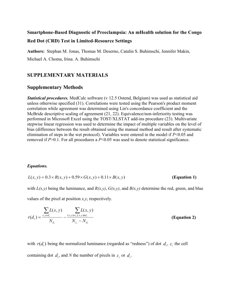
Equations.
L ( x , y )
0 .
3
R ( x , y )
0 .
59
G ( x , y )
0 .
11
B ( x , y ) (Equation 1) with L(x,y) being the luminance, and R(x,y) , G(x,y) , and B(x,y) determine the red, green, and blue values of the pixel at position x,y , respectively. r ( d i
)
x
, y
d i
L ( x , y )
N d i
( x , y
N
)
c i
x ,( c i
L ( x ,
, y )
d i
N d i y )
(Equation 2) with r ( d i
) being the normalized luminance (regarded as “redness”) of dot d i
, c i the cell containing dot d i
, and N the number of pixels in c i or d i
.
Supplementary Tables and Figures
Table S1. Handling issues encountered in stage 3 of our study and solutions
Issue * Imaging issue Effect Remedy
1 Extreme viewpoint or perspective
2
3
Sheet not completely captured on image
Non-uniform illumination and shadowing
Linear interpolation fails; wrong localization of patient cells
Warning message if opposing borders vary more than 10% (15º viewing angle); changed acquisition protocol to inclined background
Corners detection impossible
Misclassification because of dark foreground
Error message upon missing corner; template position and orientation overlay on camera view
Warning message if high variance in illumination is detected
4 Reflection of water on, or near the sheet
Boundary and corner detection fails
Warning message suggesting inclined photographic surface to avoid puddle formation
5 Blurriness/Out-offocus
The sheet is blurred Blurring was neglected due to minor impact
(tolerated up to Gaussian blur with sigma=5)
* listed in the order of decreasing frequency with which they were observed
Fig. S1. True to size sample positioning template
As we cannot control for different printer settings, please check the dimensions before using the template.
