Anterior Cruciate Ligament Reconstruction in Men and Women:
advertisement
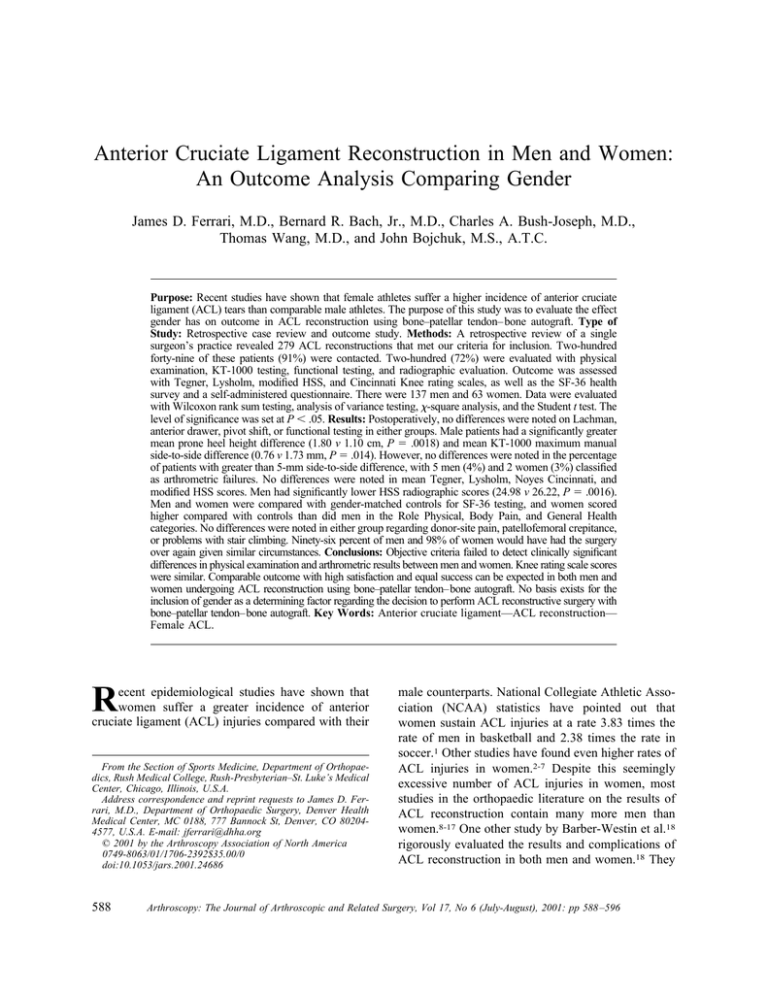
Anterior Cruciate Ligament Reconstruction in Men and Women: An Outcome Analysis Comparing Gender James D. Ferrari, M.D., Bernard R. Bach, Jr., M.D., Charles A. Bush-Joseph, M.D., Thomas Wang, M.D., and John Bojchuk, M.S., A.T.C. Purpose: Recent studies have shown that female athletes suffer a higher incidence of anterior cruciate ligament (ACL) tears than comparable male athletes. The purpose of this study was to evaluate the effect gender has on outcome in ACL reconstruction using bone–patellar tendon– bone autograft. Type of Study: Retrospective case review and outcome study. Methods: A retrospective review of a single surgeon’s practice revealed 279 ACL reconstructions that met our criteria for inclusion. Two-hundred forty-nine of these patients (91%) were contacted. Two-hundred (72%) were evaluated with physical examination, KT-1000 testing, functional testing, and radiographic evaluation. Outcome was assessed with Tegner, Lysholm, modified HSS, and Cincinnati Knee rating scales, as well as the SF-36 health survey and a self-administered questionnaire. There were 137 men and 63 women. Data were evaluated with Wilcoxon rank sum testing, analysis of variance testing, -square analysis, and the Student t test. The level of significance was set at P ⬍ .05. Results: Postoperatively, no differences were noted on Lachman, anterior drawer, pivot shift, or functional testing in either groups. Male patients had a significantly greater mean prone heel height difference (1.80 v 1.10 cm, P ⫽ .0018) and mean KT-1000 maximum manual side-to-side difference (0.76 v 1.73 mm, P ⫽ .014). However, no differences were noted in the percentage of patients with greater than 5-mm side-to-side difference, with 5 men (4%) and 2 women (3%) classified as arthrometric failures. No differences were noted in mean Tegner, Lysholm, Noyes Cincinnati, and modified HSS scores. Men had significantly lower HSS radiographic scores (24.98 v 26.22, P ⫽ .0016). Men and women were compared with gender-matched controls for SF-36 testing, and women scored higher compared with controls than did men in the Role Physical, Body Pain, and General Health categories. No differences were noted in either group regarding donor-site pain, patellofemoral crepitance, or problems with stair climbing. Ninety-six percent of men and 98% of women would have had the surgery over again given similar circumstances. Conclusions: Objective criteria failed to detect clinically significant differences in physical examination and arthrometric results between men and women. Knee rating scale scores were similar. Comparable outcome with high satisfaction and equal success can be expected in both men and women undergoing ACL reconstruction using bone–patellar tendon–bone autograft. No basis exists for the inclusion of gender as a determining factor regarding the decision to perform ACL reconstructive surgery with bone–patellar tendon–bone autograft. Key Words: Anterior cruciate ligament—ACL reconstruction— Female ACL. R ecent epidemiological studies have shown that women suffer a greater incidence of anterior cruciate ligament (ACL) injuries compared with their From the Section of Sports Medicine, Department of Orthopaedics, Rush Medical College, Rush-Presbyterian–St. Luke’s Medical Center, Chicago, Illinois, U.S.A. Address correspondence and reprint requests to James D. Ferrari, M.D., Department of Orthopaedic Surgery, Denver Health Medical Center, MC 0188, 777 Bannock St, Denver, CO 802044577, U.S.A. E-mail: jferrari@dhha.org © 2001 by the Arthroscopy Association of North America 0749-8063/01/1706-2392$35.00/0 doi:10.1053/jars.2001.24686 588 male counterparts. National Collegiate Athletic Association (NCAA) statistics have pointed out that women sustain ACL injuries at a rate 3.83 times the rate of men in basketball and 2.38 times the rate in soccer.1 Other studies have found even higher rates of ACL injuries in women.2-7 Despite this seemingly excessive number of ACL injuries in women, most studies in the orthopaedic literature on the results of ACL reconstruction contain many more men than women.8-17 One other study by Barber-Westin et al.18 rigorously evaluated the results and complications of ACL reconstruction in both men and women.18 They Arthroscopy: The Journal of Arthroscopic and Related Surgery, Vol 17, No 6 (July-August), 2001: pp 588 –596 ACL RECONSTRUCTION IN MEN AND WOMEN studied men and women who were matched for age, time interval from injury to surgery, preoperative activity level, months of follow-up, condition of the articular cartilage, and number of prior operative procedures on the knee. In this study there was no difference found between men and women with regard to failure rate, reoperation, and motion problems. No outcomes analysis was used. The purpose of our study was to determine whether differences existed between men and women in the outcome of ACL reconstruction using autogenous bone–patellar tendon– bone graft. METHODS The study group consisted of all patients who underwent arthroscopically assisted or endoscopic ACL reconstruction using bone–patellar tendon– bone autograft without extra-articular augmentation between June 1987 and April 1994. Between June 1987 and September 1991 patients underwent arthroscopically assisted 2-incision ACL reconstruction and between October 1991 and April 1994 patients underwent endoscopic single-incision ACL reconstruction. The results of these 2 groups of patients have been previously reported.9,10 Patients were identified from a computerized database maintained by the senior author (B.R.B.), who performed all the surgical procedures. All patients were retrospectively reviewed. The 2 groups were combined in order to increase statistical power and to simplify reporting of data. Before combining the 2 groups, they were compared for preoperative and postoperative physical findings, demographics, and rating scales. The only significant difference was length of follow-up (79 v 36 months). There were no significant differences with regard to physical examination findings (Lachman, anterior drawer, pivot shift), functional testing, outcome measures (Tegner, Lysholm, HSS, Noyes rating scales), satisfaction, and KT-1000 testing. This was consistent with other studies.16,19-21 Exclusionary criteria for this study included bilateral reconstructions, concomitant extra-articular reconstruction, multiligament reconstruction, associated posterolateral reconstruction, combined high tibial osteotomy and ACL reconstructions, or medical illness that precluded follow-up evaluation. Two-hundred seventy-five patients met the inclusionary criteria and of these, 200 patients (73%) were evaluated. Fortynine patients (18%) were contacted but were unable to be evaluated because of geographic constraints. 589 Twenty-six patients (9%) were lost to follow-up. There were 137 men and 63 women. Surgical Technique The surgical technique for both groups has been previously described.22,23 For both groups, a mid-third 10- or 11-mm wide patellar tendon graft with 25-mm length tibial and patellar bone plugs was used. Interference screw fixation was used in all knees. In the 2-incision group, 9 ⫻ 25 mm screws were used for both femoral and tibial fixation, and in the endoscopic 1-incision group, 7 ⫻ 25 mm screws were used for femoral fixation and 9 ⫻ 20 mm screws were used for tibial fixation. Tibial fixation was performed with the knee in full extension with the screws placed anteriorly on the cortical surface of the tibial bone plug, which had been externally rotated to achieve this orientation. In less than 10% of cases, the tibial fixation was supplemented with a staple or screw-and-post technique. All meniscal repairs and partial meniscectomies were performed concurrently. Postoperative Rehabilitation The postoperative protocol evolved during the study period. In general, patients who had a 2-incision technique were hospitalized for 2 to 3 days, placed on continuous passive motion machines, and were gradually brought to full weight bearing over the course of the first 6 postoperative weeks. Emphasis was not placed on obtaining early full hyperextension until after 1989.24 Patients who underwent an endoscopic technique were hospitalized an average of 1.3 days, full weight bearing was begun immediately, and emphasis was placed on obtaining early full hyperextension. In both groups, a hinged knee brace was used for 6 weeks postoperatively to protect the donor site. A custom ACL orthosis was worn from 6 weeks to 6 months for activities of daily living and was used for sports from 6 months to 1 year after surgery. No modifications were made for patients who had meniscal repairs. The formal physical therapy program used has been previously described.25 Physical Examination The follow-up physical examination of both knees was performed by a sports medicine fellow independent of the operative surgeon (B.R.B.). The evaluation consisted of supine range of motion measurements with a goniometer, prone heel height differences measured to the nearest centimeter,26 thigh circumference measurement differences, and evaluation of the patel- 590 J. D. FERRARI ET AL. TABLE 1. Patient Demographics Gender Age at Surgery (yr) Time to Surgery (d) Follow-up (mo) Male Female P Value 30.25 (SD 7.64) range, 15-56 27.30 (SD 9.09) range, 15-55 .0181 37.84 (SD 52.8) range, 14-249 22.19 (SD 45.16) range, 14-240 .0091 59.54 (SD 25.4) range, 24-113 51.94 (SD 23.78) range, 24-102 .0462 lofemoral compartment crepitation. Crepitation was graded as 0 (absent), 1⫹ (mild), 2⫹ (moderate), and 3⫹ (severe). Varus-valgus stability at 0° and 30°, Lachman, anterior drawer, posterior drawer, and pivot shift tests were also performed. Ligamentous laxity was graded as 0 (absent), 1⫹ (0 to 5 mm), 2⫹ (6 to 10 mm), and 3⫹ (⬎10 mm). The pivot shift phenomenon was graded as 0 (absent), 1⫹ (slide), 2⫹ (jump), and 3⫹ (transient lock). This was performed in the position of thigh abduction and external rotation, which maximizes the pivot shift phenomenon.27 mined as well.33 The scores of male and female patients were compared with standard means of males and females and the mean differences compared. Functional Examination Subjective patient satisfaction was evaluated using several methods. Patients were asked to respond with a simple yes or no if they would consider having the procedure performed on the opposite knee if faced with similar circumstances. They were asked to categorize their satisfaction level as completely, mostly, or somewhat satisfied, or dissatisfied. Finally, patients graded their satisfaction on a 10-point visual analog scale. All patients underwent bilateral knee functional testing at the follow-up examination by an experienced athletic trainer (J.B.). The functional indices recorded included timed single-leg 6-m hop, measured single-leg hop, and single-leg vertical jump. Patients underwent 3 trials of each functional test on each leg, with the trials averaged and reported as side-to-side percentage differences. A negative percentage denoted a greater time or less distance on the operative side. Arthrometric Examination Each knee was tested preoperatively and postoperatively with the KT-1000 arthrometer (MEDmetric, San Diego, CA) by an experienced and independent examiner (J.B.). Testing was performed as described by Daniel et al.28,29 Testing was performed at 15 and 20 lb and at manual maximum. Side-to-side differences were calculated and an arthrometric failure was defined as a manual maximum side-to-side difference of greater than 5 mm. Questionnaire A detailed questionnaire was developed so that the modified Hospital for Special Surgery (HSS) knee ligament questionnaire, Lysholm rating scale, Tegner rating scale, and Noyes Cincinnati rating scales could be determined.30-32 The questionnaire was self-administered to eliminate interviewer bias and was done at the time of the evaluation. SF-36 scores were deter- Radiographic Analysis Radiographs taken at final follow-up were evaluated using the HSS radiographic scale.34 Scores were performed by an independent orthopaedic surgeon (C.A.B-J.) Subjective Assessment Data Acquisition and Analysis To eliminate surgeon bias, review of the chart and recording of the data were performed independently by sports medicine fellows. Preoperative, intraoperative, and postoperative data were obtained to supplement the follow-up evaluation. Data were recorded on computerized scantron sheets so that the data could be automatically input into a computer program. Descriptive statistics, analysis of variance testing, -square analysis, linear regression analysis, and Wilcoxon rank sum testing were performed where applicable, depending on the variables and their distribution. Statistical analysis was performed using the SPSSX software package (SPSS, Chicago, IL). Statistical significance was established at P ⬍ .05. RESULTS Patient demographic data are shown in Table 1. Statistically significant differences were noted in age at surgery, time to surgery, and length of follow-up. The men were older than the women (30.25 v 27.30 ACL RECONSTRUCTION IN MEN AND WOMEN 591 TABLE 2. Preoperative Examination Under Anesthesia: Physical Findings Test Gender Grade 0 Grade 1 Grade 2 Grade 3 NR M F M F M F 0 0 17 (12%) 10 (8%) 1 (1%) 1 (2%) 3 (2%) 3 (5%) 84 (61%) 40 (63%) 30 (22%) 13 (21%) 78 (57%) 38 (60%) 29 (21%) 8 (13%) 72 (53%) 32 (51%) 51 (37%) 21 (33%) 2 (1%) 3 (5%) 27 (20%) 14 (22%) 5 (4%) 1 (2%) 5 (4%) 2 (3%) 7 (5%) 3 (5%) Lachman Ant drawer Pivot shift P .42 .39 .79 Abbreviation: NR, not recorded. years, respectively, P ⫽ .0181), had a longer time to surgery (37.84 v 22.19 days, P ⫽ .0091), and had longer follow-up (59.54 v 51.94 months, P ⫽ .0462). No patient had surgery earlier than 14 days from the injury; 112 men (82%) and 45 women (71%) had their knees reconstructed 6 weeks or more after the initial injury. Associated Procedures and Surgical Findings There were 73 male right knees and 64 left knees, and 27 female right knees and 36 left knees. Twenty men (15%) and 6 women (10%) underwent medial meniscal repair (P ⫽ .31), and 10 men (7%) and 2 women (3%) underwent lateral meniscal repair (P ⫽ .35). Twenty-nine men (18%) and 7 women (11%) had a partial medial meniscectomy (P ⫽ .074), and 28 men (17%) and 10 women (16%) had a partial lateral meniscectomy (P ⫽ .39). Physical Examination Preoperative: Data regarding the physical examination performed at the time of surgery with the patient under anesthesia are listed in Table 2. No significant differences were noted regarding the preoperative Lachman, anterior drawer, and pivot shift grades between men and women. Postoperative: Postoperative physical examination findings for Lachman, anterior drawer, and pivot shift testing are listed in Table 3. No significant differences were noted with regard to the percentage of TABLE 3. Postoperative Physical Examination Findings Test Lachman Ant drawer Pivot shift Gender Grade 0 Grade 1 Grade 2 M F M F M F 92 (67%) 42 (67%) 119 (87%) 56 (88%) 118 (86%) 57 (90%) 41 (30%) 21 (33%) 17 (12%) 7 (12%) 19 (14%) 6 (10%) 4 (3%) 0 1 (1%) 0 0 0 P .95 .87 .39 patients with grade 0, 1, or 2 findings. Four men and no women had grade 2 Lachman examinations, and no patient in either group had a grade 2 pivot shift. Range of motion measurements showed that men had less hyperextension than women (0.09° ⫾ 2.9° v ⫺1.2° ⫾ 2.9°, P ⫽ .0008), but no difference was detected in flexion (136.3° ⫾ 7.4° v 137.9° ⫾ 8.5°, P ⫽ .102). No differences were noted in thigh circumference difference (0.97 ⫾ 1.0 cm for men v 0.87 ⫾ 0.90 cm for women, P ⫽ .71). Mean prone heel height difference for men was significantly higher by statistical means at 1.80 cm (SD, 1.52 cm; range, 0 to 10 cm) versus 1.10 cm for women (SD, 1.23 cm; range, 0 to 6 cm) (P ⫽ .0018). Patients were also separated into having 1 cm or less difference or greater than 1 cm difference. Thirty-one men (48%) and 32 women (84%) had 1 cm or less difference (P ⫽ .00031). The differences between the preoperative and postoperative Lachman and pivot shift results for both men and women were significant (P ⬍ .0005). Preoperative KT-1000 Results Mean manual maximum side-to-side difference was 6.95 mm (SD, 3.5 mm; range, 1 to 15 mm) for men and 7.15 mm (SD, 3.0 mm; range, 0.5 to 13 mm) for women (P ⫽ .67). Postoperative KT-1000 Results Overall KT-1000 results are shown in Table 4. The mean manual maximum side-to-side difference for men was 0.76 mm (range, ⫺11.5 to 7.0 mm; SD, 2.8 mm) and 1.73 mm (range, ⫺3 to 7 mm; SD, 2.2 mm) for women, which was statistically significantly lower (P ⫽ .014). However, no differences were found in the percentage of patients with greater than 5 mm side-to-side difference, with 5 men (4%) and 2 women (3%) being classified as arthrometric failures. Thirtyseven men (27%) and 13 women (21%) had a manual maximum difference denoting a tighter knee on the 592 J. D. FERRARI ET AL. TABLE 4. KT-1000 Results Gender Mean MMD ⬍3 mm 3.5 to 5.0 mm ⬎5 mm Male Female 0.77 mm (⫺11.5 to 7.0 mm) SD 2.77 1.73 mm (⫺3 to 7 mm) SD 2.16 115 (84%) 50 (79%) 17 (12%) 11 (17%) 5 (4%) 2 (3%) NOTE. Comparison of means: P ⫽ .014 (Wilcoxon rank sum). Comparison of number of patients with ⱕ3 mm, 3.5-5.0 mm, or ⬎5 mm side-to-side difference: P ⫽ .63, -square). Abbreviation: MMD, manual maximum difference. operative leg (P ⫽ .33). The changes from preoperative to postoperative were significant for both men and women (P ⬍ .0005). Radiographic Scores The mean radiographic score for men was 24.98 (range, 8-28; SD, 3.3) and for women was 26.22 (range, 17-28; SD, 2.8) (P ⫽ .0016, Wilcoxon rank sum). Functional Testing There were no significant differences among the 3 parameters for percentage difference between sides. The mean percentage difference for the timed 6-m hop for men was 2.6% (range, ⫺56.3% to 28.0%; SD, 11.0%) and ⫺4.4% for women (range, ⫺53.9% to 12.8%; SD, 10.2%) (P ⫽ .16). The mean percentage difference for the single-leg hop for distance was 2.6% for men (range, ⫺46.8% to 32.5%; SD, 11.0%) and 3.5% for women (range, ⫺25.7% to 32.7%; SD, 8.6%) (P ⫽ .53). The mean percentage difference for the single-leg vertical jump was 1.8% for men (range, ⫺166.7% to 42.9%; SD, 21.0%) and 4.5% for women (range, ⫺72.7% to 45.8%; SD, 18.4%) (P ⫽ .40). (P ⫽ .0003) and postinjury (P ⫽ .03), but not at final follow-up (P ⫽ .10). The decreases in Tegner score from preinjury to postinjury between men and women was not significant (P ⫽ .86). Men improved an average of 2.85 points (SD, 2.2) from postinjury to postoperative and women improved an average of 3.03 points (SD, 1.6) (P ⫽ .72). Donor-Site Morbidity No significant differences were found with regard to patellar pain, crepitus, and difficulties ascending and descending stairs. Thirty-eight percent of men and 29% of women had pain categorized as the inability to kneel longer than 10 minutes (P ⫽ .20). Eighteen percent of men and 14% of women had mild or moderate difficulty ascending and descending stairs (P ⫽ .49). No patients had severe pain. Six percent of men and 3% of women had greater than grade 1 crepitus (P ⫽ .70). SF-36 Scores SF-36 results are presented in Table 6. Because men and women will score differently in the SF-36 scores, men and women were compared with gender-matched Rating Scales The results of the rating scales for Tegner, Lysholm, modified HSS, and Noyes Cincinnati Knee Score are seen in Table 5. No significant differences were noted in any scoring scale. Significant improvements were seen in the Tegner scores from postinjury to postoperative in both groups for both men and women. The mean male retrospective preinjury Tegner score was 7.3 (SD, 1.0), and postinjury was 3.7 (SD, 1.6) (P ⬍ .0005). The improvement from postinjury to postoperative at 6.5 was significant as well (P ⬍ .0005). The female retrospective preinjury Tegner score was 6.7 (SD, 1.0) and decreased significantly postinjury to 3.1 (SD, 1.2; P ⬍ .0005), but improved significantly postoperatively to 6.1 (P ⬍ .0005). Significant differences were found between men and women at preinjury TABLE 5. Functional Scoring Scales Scale Tegner Lysholm HSS Noyes ADL Noyes function sports Noyes problems with sports Noyes sports activity scale Gender Mean M F M F M F M F M F M F M F SD Range P 6.52 1.6 2-9 .10 6.13 1.5 3-9 88.0 12.2 34-100 .52 88.49 8.6 63-100 88.62 7.3 72-100 .81 88.95 7.1 72-100 36.42 3.7 20-40 .76 36.53 3.8 23-40 89.74 12.8 27-100 .89 89.43 13.0 33-100 4.24 0.9 1-5 .64 4.19 0.9 1-5 10.57 2.8 0-13 .92 10.75 2.6 2-13 ACL RECONSTRUCTION IN MEN AND WOMEN 593 TABLE 6. SF-36 Results Category Gender Mean Range SD Control33 P Physical function M F M F M F M F M F M F M F M F 90.85 90.34 92.07 94.83 77.06 81.88 79.63 83.22 69.75 66.81 90.16 90.47 90.59 92.66 70.10 66.46 15-100 50-100 0-100 50-100 32-100 32-100 20-100 45-100 30-100 20-95 25-100 38-100 0-100 0-100 16-84 20-80 12.9 10.9 22.1 13.9 17.7 17.8 17.5 13.9 14.4 16.9 16.2 17.0 23.9 23.2 10.2 12.8 87.18 81.47 86.61 77.77 76.88 73.59 73.48 70.61 63.59 58.43 85.23 81.54 83.28 79.47 76.37 73.25 .16 Role physical Body pain General health Vitality Social function Role emotional Mental health .035 .032 .053 .51 .27 .27 .86 NOTE. Differences were computed between each score and the gender-specific norm. The average male difference was compared with the average female difference to arrive at significance. controls.33 Differences were computed between each score and the gender-specific norm. The average male difference was compared with the average female difference to arrive at significance. Women scored relatively higher in the Role Physical, Body Pain, and General Health categories. No statistical differences were seen in the Physical Function, Vitality, Social Function, Role Emotional, and Mental Health categories. Complications and Reoperations There were no infections, patellar fractures, or neurovascular complications. One patient, a police officer, suffered a traumatic patellar tendon rupture 6 weeks postoperatively and required a repair with hamstring augmentation, but was able to return to work at 6 months.35 Two men and 4 women required tibial screw removal, 5 men and 3 women suffered de novo meniscal tears, and 4 men and 2 women tore previously repaired meniscal tears. Fifteen men (11%) and 3 women (5%) required arthroscopy for removal of intercondylar notch fibrosis or “cyclops” lesions (P ⫽ .16). Subjective Satisfaction Ninety-six percent of men and 98% of women would have had the surgery over again given similar circumstances (P ⫽ .42). For men, 61% were completely satisfied, 33% were mostly satisfied, 6% were somewhat satisfied, and no patients were dissatisfied. For women, 67% were completely satisfied, 28% were mostly satisfied, 3% were somewhat satisfied, and 2% were dissatisfied. Ninety-seven percent of women said that they would undergo the procedure again given similar circumstances. No significant differences were noted between men and women in the percentage of patients with completely, mostly, somewhat, or dissatisfied responses (P ⫽ .34). The 10-point visual analog scale score for men was 7.7 (range, 2-10; SD, 1.6) and for women was 8.0 (range, 3-10; SD, 1.6) (P ⫽ .25). DISCUSSION Although it is clearly evident that women suffer a greater incidence of ACL tears compared with their male counterparts,1-6 little has been presented in the literature on surgical success comparing genders. Barber-Westin et al.18 compared men and women with regard to physical examination findings, arthrometry at 20 and 30 lb, isokinetic testing, complications, and subjective assessment regarding symptoms with athletic endeavors. This study differed from ours in several respects. Barber-Westin et al. matched women with men for age, chronicity of injury, preoperative sports level, articular cartilage integrity, and months of follow-up. KT-1000 testing was done at 20 and 30 lb and not at manual maximum, and no postoperative rating scales were used. The surgical technique used was a 2-incision autogenous bone–patellar tendon– 594 J. D. FERRARI ET AL. bone reconstruction. Our study included all patients who underwent a rigorous evaluation, and we used outcome analysis to assess our results. Important features of outcome analysis include objective clinical results, subjective patient satisfaction, quality of life improvements, and cost. Barber-Westin et al. did not include subjective patient satisfaction or quality of life improvements in their analysis, but did address cost only by evaluating the number of physical therapy visits that were made by a small subset of their patients who received physical therapy at their outpatient facility. We did not address costs for several reasons. First, tracking of costs would be difficult and, during the study period, our approach to length of stay after surgery changed, which affects cost and charges considerably.36 Currently, all of our ACL surgery is performed on an outpatient basis with a fixed facility charge. Tracking costs for physical therapy would be difficult as well because multiple independent physical therapy facilities are used, and the number of visits for physical therapy is dictated to an extent by insurance carriers, rather than clinical need. In our opinion, our current study represents an outcome study. Several salient features of the study by BarberWestin et al. are mirrored in ours. We found no difference between men and women with regard to objective physical examination and arthrometric failure rates and complication rate. Although we found a significant difference in the mean manual maximum side-to-side KT-1000 difference between men and women (0.77 v 1.73 mm, P ⫽ .014), no significant difference was noted for differences for patients with ⱕ3 mm side-to-side difference (84% v 79%, P ⫽ .63). Only 4% of men and 3% of women had arthrometric failures. A trend was seen that more men had a “tighter” knee (a negative side-to-side difference) on the operative side (27% v 21%, P ⫽ .33), which may have contributed to the significantly lower mean value. No differences were noted on Lachman and anterior drawer testing, and no patients had a grade II pivot shift either. Our failure rate was similar to that of Barber-Westin et al. (4% male, 6% female). Significant differences were noted with regard to prone heel height difference (HHD) measurements. The mean HHD was significantly greater in men compared with women (1.80 v 1.23 cm, P ⫽ .0018). Furthermore, women constituted a significantly greater percentage of patients with HHD ⱕ1 cm (84% v 48%, P ⫽ .00031). As Sachs et al.26 have shown, achieving full or near physiologic hyperextension contributes greatly to avoiding adverse patellofemoral symptomatology. In both groups, however, there were no significant differences regarding patellofemoral problems in kneeling, stair climbing, and crepitus. A trend was seen in the greater percentage of men who required repeat arthroscopy for the debridement of intercondylar notch fibrosis causing flexion contracture. Previous studies have addressed whether gender differences exist regarding patellofemoral problems and arthrofibrosis after ACL reconstruction.18,37-39 Aglietti et al.37 saw a greater percentage of women with anterior knee pain and crepitus compared with men (36% v 23%, P ⫽ .06), whereas Harner et al.39 reported an increased rate of arthrofibrosis in men (13% v 5%, P ⬍ .05). Barber-Westin et al.18 and Cosgarea et al.38 reported no significant differences between men and women. Certainly, previous studies from our own center9,10 have found a decreasing rate of reoperation for arthrofibrosis after ACL reconstruction, which we attribute to an accelerated rehabilitation program that emphasizes early motion and attaining full hyperextension, as well as delaying surgery until full motion is regained. It is likely that a study with larger numbers would be required to find a significant difference in the rates of arthrofibrosis and intracondylar notch fibrosis between men and women given the current low incidence. Functional testing did not reveal any differences between men and women in any of the 3 tests. This suggests that women have an equal capability to rehabilitate the operative extremity. Although we did see significant differences in radiographic score, we view this difference as clinically insignificant given the low 1.24-point difference, the near normal scores (out of 28 points), and the fact that no differences were noted in Tegner, Lysholm, HSS, and Noyes Cincinnati rating scales. The SF-36 is a generic measure of a patient’s assessment of his or her physical functioning, subjective well-being, and general health perception.33 In ACL reconstructive surgery, the most important parameters are likely the Physical Function, Role Physical, and Bodily Pain categories.40 Because we did not have preoperative SF-36 data, we chose to compare our scores with gender-matched controls. We specifically did not compare men and women, as scores differ in normal populations. In our study, we found that women scored higher compared with female norms than did men compared with male norms in the Role Physical, Body Pain, and General Health categories. Patients of both genders in both groups were highly satisfied with the procedure, enough so that they would undergo the procedure again given similar circumstances. We did see a significant difference in the ACL RECONSTRUCTION IN MEN AND WOMEN visual analog scale satisfaction score in the endoscopic 1-incision group, with women scoring higher than men, but this was a small difference (0.59 points). We agree with the findings of Barber-Westin et al.18 that no differences exist between men and women undergoing ACL reconstruction for objective stability and clinical outcome and complication rates. Furthermore, we do not find any differences in patient satisfaction and quality of life improvements. With these results in mind, we question suggestions by some that alternative graft sources, such as hamstring grafts, may be a more appropriate choice for women.41 A previous study by Corry et al.42 found no differences in mean KT-1000 manual maximum side-to-side differences between men and women for autogenous patellar tendon grafts, but a significant difference was reported for quadrupled hamstring grafts (0.87 v 2.46 mm). The authors speculated that differences in proximal tibia bone mineral density may play a role in graft incorporation into host bone as well as overall stability. Hartzog et al.43 compared semitendinosus reconstruction in men and women and found a higher percentage of female patients with grade 1 Lachman examinations and 1⫹ pivot shifts. These authors speculated that because women may be more dependent on their hamstrings for proprioception,7 a greater loss of protection and proprioception may be taking place in women when autogenous hamstring tendon is used for reconstruction. Given the results we have achieved in terms of stability, subjective satisfaction, and low donor-site morbidity, we endorse the use of autogenous bone–patellar tendon– bone grafts for the use of ACL reconstruction in men and women. The limitations of our study are its noncontrolled, retrospective nature and lack of 100% follow-up. The SF-36 data undoubtedly would have been more meaningful if we had obtained preoperative scores. In conclusion, our study shows that equal objective and subjective success can be obtained with autogenous bone–patellar tendon– bone autograft for reconstruction of the ACL-deficient knee in both men and women. No difference should be expected in failure rates as defined by physical examination or arthrometry. Equal functional scores and an overall high subjective satisfaction level can be expected for both men and women. We found no difference in postoperative patellofemoral problems or donor-site morbidity between men and women. No bias should exist for the use of gender as a selection criteria for ACL reconstruction, nor should a bias exist for the exclusion of 595 bone–patellar tendon– bone autograft as an appropriate graft choice for women. Acknowledgment: The authors acknowledge the assistance of former sports medicine fellows, Matthew Levy, M.D. and Steven Tradonsky, M.D., who participated in the clinical evaluations of these patients. We would also like to thank Sue Leurgans, Ph.D., Director of the Section of Biostatistics, Department of Preventive Medicine, Rush Medical College, and Rema Raman, M.A. who assisted in the statistical analysis of the data. REFERENCES 1. Hutchinson MR, Ireland ML. Knee injuries in female athletes. Sports Med 1995;19:288-302. 2. Arendt E, Dick R. Knee injury patterns among men and women in collegiate basketball and soccer. Am J Sports Med 1995;23:694-701. 3. Garrick J, Requa R. Girls’ sports injuries in high school athletics. JAMA 1978;239:2245-2248. 4. Gray J, Taunton JE, McKenzie DC, et al. A survey of injuries to the anterior cruciate ligament of the knee in female basketball players. Int J Sports Med 1985;6:314-316. 5. Lindenfeld TN, Schmitt DJ, Hendy MP, et al. Incidence of injury in indoor soccer. Am J Sports Med 1994;22:364-371. 6. Malone TR, Hardaker WT, Garrett WE, et al. Relationship of gender to anterior cruciate ligament injuries in intercollegiate basketball players. J South Orthop Assoc 1993;2:36-39. 7. Huston L, Wojtys E. Neuromuscular performance characteristic in elite female athletes. Am J Sports Med 1996;24:427-436. 8. Bach BR Jr, Jones GT, Sweet FA, Hager CA. Arthroscopyassisted anterior cruciate ligament reconstruction using patellar tendon substitution. Two to four year follow-up results. Am J Sports Med 1994;22:758-767. 9. Bach BR Jr, Levy ME, Bojchuck J, Tradonsky S, Bush-Joseph CA, Khan NH. Single incision endoscopic anterior cruciate ligament reconstruction using patellar tendon autograft—Minimum two year follow up evaluation. Am J Sports Med 1998; 26:30-40. 10. Bach BR Jr, Tradonsky S, Bojchuck J, Levy ME, Bush-Joseph CA, Khan NH. Arthroscopy assisted anterior cruciate ligament reconstruction using patellar tendon autograft—Five to nine year follow-up evaluation. Am J Sports Med 1998;26:20-29. 11. Buss DD, Warren RF, Wickiewicz TJ, Galinat BJ, Panariello R. Arthroscopically assisted reconstruction of the anterior cruciate ligament with the use of autogenous patellar-ligament grafts. Results after twenty-four to forty-two months. J Bone Joint Surg Am 1993;75:1346-1355. 12. Daniel DM, Stone ML, Dobson BE, et al. Fate of the ACLinjured patient: a prospective outcome study. Am J Sports Med 1994;22:632-644. 13. Marder RA, Raskind JR, Carroll M. Prospective evaluation of arthroscopically assisted anterior cruciate ligament reconstruction. Patellar tendon versus semitendinosus and gracilis tendons. Am J Sports Med 1991;19:478-484. 14. Novak PJ, Bach BR Jr, Hager CA. Clinical and functional outcome of anterior cruciate ligament reconstruction in the recreational athlete over the age of 35. Am J Knee Surg 1996;9:111-116. 15. O’Brien SJ, Warren RF, Pavlov H, Panariello R, Wickiewicz TL. Reconstruction of the chronically insufficient anterior cruciate ligament with the central third of the patellar ligament. J Bone Joint Surg Am 1991;73:278-286. 16. O’Neill DB. Arthroscopically assisted reconstruction of the 596 17. 18. 19. 20. 21. 22. 23. 24. 25. 26. 27. 28. 29. J. D. FERRARI ET AL. anterior cruciate ligament. J Bone Joint Surg Am 1996;78:803813. Shelbourne KD, Klootwyk TE, Wilckens JH, DeCarlo MS. Ligament stability two to six years after anterior cruciate ligament reconstruction with autogenous patellar tendon graft and participation in accelerated rehabilitation program. Am J Sports Med 1995;23:575-579. Barber-Westin SD, Noyes FR, Andrews M. A rigorous comparison between the sexes of results and complications after anterior cruciate ligament reconstruction. Am J Sports Med 1997;25:514-526. Arciero RA, Scoville CR, Snyder RJ, Uhorchak JM, Taylor DC, Huggard DJ. Single versus two-incision arthroscopic anterior cruciate ligament reconstruction. Arthroscopy 1996;12: 462-469. Harner CD, Marks PH, Fu FH, Irrgang JJ, Silby MB, Mengato R. Anterior cruciate ligament reconstruction: Endoscopic versus two-incision technique. Arthroscopy 1994;10:502-512. Sgaglione NA, Schwartz RE. Arthroscopically assisted reconstruction of the anterior cruciate ligament: Initial clinical experience and minimal 2-year follow-up comparing endoscopic transtibial and two-incision techniques. Arthroscopy 1997;13: 156-165. Bach BR Jr. Arthroscopy assisted patellar tendon substation for anterior cruciate ligament reconstruction. Am J Knee Surg 1989;2:3-20. Hardin GT, Bach BR Jr, Bush-Joseph CA, Farr J. Endoscopic single-incision anterior cruciate ligament reconstruction using patellar tendon autograft. Am J Knee Surg 1992;5:144-55. Rubinstein RA Jr, Shelbourne KD, VanMeter CD, et al. Effect on knee stability if full hyperextension is restored immediately after autogenous bone-patellar tendon-bone anterior cruciate ligament reconstruction. Am J Sports Med 1995;23:365-368. Williams JS Jr, Bach BR Jr. Operative and non-operative rehabilitation of the ACL-injured knee. Sports Med Arthrosc Rev 1996;4:69-82. Sachs RA, Daniel DM, Stone ML, et al. Patellofemoral problems after anterior cruciate ligament surgery. Am J Sports Med 1989;17:760-765. Bach BR Jr, Warren RF, Wickiewicz TL. The pivot shift phenomenon: Results and description of a modified clinical test for anterior cruciate ligament insufficiency. Am J Sports Med 1988;16:571-576. Bach BR Jr, Warren RF, Flynn WM, et al. Arthrometric evaluation of knees that have a torn anterior cruciate ligament. J Bone Joint Surg Am 1990;72:1299-1306. Daniel DM, Malcom LL, Losse G, et al. Instrumented measurement of anterior laxity of the knee. J Bone Joint Surg Am 1985;67:720-726. 30. Lysholm J, Gillquist J. Evaluation of knee ligament surgery results with special emphasis on use of scoring scale. Am J Sports Med 1982;10:150-153. 31. Noyes FR, ed. The Noyes knee rating system. Cincinnati: Cincinnati Sports Medicine and Research and Education Foundation, 1995. 32. Tegner Y, Lysholm J. Rating systems in the evaluation of knee ligament injuries. Clin Orthop 1985;198:43-49. 33. Ware JE Jr. SF-36 health survey. Manual and interpretation Guide. Boston: Nimrod, 1997. 34. Sherman MF, Warren RF, Marshall JL, et al. A clinical and radiographical analysis of 127 anterior cruciate insufficient knee. Clin Orthop 1988;227:229-237. 35. Bach BR Jr, Hardin GT. Distal rupture of the infrapatellar tendon after use of its central third for anterior cruciate ligament reconstruction. Am J Knee Surg 1992;5:140-143. 36. Novak PJ, Bach BR Jr, Bush-Joseph CA, Badrinath S. Cost containment: A charge comparison of anterior cruciate ligament reconstruction. Arthroscopy 1996;12:160-164. 37. Aglietti P, Buzzi R, D’Andria S, et al. Patellofemoral problems after intraarticular anterior cruciate ligament reconstruction. Clin Orthop 1993;288:195-204. 38. Cosgarea AJ, Sebastianelli WJ, DeHaven KE. Prevention of arthrofibrosis after anterior cruciate ligament reconstruction using the central third patellar tendon autograft. Am J Sports Med 1995;23:87-92. 39. Harner CD, Irrgang JJ, Paul J, et al. Loss of motion after anterior cruciate ligament reconstruction. Am J Sports Med 1992;20:499-506. 40. Shapiro IT, Richmond JC, Rockett SE, et al. The use of a generic, patient-based health assessment (SF-36) for evaluation of patients with anterior cruciate ligament injuries. Am J Sports Med 1996;24:196-200. 41. Fowler PJ. Surgical options. In: Facts and fallacies of ACL injuries in women. Presented at the Annual Meeting of the American Academy of Orthopaedic Surgeons, Anaheim, CA, February 4, 1999. 42. Corry IS, Webb JM, Clingeleffer AJ, Pinczewski LA. Arthroscopic reconstruction of the anterior cruciate ligament. A comparison of patellar tendon autograft and four-strand hamstring tendon autograft. Am J Sports Med 1999;27:444-454. 43. Hartzog CW, Barrett GR, Ruff CG, Nash CR. Clinical comparison of intra-articular anterior cruciate ligament reconstruction using autogenous semitendinosus tendon in males versus females. Presented at the Annual Meeting of the American Orthopaedic Society for Sports Medicine, Sun Valley, ID, 1997.
