LETTER
doi:10.1038/nature11215
Structured spheres generated by an in-fibre fluid
instability
Joshua J. Kaufman1, Guangming Tao1, Soroush Shabahang1, Esmaeil-Hooman Banaei1,2, Daosheng S. Deng3, Xiangdong Liang4,
Steven G. Johnson4, Yoel Fink5 & Ayman F. Abouraddy1
From drug delivery1,2 to chemical and biological catalysis3 and
cosmetics4, the need for efficient fabrication pathways for particles
over a wide range of sizes, from a variety of materials, and in many
different structures has been well established5. Here we harness the
inherent scalability of fibre production6 and an in-fibre Plateau–
Rayleigh capillary instability7 for the fabrication of uniformly
sized, structured spherical particles spanning an exceptionally
wide range of sizes: from 2 mm down to 20 nm. Thermal processing
of a multimaterial fibre8 controllably induces the instability9,
resulting in a well-ordered, oriented emulsion10 in three dimensions. The fibre core and cladding correspond to the dispersed and
continuous phases, respectively, and are both frozen in situ on
cooling, after which the particles are released when needed. By
arranging a variety of structures and materials in a macroscopic
scaled-up model of the fibre, we produce composite, structured,
spherical particles, such as core–shell particles, two-compartment
‘Janus’ particles11, and multi-sectioned ‘beach ball’ particles.
Moreover, producing fibres with a high density of cores allows
for an unprecedented level of parallelization. In principle, 108
50-nm cores may be embedded in metres-long, 1-mm-diameter
fibre, which can be induced to break up simultaneously throughout
its length, into uniformly sized, structured spheres.
Bottom-up approaches to particle synthesis—through nucleation, chemical reactions or self-assembly12,13—yield nanometre-scale
particles from a variety of materials. The particles produced using such
approaches are typically characterized by a large dispersion in the size
distribution, and are hampered by coalescence and agglomeration
during particle growth. Producing complex multimaterial structures
or mono-disperse micro-sized particles is technologically challenging
in bottom-up synthesis approaches. On the other hand, top-down
approaches14—such as microfluidics15,16, lithography17,18 and imprint
lithography19—typically yield larger, mono-disperse particles, but
are each suited to a specific material and size range determined
by the underlying kinetics of the process. Lithography and imprint
lithography produce nanoparticles at low rates, because the produced
volume scales with particle size in two-dimensional processes.
Furthermore, fabricating structured spherical particles requires nontrivial modifications to these approaches, which ultimately impose
constraints on the number of geometric features and types of material
combined in a particle. Our new, scalable process exploits a fluid
instability occurring within a multimaterial fibre to produce a necklace
of spherical particles inside the fibre, with complex geometries
combining multiple materials, made with the same ease as producing
single-material particles.
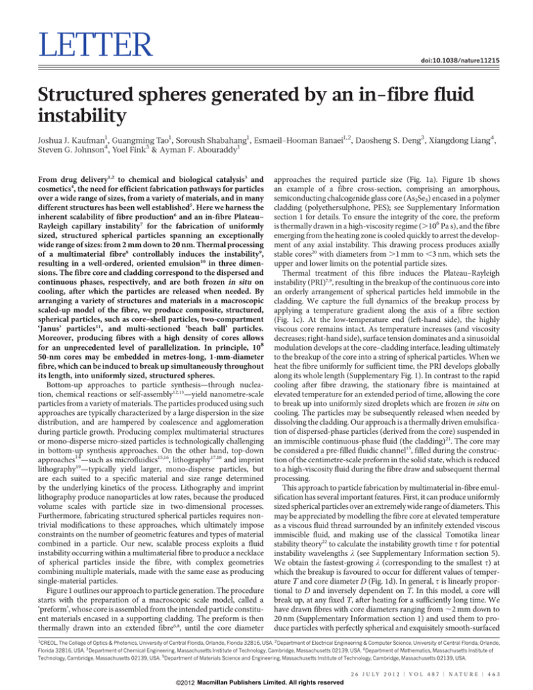
Figure 1 outlines our approach to particle generation. The procedure
starts with the preparation of a macroscopic scale model, called a
‘preform’, whose core is assembled from the intended particle constituent materials encased in a supporting cladding. The preform is then
thermally drawn into an extended fibre6,8, until the core diameter
approaches the required particle size (Fig. 1a). Figure 1b shows
an example of a fibre cross-section, comprising an amorphous,
semiconducting chalcogenide glass core (As2Se3) encased in a polymer
cladding (polyethersulphone, PES); see Supplementary Information
section 1 for details. To ensure the integrity of the core, the preform
is thermally drawn in a high-viscosity regime (.106 Pa s), and the fibre
emerging from the heating zone is cooled quickly to arrest the development of any axial instability. This drawing process produces axially
stable cores20 with diameters from .1 mm to ,3 nm, which sets the
upper and lower limits on the potential particle sizes.
Thermal treatment of this fibre induces the Plateau–Rayleigh
instability (PRI)7,9, resulting in the breakup of the continuous core into
an orderly arrangement of spherical particles held immobile in the
cladding. We capture the full dynamics of the breakup process by
applying a temperature gradient along the axis of a fibre section
(Fig. 1c). At the low-temperature end (left-hand side), the highly
viscous core remains intact. As temperature increases (and viscosity
decreases; right-hand side), surface tension dominates and a sinusoidal
modulation develops at the core–cladding interface, leading ultimately
to the breakup of the core into a string of spherical particles. When we
heat the fibre uniformly for sufficient time, the PRI develops globally
along its whole length (Supplementary Fig. 1). In contrast to the rapid
cooling after fibre drawing, the stationary fibre is maintained at
elevated temperature for an extended period of time, allowing the core
to break up into uniformly sized droplets which are frozen in situ on
cooling. The particles may be subsequently released when needed by
dissolving the cladding. Our approach is a thermally driven emulsification of dispersed-phase particles (derived from the core) suspended in
an immiscible continuous-phase fluid (the cladding)21. The core may
be considered a pre-filled fluidic channel15, filled during the construction of the centimetre-scale preform in the solid state, which is reduced
to a high-viscosity fluid during the fibre draw and subsequent thermal
processing.
This approach to particle fabrication by multimaterial in-fibre emulsification has several important features. First, it can produce uniformly
sized spherical particles over an extremely wide range of diameters. This
may be appreciated by modelling the fibre core at elevated temperature
as a viscous fluid thread surrounded by an infinitely extended viscous
immiscible fluid, and making use of the classical Tomotika linear
stability theory22 to calculate the instability growth time t for potential
instability wavelengths l (see Supplementary Information section 5).
We obtain the fastest-growing l (corresponding to the smallest t) at
which the breakup is favoured to occur for different values of temperature T and core diameter D (Fig. 1d). In general, t is linearly proportional to D and inversely dependent on T. In this model, a core will
break up, at any fixed T, after heating for a sufficiently long time. We
have drawn fibres with core diameters ranging from ,2 mm down to
20 nm (Supplementary Information section 1) and used them to produce particles with perfectly spherical and exquisitely smooth-surfaced
1
CREOL, The College of Optics & Photonics, University of Central Florida, Orlando, Florida 32816, USA. 2Department of Electrical Engineering & Computer Science, University of Central Florida, Orlando,
Florida 32816, USA. 3Department of Chemical Engineering, Massachusetts Institute of Technology, Cambridge, Massachusetts 02139, USA. 4Department of Mathematics, Massachusetts Institute of
Technology, Cambridge, Massachusetts 02139, USA. 5Department of Materials Science and Engineering, Massachusetts Institute of Technology, Cambridge, Massachusetts 02139, USA.
2 6 J U LY 2 0 1 2 | VO L 4 8 7 | N AT U R E | 4 6 3
©2012 Macmillan Publishers Limited. All rights reserved
RESEARCH LETTER
b
d
100
2
10
1
0
Preform
0.1
P
500 μm
Fibre
c
0.01
external morphology over the entire range of diameters (Fig. 1e, f),
confirmed by scanning electron microscope (SEM) imaging after dissolving the polymer cladding using dimethylacetamide23. This range
corresponds to five orders of magnitude in linear dimension—fifteen
in volume, from ,8 mm3 to ,8,000 nm3. The polydispersity of the
particle distribution for targeted micro- and nanoparticle sizes was
determined using dynamic light scattering, and the standard deviation
normalized with respect to the mean of the size distribution was found
to be ,10% (see Supplementary Fig. 2 for details).
The second key aspect of the in-fibre process is its scalability—that
is, the ability to produce large numbers of particles by parallelizing the
simultaneous breakup of a high density of cores occupying the same
long fibre. Starting from a macroscopic rod, one may in principle
convert its entirety into particles of prescribed size. Using a stackand-draw approach, we have produced fibres of 1 mm outer diameter,
containing 12 20-mm cores (Fig. 2a and Supplementary Fig. 3), 4,000
500-nm cores, or 27,000 200-nm cores (Fig. 2d, Supplementary Figs 4, 5;
Supplementary Table 1). In principle, one may combine 108 50-nm
cores in such a fibre with 25% fill factor. This far exceeds the current
parallelization capabilities of microfluidics-based approaches24.
Furthermore, the resulting spatial distribution of particles held immobilized in the scaffold is well-ordered in three dimensions (Fig. 2b,
Supplementary Fig. 6). In the axial direction the particles are ordered
because the instability growth is dominated by a single wavelength. In
the transverse dimensions, order is imposed on the cores during the
stacking process (Supplementary Figs 3–6).
The third characteristic is the ease by which this top-down process
may be configured to produce structured particles. Because the preform
is constructed at the centimetre scale, complex preform geometries may
be readily designed and realized, so that the PRI-driven breakup in the
drawn fibre produces a desired particle structure. We demonstrate
here the size-controllable fabrication of spherical core–shell particles
(Fig. 3) and ‘Janus’ particles (Fig. 4). The preform used to produce the
core–shell particles (corresponding to a double emulsion15) consists of
a polymer core (diameter D1) and glass cladding (diameter
D2 < 2.5 3 D1), surrounded by a polymer matrix (Fig. 3a; crosssections shown in Fig. 3b, c). The polymer core and glass shell undergo
a correlated PRI-driven breakup that results in core–shell particles,
observed experimentally (Fig. 3d, e) and confirmed through simulations (Fig. 3f). To confirm that the PRI-driven breakup produces the
4
280
log [τ (s)]
G
1,000
D (μm)
a
310
340
T (°C)
200 μm
G
P
T
Fib
re
Pa
rtic
les
e
500 μm
100 μm
10 μm
1μm
f
500 nm
300 nm
50 nm
20 nm
Figure 1 | Fluid capillary instabilities in multimaterial fibres as a route to
size-tunable particle fabrication. a, A macroscopic preform is thermally
drawn into a fibre. Subsequent thermal processing of the fibre induces the PRI,
which results in the breakup of the intact core into spherical droplets that are
frozen in situ on cooling. b, Reflection optical micrograph of a fibre crosssection with 20-mm-diameter core; inset shows the core (scale bar, 20 mm). The
fibre consists of an As2Se3 glass core (G), encased in a PES polymer cladding
(P). c, Transmission optical micrograph of the fibre side-view in b after a
temperature (T) gradient is applied along the axis to induce the PRI at the core–
cladding interface. d, Calculated instability time, t, for various temperatures T
and core diameters D (see Supplementary Information). e, SEM images of
microparticles with diameters of ,1.4 mm, 200 mm, 18 mm and 2.7 mm. f, SEM
images of nanoparticles with diameters of ,920, 560, 62 and 20 nm.
a
b
500 μm
G
c
150 μm
200 μm
G
P
P
d
e
10 μm
f
10 μm
500 nm
G
Figure 2 | Scalable fabrication of micro- and nano-scale spherical particles.
a, SEM micrograph of 12 20-mm intact glass cores (G, As2Se3), exposed from a
1-mm-diameter fibre after dissolving the polymer cladding (P, PES). An SEM
micrograph of the fibre cross-section is shown in Supplementary Fig. 3.
b, Transmission optical micrograph of the fibre side-view, showing the cores
after global heating of the fibre, which results in the simultaneous breakup of
the cores into an ordered distribution of particles in three dimensions held in
the polymer cladding. c, SEM micrograph of a large number of 40-mm (average
diameter) glass particles released from the fibre in b by dissolving the polymer
cladding. d, SEM micrograph of 27,000 200-nm-diameter intact glass cores
exposed from a 1-mm-diameter fibre. An SEM micrograph of the fibre crosssection is shown in Supplementary Figs 4, 5. e, SEM micrograph of a large
number of 400-nm (average diameter) glass particles. f, SEM micrograph of a
few particles from e. See Supplementary Fig. 2 for the particle-size distribution.
4 6 4 | N AT U R E | V O L 4 8 7 | 2 6 J U LY 2 0 1 2
©2012 Macmillan Publishers Limited. All rights reserved
LETTER RESEARCH
P
G
a
P
b
c
200 μm
20 μm
P G P
f
λ
d
D1 D2
e
2 μm
Top view
g
G
G
G
G
P
P
P
P
Front view
h
G
20 μm
G
P
3 μm
G
G
P
P
500 nm
P
200 nm
Figure 3 | Polymer-core/glass-shell spherical particle fabrication.
a, Schematic of the fibre structure (P, G as in Figs 1, 2). b, c, SEM images of fibre
cross-sections. d, SEM image of the glass-shell outer surface, showing the
modulation characteristic of the PRI. e, SEM image of the structure in d after
sectioning off half of the glass shell using a focused ion beam (FEI 200 THP;
current ,10–100 pA), revealing the correlated modulations on the two
interfaces (inner polymer/glass and outer glass/polymer interfaces), and
resulting ultimately in two concentric spherical surfaces as shown in g and
h. f, Three snapshots from a three-dimensional simulation of the Stokes
equations using a representative fibre structure (full movie available online; see
Supplementary Information), illustrating the full breakup process. Time
progresses from top to bottom. Scale bar, 50 mm. Dark green, polymer core;
light green, glass shell; the outer polymer scaffold cladding is made transparent
for clarity. g, Top and h, front (tilted) SEM views of four differently sized core–
shell particles (outer diameters 34 mm, 7 mm, 1.2 mm and 650 nm, respectively).
Scale bars in the corresponding top and front views are the same length.
expected structure, we use a focused ion beam (FIB) to ‘slice’ the particle
down the middle by raster-scanning the FIB across a box with an edge
lying through the particle. The FIB etches the semiconducting glass
shell, with its higher electrical conductivity, more effectively than the
insulating polymer core. Figure 3g, h shows SEM images of particles
with outer diameters from 35 mm to 600 nm (the FIB damages the
smaller particles that we produced), showing the intact polymer core
protruding from the remaining glass half-shell. Figure 3g, h confirms
the smooth core/shell interface, the particles’ concentric spherical
surfaces, and that the expected core/shell diameter ratio is
D19/D29 5 (D1/D2)2/3, as dictated by conservation of volume (where
D19 and D29 are the particle core and shell diameters, respectively).
Since D1/D2 5 0.4 (Fig. 3b, c), we expect D19/D29 < 0.543, in close
agreement with the measured value of ,0.575 (Fig. 3g).
We quantitatively analyse the breakup process in this nested,
cylindrical, multi-fluid structure using linear stability analysis25 to
determine the exponential growth rates of small sinusoidal perturbations. The dominant breakup wavelength is plotted as a function of the
core/shell viscosity ratio in Supplementary Fig. 7. The predicted and
measured breakup length scales are consistent, within the experimental
uncertainties in D1 and viscosity contrast. To study the dynamics
of the full breakup process, we performed full three-dimensional
simulations of the Stokes equations (valid here because the
Reynolds number is low26), using a level-set/spectral method25. For
illustration purposes we used equal core and shell viscosities (105 Pa s,
corresponding to T < 270 uC) and an initial diameter D1 5 23 mm.
Three snapshots of the simulation, starting from white-noise initial
perturbations, are shown in Fig. 3f (full movie available online). The
inner interface breaks up first, as predicted from stability analysis and
observed experimentally (Fig. 3d, e), and we also occasionally observe
small ‘satellite’ droplets forming among the larger droplets.
The second structured particle we produce is a broken-symmetry,
spherical Janus particle, comprising two hemispheres of different
optical glasses (Fig. 4). The preform core is constructed of two half
cylinders, each of a different semiconducting glass with distinct complex refractive index: G1 (As2S3) and G2 ((As2Se3)99Ge1) (Fig. 4a–c;
Supplementary Information section 7). The induced breakup produces
spherical Janus particles held immobilized with the same orientation
in the cladding (Fig. 4d). Figure 4e shows a reflection optical
micrograph of a single Janus particle removed from the cladding.
We confirm the three-dimensional structure of the particle by optically
imaging multiple parallel planes cutting through a particle still
embedded in the polymer cladding (Fig. 4f), and correlating the optical
images with energy-dispersive X-ray diffraction (EDX) spectral images
of the particle cross-section (Fig. 4g) that identify arsenic and sulphur.
The measurements confirm the three-dimensional, two-compartment
structure of the Janus particle.
Modelling Janus-particle formation is difficult because it involves a
point where three fluids meet, so that sophisticated level-set techniques
are required to describe the interfaces27. The physics of such a contact
point is not well understood28, although it is likely to be less relevant in
the Stokes regime29,30. Nevertheless, energy considerations yield some
qualitative predictions. A large glass–glass surface tension, compared
to that between glass and polymer, would make it energetically favourable for the Janus particles to pinch in the centre. On the other hand,
for negligible glass–glass tension, if the glass–polymer surface tension
were very different for the two glasses, energy would be lowered if one
glass were to flow to envelop the other. As neither of these scenarios is
observed experimentally (Fig. 4d, e), we can conclude that the observed
breakup process is consistent with low glass–glass surface tension and
similar glass–polymer tensions. These considerations indicate a general
strategy for the construction of particles with even more complex geometry. Furthermore, to form two-component particles, the viscosities of
the two materials must be matched; we identify pairs of compatible
materials by looking for overlapping softening temperatures.
The two particle structures considered above, the core–shell and
two-compartment Janus particles, are prototypical structures from
which more complex geometries may be constructed. For example,
multilayer particles may be produced using a core consisting of nested
cylindrical shells of appropriate thicknesses, and additional azimuthal
compartments in the particle result from a core appropriately prepared
with azimuthal sections. Furthermore, these two prototypical structures may be combined in the same particle. The power of this
approach is highlighted in Fig. 4h–k, which shows the fabrication of
a ‘beach ball’ particle, consisting of six equally sized wedge-shaped
sections of alternating materials (G1 and G2). The preform consists
of a cylindrical core with six equally sized segments, each subtending a
60u polar angle. More complex particle structures may be produced by
judiciously structuring the core.
This process uses thermally compatible material systems dominated
by viscous forces and surface tension, such as glasses, polymers, metals
above their melting temperature, and liquids. An example of breakup
in an all-polymer fibre is shown in Supplementary Fig. 10. Moreover,
drawing multimaterial fibres with crystalline semiconductor cores31
(silicon, germanium and III–V binary compounds) and the synthesis
of new materials during fibre drawing32 indicate the possibility of
extending our methodology to a wider range of materials. Finally,
fibre fabrication technology produces kilometres of fibre in a few
hours6, with a total core mass of 1 kg that is potentially converted
entirely into particles, with each metre of fibre containing up to
2 6 J U LY 2 0 1 2 | VO L 4 8 7 | N AT U R E | 4 6 5
©2012 Macmillan Publishers Limited. All rights reserved
RESEARCH LETTER
c
b
G2
a
G2
100 μm
G1
G1
G1
P
G2
d 200 μm
T
P
e
f
g
10 μm
5 μm
G2
G1
h
G2
G1
i
j
As
S
As
S
10 μm
P
k
P
G2
G1
G2
G1
Figure 4 | Broken-symmetry Janus particle and ‘beach ball’ particle
fabrication. a, Schematic of the Janus preform. G1, As2S3; G2, (As2Se3)99Ge1; P,
PES. b, Reflection optical micrograph of a Janus fibre cross-section; scale bar
20 mm. c, Transmission optical micrograph of the fibre side view. d, Transmission
optical micrograph showing PRI growth, leading to breakup of the Janus
particles. e, Reflection optical micrograph of an individual Janus particle after
removal from the fibre. f, Optical micrographs of multiple sections at different
depths within a single Janus particle embedded in the fibre, exposed sequentially
by polishing. The particle symmetry plane is tilted with respect to the direction of
polishing, and the tilt is similar to that in the particle shown in e. g, EDX spectral
images (for arsenic, As, and sulphur, S) of an exposed Janus particle cross-section,
corresponding to a section from f. The dashed blue circle and line are visual aids.
See Supplementary Fig. 8 for full EDX spectrum, Supplementary Fig. 9a–c for
another example of EDX spectral imaging, and Supplementary Fig. 9d–f for a
demonstration of Janus particle size-control. h, Schematic of the preform to
produce ‘beach ball’ particles; G1, G2 and P as above. i, Reflection optical
micrograph of a ‘beach ball’ fibre cross-section; scale bar, 20 mm. j, EDX spectral
images (as in g) of an exposed ‘beach ball’ particle cross-section. k, Transmission
optical micrographs of the cross-sections of a 40-mm-diameter particle
immobilized in the polymer matrix in the fibre; scale bar 20 mm.
1014 100-nm-diameter particles, well ordered in three dimensions.
Particles are produced at the same volume rate regardless of the
particle size, as it is inherently a three-dimensional process that relies
only on the fibre fill factor.
Further control over the preform construction will result in particles
with even more complex structures. This scalable process, in which we
assemble disparate components that ‘fit’ together in size and shape
macroscopically for the scalable production of size-tunable structured
particles, enables a large range of applications. The well ordered,
oriented and immobilized three-dimensional particle distribution in
a scaffold (Supplementary Fig. 6) could potentially be used as threedimensional optical and acoustic meta-materials; the surface-tensiondriven smooth spherical surface morphology of the particles enables
optical-resonance-based sensitive detection of chemical species and
pathogens; and three-dimensional structural control over particles
impregnated with drugs could help realize sophisticated controlledrelease drug delivery systems.
2.
3.
4.
5.
6.
7.
8.
9.
10.
11.
12.
13.
14.
Received 20 December 2011; accepted 2 May 2012.
15.
Published online 18 July 2012.
1.
Timko, B. P. et al. Advances in drug delivery. Annu. Rev. Mater. Res. 41, 1–20
(2011).
16.
Wang, J., Byrne, J. D., Napier, M. E. & DeSimone, J. M. More effective nanomedicines
through particle design. Small 7, 1919–1931 (2011).
Bell, A. T. The impact of nanoscience on heterogeneous catalysis. Science 299,
1688–1691 (2003).
Souto, E. B. & Müller, R. H. Cosmetic features and applications of lipid
nanoparticles. Int. J. Cosmet. Sci. 30, 157–165 (2008).
Rotello, V. Nanoparticles: Building Blocks for Nanotechnology (Springer, 2003).
Li, T. (ed.) Optical Fiber Communications Vol. 1, Fiber Fabrication (Academic, 1985).
Eggers, J. & Villermaux, E. Physics of liquid jets. Rep. Prog. Phys. 71, 036601
(2008).
Abouraddy, A. F. et al. Towards multimaterial multifunctional fibres that see, hear,
sense and communicate. Nature Mater. 6, 336–347 (2007).
Shabahang, S., Kaufman, J. J., Deng, D. S. & Abouraddy, A. F. Observation of the
Plateau-Rayleigh capillary instability in multi-material optical fibers. Appl. Phys.
Lett. 99, 161909 (2011).
Sjöblom, J. Encyclopedic Handbook of Emulsion Technology (Marcel Dekker, 2001).
Walther, A. & Müller, A. H. E. Janus particles. Soft Matter 4, 663–668 (2008).
Cao, G. Nanostructures and Nanomaterials: Synthesis, Properties and Applications
(Imperial College Press, 2004).
Vollath, D. Nanomaterials: An Introduction to Synthesis, Properties and Application
(Wiley-VCH, 2008).
Merkel, T. J. et al. Scalable shape-specific, top-down fabrication methods for the
synthesis of engineered colloidal microparticles. Langmuir 26, 13086–13096
(2010).
Utada, A. S. et al. Monodisperse double emulsions generated from a microcapillary
device. Science 308, 537–541 (2005).
Dendukuri, D. & Doyle, P. S. The synthesis and assembly of polymeric
microparticles using microfluidics. Adv. Mater. 21, 4071–4086 (2009).
4 6 6 | N AT U R E | V O L 4 8 7 | 2 6 J U LY 2 0 1 2
©2012 Macmillan Publishers Limited. All rights reserved
LETTER RESEARCH
17. Dendukuri, D., Pregibon, D. C., Collins, J., Hatton, T. A. & Doyle, P. S. Continuous-flow
lithography for high-throughput microparticle synthesis. Nature Mater. 5,
365–369 (2006).
18. Hernandez, C. J. & Mason, T. G. Colloidal alphabet soup: Monodisperse dispersions
of shape-designed LithoParticles. J. Phys. Chem. C 111, 4477–4480 (2007).
19. Rolland, J. P. et al. Direct fabrication and harvesting of monodisperse, shape
specific nano-biomaterials. J. Am. Chem. Soc. 127, 10096–10100 (2005).
20. Kaufman, J. J. et al. Thermal drawing of high-density macroscopic arrays of wellordered sub-5-nm-diameter nanowires. Nano Lett. 11, 4768–4773 (2011).
21. Nie, Z. H. et al. Emulsification in a microfluidic flow-focusing device: Effect of the
viscosities of the liquids. Microfluidics and Nanofluidics 5, 585–594 (2008).
22. Tomotika, S. On the instability of a cylindrical thread of a viscous liquid surrounded
by another viscous fluid. Proc. R. Soc. Lond. A 150, 322–337 (1935).
23. Deng, D. S. et al. In-fiber nanoscale semiconductor filament arrays. Nano Lett. 8,
4265–4269 (2008).
24. Nisisako, T. & Torii, T. Microfluidic large-scale integration on a chip for mass
production of monodisperse droplets and particles. Lab Chip 8, 287–293 (2008).
25. Liang, X., Deng, D. S., Nave, J.-C. & Johnson, S. G. Linear stability analysis of capillary
instabilities for concentric cylindrical shells. J. Fluid Mech. 683, 235–262 (2011).
26. Deng, D. S., Nave, J.-C., Liang, X., Johnson, S. G. & Fink, Y. Exploration of in-fiber
nanostructures from capillary instability. Opt. Express 19, 16273–16290 (2011).
27. Smith, K. A., Solis, F. J. & Chopp, D. L. A projection method for motion of triple
junctions by levels sets. Interfaces Free Bound. 4, 263–276 (2002).
28. Dussan, V. E. B. On the spreading of liquids on solid surfaces: static and dynamic
contact lines. Annu. Rev. Fluid Mech. 11, 371–400 (1979).
29. de Gennes, P. G. Wetting: statics and dynamics. Rev. Mod. Phys. 57, 827–863
(1985).
30. Israelachvili, J. N. Intermolecular and Surface Forces (Academic, 1992).
31. Ballato, J. et al. Advancements in semiconductor core optical fiber. Opt. Fiber
Technol. 16, 399–408 (2010).
32. Orf,N. D.et al. Fiber draw synthesis.Proc. Natl Acad. Sci. USA108, 4743–4747 (2011).
Supplementary Information is linked to the online version of the paper at
www.nature.com/nature.
Acknowledgements Work at UCF was supported by the US National Science
Foundation (award number ECCS-1002295), a Ralph E. Powe Junior Faculty
Enhancement Award from the Oak Ridge Associated Universities (ORAU), in part by the
US Air Force Office of Scientific Research (AFOSR) under contract FA-9550-12-1-0148,
and by CREOL, The College of Optics & Photonics. Work at MIT was supported in part by
the Materials Research Science and Engineering Program of the US NSF under award
number DMR-0819762, and also in part by the US Army Research Office through the
Institute for Soldier Nanotechnologies under contract number W911NF-07-D-0004.
We thank Sasha Stolyarov, J. Manuel Perez, Sudipta Seal and Kirk Scammon for
assistance. We especially thank M. J. Soileau, B. E. A. Saleh, D. N. Christodoulides and
M. Z. Bazant for encouragement and support.
Author Contributions J.J.K., Y.F. and A.F.A. developed and directed the project. S.S. first
observed the PRI phenomenon, developed the fibre tapering process and the particle
extraction approach, and demonstrated the scale invariance of the PRI and particle
extraction strategies. G.T. prepared and characterized all the glasses, carried out the
preform extrusions, and produced the ‘beach ball’ fibre. J.J.K. produced the other
preforms and fibres, performed PRI breakup and particle extraction experiments, and
carried out the SEM, EDX, FIB and optical imaging and characterization. E.-H.B. aided in
choice and characterization of materials and in preparation of the polymers. D.S.D., X.L.
and S.G.J. carried out the theoretical calculations and performed the simulations. J.J.K.,
D.S.D., Y.F. and A.F.A. wrote the paper. All authors contributed to the interpretation of the
results.
Author Information Reprints and permissions information is available at
www.nature.com/reprints. The authors declare no competing financial interests.
Readers are welcome to comment on the online version of this article at
www.nature.com/nature. Correspondence and requests for materials should be
addressed to A.F.A. (raddy@creol.ucf.edu).
2 6 J U LY 2 0 1 2 | V O L 4 8 7 | N AT U R E | 4 6 7
©2012 Macmillan Publishers Limited. All rights reserved
CORRECTIONS & AMENDMENTS
ERRATUM
doi:10.1038/nature11454
Erratum: Structured spheres
generated by an in-fibre fluid
instability
Joshua J. Kaufman, Guangming Tao, Soroush Shabahang,
Esmaeil-Hooman Banaei, Daosheng S. Deng, Xiangdong Liang,
Steven G. Johnson, Yoel Fink & Ayman F. Abouraddy
Nature 487, 463–467 (2012); doi:10.1038/nature11215
In this Letter, the received date was incorrectly listed as 20 December
2012 instead of 20 December 2011; this has been corrected in the
HTML and PDF versions of the manuscript.
0 0 M O N T H 2 0 1 2 | VO L 0 0 0 | N AT U R E | 1
©2012 Macmillan Publishers Limited. All rights reserved


