Respiratory passages
advertisement
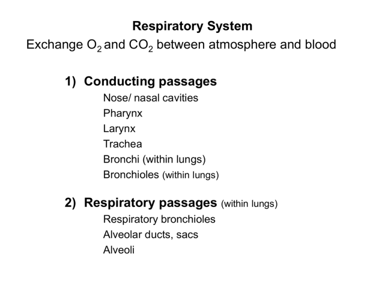
Respiratory System Exchange O2 and CO2 between atmosphere and blood 1) Conducting passages Nose/ nasal cavities Pharynx Larynx Trachea Bronchi (within lungs) Bronchioles (within lungs) 2) Respiratory passages (within lungs) Respiratory bronchioles Alveolar ducts, sacs Alveoli Nose – Nasal Cavities Function 1) Cleanse 2) Warm 3) Humidify air 4) Olfaction Structure Bone Cartilage Mucous membrane = mucosa 1) Epithelium 2) Underlying connective tissue Mucous membrane = mucosa 1) Epithelium Pseudostratified columnar epithelium with cilia and goblet cells Goblet cells are mucus secreting cells shaped like goblets due to apical region filled with mucus Pharynx Lacks anterior wall; opens to nose, mouth, and larynx anteriorly 1) Nasal (naso-)pharynx 2) Oral (oro-)pharynx 3) Laryngeal (laryngo-)pharynx (hypophaynx) Larynx: Skeleton Major Movements of Larynx 1) Thyroid cartilage hinges on cricoid cartilage Tenses or loosens the vocal cords 2) Arytenoid cartilages pivot on cricoid cartilage Open and close the glottis (space between the vocal cords) Trachea 10 to 12 cm (4-5”) C6 to T4 2 to 2 ½ cm diameter Primary Bronchi 1) Right bronchus is shorter Trachea is slightly to right of aorta 2) Right bronchus is wider Right lung is larger (heart is on the left) 3) Right bronchus has more direct path (more vertical) 1) Primary bronchi Trachea bifurcates to 2; Right and left One / lung Branch into: 2) Secondary (lobar) bronchi One / lobe of lung 3 on right, 2 on left Branch into: 3) Tertiary (segmental) bronchi One / bronchopulmonary segments Bronchopulmonary Segments Figure 21.15 (1 of 2) Phrenic nerve (C3,4,5) from cervical plexus innervates diaphragm 1) Extrapulmonary bronchi Same structure as trachea 2) Intrapulmonary bronchi Cartilage in spirals and plaques Layer of smooth muscle internal to cartilage Layers of bronchial wall 1) 2) 3) 4) 5) Mucosa – same Layer of smooth muscle Submucosa – CT Cartilage in plaques Elastic fibers in submucosa Bronchi get progressively smaller As decrease size of bronchi > 1) Increase in smooth muscle 2) Decrease in cartilage When cartilage gone = bronchiole Bronchioles No cartilage Complete ring of smooth muscle, also elastic fibers Epithelium = simple columnar > simple cuboidal, no goblet cells Smallest = Terminal bronchioles Respiratory Passages Respiratory bronchioles Alveoli along wall Alveolar ducts Alveoli increase in density until solid wall of alveoli Alveolar sac Blind end of passages Totally lined by alveoli Alveolus 300 million alveoli 70 to 80 square meters of surface area Thin walled sacs (<1 micron) Back to back with capillary network Alveolar wall 1) Type I pulmonary epithelial cells 2) Type II pulmonary cell = great alveolar cells = septal cells Dust cells Alveolar wall 1) Type I pulmonary epithelial cells Simple squamous epithelial cells Alveolar wall 2) Type II pulmonary cell = great alveolar cells = septal cells Produce surfactant – decreases surface tension to ease work of distending lungs Alveolar wall 3) Dust cells (does not form wall) Wandering macrophages, within lumen Diffusion barrier (.5 micron) 1) Pulmonary cell 2) Basement membrane 3) Endothelial cells of capillary
