Cow Eye Dissection Lab: Anatomy & Procedure
advertisement
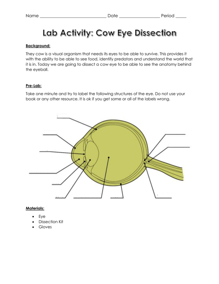
Name _______________________________ Date ___________________ Period _____ Background: They cow is a visual organism that needs its eyes to be able to survive. This provides it with the ability to be able to see food, identify predators and understand the world that it is in. Today we are going to dissect a cow eye to be able to see the anatomy behind the eyeball. Pre-Lab: Take one minute and try to label the following structures of the eye. Do not use your book or any other resource. It is ok if you get some or all of the labels wrong. Materials: Eye Dissection Kit Gloves Name _______________________________ Date ___________________ Period _____ Procedure: 1. Examine the outside of the eye. See how many parts of the eye you can identify. You should be able to find the whites (or sclera), the tough, outer covering of the eyeball. You should also be able to identify the fat and muscle surrounding the eye. You should be able to find the covering over the front of the eye (the cornea). When the cow was alive, the cornea was clear. In your cow’s eye, the cornea may be cloudy. You may be able to look through the cornea and see the iris, the colored part of the eye, and the pupil, the dark oval in the middle of the iris. 2. Cut away the fat and muscle. However be careful not to cut the optic nerve. 3. Use a scalpel to make an incision across the cornea. Cut until the clear liquid under the cornea is released. That clear liquid is the aqueous humor. It’s made of mostly of water and keeps the shape of the cornea. 4. Using your scalpel, now make an incision along the coronal plane of the eye. This incision should separate the lens, iris and cornea from the retina and the optic nerve 5. Now, use your scissors to cut around the middle of the eye, cutting the eye in half. You’ll end up with two halves. On the front half will be the cornea. Once you have removed the cornea, place it on the board (or cutting surface) and cut it with your scalpel or razor. Listen. Hear the crunch? That’s the sound of the scalpel crunching through layers of clear tissue. The cow’s cornea has many layers to make it thick and strong. 6. The next step is to pull out the iris. The iris is between the cornea and the lens. It may be stuck to the cornea or it may have stayed with the back of the eye. Find the iris and pull it out. It should come out in one piece. You can see that there’s a hole in the center of the iris. That’s the pupil, the hole that lets light into the eye. The iris contracts or expands to change the size of the pupil. In dim light, the pupil opens wide to let light in. In bright light, the pupil shuts down to block light out. 7. The back of the eye is filled with a clear jelly. That’s the vitreous humor, a mixture of protein and water. It’s clear so light can pass through it. It also helps the eyeball maintain its shape. 8. Now you want to remove the lens. It’s a clear lump about the size and shape of a squashed marble. The lens of the cow’s eye feels soft on the outside and hard in the middle. Hold the lens up and look through it. What do you see? Name _______________________________ Date ___________________ Period _____ 9. Put the lens down on a spare piece of paper with words written on the paper. What do you see? 10. Now take a look at the rest of the eye. If the vitreous humor is still in the eyeball, empty it out. On the inside of the back half of the eyeball, you can see some blood vessels that are part of a thin fleshy film. That film is the retina. Before you cut the eye open, the vitreous humor pushed against the retina so that it lay flat on the back of the eye. It may be all pushed together in a wad now. 11. Use your finger to push the retina around. The retina is attached to the back of the eye at just one spot. Can you find that spot? That’s the place where nerves from all the cells in the retina come together. All these nerves go out the back of the eye, forming the optic nerve, the bundle of nerves that carries messages from the eye to the brain. The brain uses information from the retina to make a mental picture of the world. The spot where the retina is attached to the back of the eye is called the blind spot. Because there are no light-sensitive cells at that spot, you can’t see anything that lands in that place on the retina. 12. Under the retina, the back of the eye is covered with shiny, blue-green stuff. This is the tapetum. It reflects light from the back of the eye. 13. Look at the other side of the back of the eye. Can you find the optic nerve? To see the separate fibers that make up the optic nerve, pinch the nerve with a pair of scissors or your fingers. If you squeeze the optic nerve, you may get some white goop. That is myelin, the fatty layer that surrounds each fiber of the nerve. 14. Use the remaining time to clean up. Wash down all of your lab equipment, lab table and any other areas that may have gotten dissection materials on them. 15. Return all equipment to its original location. Name _______________________________ Date ___________________ Period _____ Analysis Questions: 1. What did you see when you looked through the lens of the eye? What did you see when the lens was put on the scrap paper? 2. Describe the cornea. What about a cow might have led to the cornea having been so tough? 3. Describe what the iris felt like. What was the reason that the iris felt like this? 4. The back of a cow’s eye is reflective. Since a human’s eye is not reflective, what part of the cow eye do we not have? 5. Look at your pre-lab. Which labels did you get correct? Which labels did you get incorrect? Use the next page and get all of them correct. Name _______________________________ Date ___________________ Period _____

