Integumentary System
advertisement
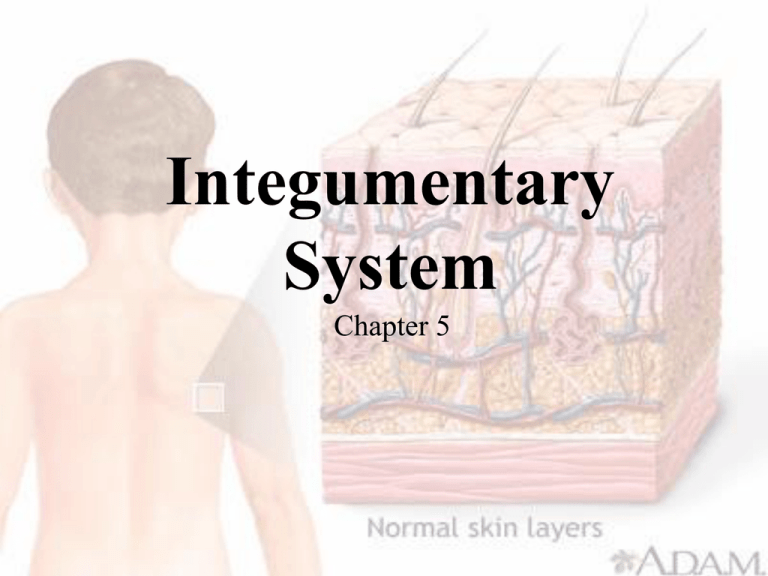
Integumentary System Chapter 5 Objectives • Classify, compare the structure of, and give examples of each type of body membrane. • Describe the structure and function of the epidermis and dermis. • List and briefly describe each accessory organ of the skin. Objectives • List and discuss the three primary functions of the integumentary system. • Classify burns and describe how to estimate the extent of a burn injury. Terminology • Integument is another name for the skin. • Comprised of a group of simple, functional, sheet like membranes. – Found throughout the body. • Two major categories: – Epithelial membranes – Connective tissue membranes Epithelial Membranes • Composed of epithelial tissue and an underlying layer of special connective tissue. • Three types: – Cutaneous Membrane – Serous Membrane – Mucous Membrane Cutaneous Membrane • Skin • Largest and most visible organ in the body. – 16% of body weight. Serous Membrane • Simple squamous epithelium on a connective tissue basement membrane. • Two Types: – Parietal – Lines the walls of a body cavity. – Visceral – Covers the surface of organs within the cavity. Serous Membrane • Examples: – Pleura • Thoracic cavity – Peritoneum • Abdominal cavity • Secretes a thin watery lubrication fluid to help reduce friction between the organs and the walls of the cavity. Serous Membrane • Conditions: – Pleurisy • Painful inflammation of the chest cavity. – Peritonitis • Inflammation of the abdominal cavity. Mucous Membrane • Line body surfaces that open directly to the exterior. • Produce mucous, a thick secretion that keeps the membranes soft and moist. • Examples: – Respiratory – Digestive – Urinary – Reproductive Mucous Membrane • Mucocutaneous junction – Transitional area that serves as a point of “fusion” where the skin and mucous membrane meet. Connective Tissue Membranes • Do not contain epithelial components. • Produce a lubricant called synovial fluid. • Examples: – Synovial membranes lining a joint. Skin • Two primary layers: – Epidermis – Dermis • Supported by a thick layer of loose connective tissue subcutaneous layer. – Insulates from heat and cold. – Shock absorbing pad. Epidermis • Thinner, outermost layer. • Composed of several layers of stratified squamous epithelium. – Stratum germinativum • Innermost, that produces new cells – Stratum corneum • Outermost, keratin-filled cells Epidermis • Keratin – Tough waterproof protein • Soft – epidermis • Hard – hair and nails • Melanin – Brown pigment produced by melanocytes. – Absorbs harmful UV rays from damaging underlying structures. Dermis • Deeper, thicker layer. • Composed largely of connective tissue. • Upper area has parallel rows of peg-like dermal papillae – Ridges and grooves form a unique pattern. • Finger prints Dermis • Deeper areas of the dermis contain tough collagen protein material and elastic fibers. • Elasticity of the skin decreases with age forming wrinkles. Dermis • Dermis also contains network of nerves and nerve endings. – Pain, Pressure, Touch, Temperature • Muscle fibers • Hair follicles • Sudoriferous (Sweat) glands • Sebaceous (Oil) glands • Blood Vessel Appendages of the Skin • Hair – Tube like structure called a follicle is formed and hair is grown from a cluster of cells called the papilla. – Hair shaft is the visible portion, while the hair root remains hidden. – Arrector pili muscle will cause hair to stand and goose bumps to form when cold or frightened. Appendages of the Skin • Special Receptors – Meissner’s corpuscle • Detects light touch – Pacinian corpuscle • Detects pressure – Krause’s end bulbs • Low frequency vibrations Appendages of the Skin • Nails – Hardened epidermal cells. – Nail body is visible. – Crescent moon shaped area near the root is called the lunula. – Root lies hidden by cuticle. – Oxygen levels may be identified by color of nail bed. Appendages of the Skin • Sudoriferous (sweat) gland – Eccrine sweat gland • Most numerous and wide spread • Produces watery perspiration onto skin surface to regulate body heat. – Apocrine sweat gland • Axilla and groin • Produces thicker secretion and may have odor due to breakdown of secretion by bacteria. Appendages of the Skin • Sebaceous (oil) gland – Secrete an oil called sebum to lubricate hair and skin. – Regulated by sex hormones – Increased production at adolescence forming pimples or blackheads. Function of the Skin • Protection • Temperature Regulation • Sense Organ Protection • Acts as the bodies first line of defense. • Protects against: – Infection by pathogens – UV rays from the sun – Harmful chemicals – Physical trauma Temperature Regulation • Skin releases almost 3,000 calories of body heat per day. • Regulation of sweat. • Regulation of blood flow near surface of skin. Sense Organ • Skin functions as enormous sense organ keeping the body informed about its environment. • Pressure • Pain • Touch • Temperature Burns • Most serious and frequent problems of the skin. • Caused by: – Fire or hot surface – Overexposure to UV light – Chemical or acids Burns • Treatment and survival depend on the total area of the body burned and the depth of the burn. • Body surface estimated by “Rule of Nines”. • Degree of burn Rule of Nines • Body divided into 11 areas of 9% each. – 1% for groin area. Degree of Burns • First-degree burn – Burn involving only the surface layers of the epidermis. – Minor discomfort and reddening. – No blistering, but may peel. • Sunburn Degree of Burns • Second-degree burn (partial-thickness) – Burn involving deep layers of the epidermis and upper layers of dermis. – Blisters, severe pain, swelling, and damage to glands or hair follicles. – Scarring is common. Degree of Burns • Third-degree (full-thickness) burn – Complete destruction of epidermis, dermis, and even subcutaneous layers, muscle, or bone. – “Painless” burn as the nerve endings are destroyed. – Great risk of infection. Questions?

