Question - Mosaiced.org
advertisement
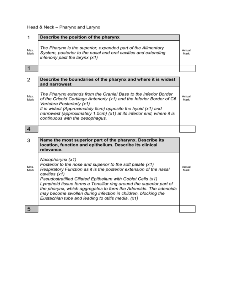
Head & Neck – Pharynx and Larynx 1 Describe the position of the pharynx Max. Mark The Pharynx is the superior, expanded part of the Alimentary System, posterior to the nasal and oral cavities and extending inferiorly past the larynx (x1) Actual Mark 1 2 Max. Mark Describe the boundaries of the pharynx and where it is widest and narrowest The Pharynx extends from the Cranial Base to the Inferior Border of the Cricoid Cartilage Anteriorly (x1) and the Inferior Border of C6 Vertebra Posteriorly (x1) It is widest (Approximately 5cm) opposite the hyoid (x1) and narrowest (approximately 1.5cm) (x1) at its inferior end, where it is continuous with the oesophagus. Actual Mark 4 3 Max. Mark 5 Name the most superior part of the pharynx. Describe its location, function and epithelium. Describe its clinical relevance. Nasopharynx (x1) Posterior to the nose and superior to the soft palate (x1) Respiratory Function as it is the posterior extension of the nasal cavities (x1) Pseudostratified Ciliated Epithelium with Goblet Cells (x1) Lymphoid tissue forms a Tonsillar ring around the superior part of the pharynx, which aggregates to form the Adenoids. The adenoids may become swollen during infection in children, blocking the Eustachian tube and leading to otitis media. (x1) Actual Mark 4 Max. Mark Name the middle part of the pharynx. Describe its location, boundaries, function and epithelium Oropharynx (x1) Posterior to the mouth (x1) Extends from the soft plate to the superior border of the epiglottis (x1) Digestive Function (x1) Stratified Squamous Epithelium non-Keratinised (x1) Actual Mark 5 5 Max. Mark Name the inferior part of the pharynx. Describe its location, boundaries and epithelium Laryngopharynx (x1) Posterior to the Larynx (x1) Ends from the superior border of the epiglottis to the inferior border of the cricoid cartilage, where it becomes continuous with the oesophagus (x1) Stratified Squamous Epithelium non-keratinised (x1) Actual Mark 4 6 Max. Mark Describe the wall of the pharynx The wall of the Pharynx consists of an incomplete outer circular muscle layer and an inner longitudinal muscle layer (x1) The muscle layer is covered internally by the Pharyngobasilar Fascia (x1), which is in turn covered by the Mucous Membrane (x1) Actual Mark 3 7 Max. Mark 4 Name the muscles involved in the outer circular muscle layer of the pharynx Superior Constrictor (x1) Middle Constrictor (x1) Inferior Constrictor (x1) Lower horizontal fibres known as Cricopharyngeus (x1) Actual Mark 8 Max. Mark Describe where the outer muscle layer attaches posteriorly and what happens to them anteriorly. Describe their function during swallowing The outer muscle layer attaches posteriorly at the midline raphe (x1) The muscles overlap each other and are incomplete anteriorly (x1) During swallowing the muscle constrict to propel the bolus of food downwards (involuntarily during the swallowing reflex) (x1) Actual Mark 3 9 Max. Mark Name the muscles involved in the inner longitudinal muscle layer of the pharynx. Describe their function during swallowing. Stylopharyngeus (x1) Palatopharyngeus (x1) Salpingopharyngeus (x1) During swallowing these muscles act to shorten and widen the pharynx (x1) Actual Mark 4 10 Max. Mark Describe the innervation of the pharynx Innervation of the Pharynx is by the Pharyngeal Plexus of nerves (x1) This is formed by branches of the Vagus (CN X) (x1) and Glossopharyngeal (CN IX) (x1) nerves along with sympathetic fibres from the Superior Cervical Ganglion (x1) Actual Mark 4 11 Describe the sensory innervation of the pharynx Max. Mark Glossopharyngeal Nerve (x1) Nasopharynx is via the Opthalmic and Maxillary divisions of the Trigeminal Nerve (CN V1+2) (x1) 2 Actual Mark 12 Describe the motor innervation of the pharynx Max. Mark Vagus Nerve (CN X) (x1) Exception to this is the Stylopharyngeus Muscle which is innervated by the glossopharyngeal nerve (CN IX) (x1) Actual Mark 2 13 Name and describe the first phase of swallowing Max. Mark Voluntary phase (x1) Tongue moves bolus to the back of the pharynx (x1) Actual Mark 2 14 Max. Mark Name and describe the second phase of swallowing Pharyngeal Phase (x1) Afferent information from pressure receptors in the palate and anterior pharynx reaches the swallowing centre in the brain stem (x1) A set of movements is triggered Inhibition of breathing Raising of the larynx – Suprahyoid and Longitudinal Muscles Closure of the glottis Opening of the upper oesophageal ‘sphincter’ (x1) for every 2 of the sets of movement Actual Mark 4 15 Max. Mark 4 Name and describe the third phase of swallowing Oesophageal Phase (x1) A wave of peristalsis sweeps down the oesophagus, propelling the bolus to the stomach in ~9 seconds (x1) Coordinated by extrinsic nerves from the swallowing centre of the brain (x1) Lower oesophageal ‘sphincter’ opens (x1) Actual Mark 16 Max. Mark Describe the different muscle in different areas of the oesophagus and what control they are under The muscle in the upper third of the oesophagus is voluntary striated muscle (x1) under somatic control (x1) The muscle of the lower two thirds is smooth muscle (x1) under control of the parasympathetic nervous system (x1) Actual Mark 4 17 Max. Mark Describe the blood supply of the pharynx. What artery do these vessels all branch off of? The blood supply of the Pharynx is via branches of the External Carotid Artery (x1) Ascending Pharyngeal Artery Lingual Artery Facial Artery Maxillary Artery Actual Mark (x1) for every 2 correct branches 3 18 Describe the venous drainage of the pharynx Max. Mark Venous drainage of the Pharynx is via the Pharyngeal Venous Plexus (x1) → Internal Jugular Vein (x1) Actual Mark 2 19 Max. Mark Describe the nerves in the afferent pathway and efferent pathway of the gag reflex Afferent - Glossopharyngeal Nerve (CN IX) (x1) Efferent - Vagus Nerve (CN X) (x1) Actual Mark 2 20 Max. Mark 1 What does the gag reflex test? Pharyngeal innervation and musculature (x1) Actual Mark 21 Max. Mark What are the adenoids? Describe their location. What happens to them during infection? What happens to them in recurrent infections? Sub-epithelial collection of lymphoid tissue (x1) Junction of roof and posterior wall of Nasopharynx (x1) Enlarge with viral / bacterial infections (x1) Recurrent infections may lead to chronically enlarge adenoids (x1) Actual Mark 4 22 Max. Mark Describe the clinical features of enlarged adenoids Nasal obstruction Mouth breathing, nasal speech Feeding difficulty (especially infants) Snoring / Obstructive Sleep Apnoea Block the opening of the Eustachian Tube Actual Mark (x1) for every 2, 3 marks for all 5 3 23 Max. Mark The Palatine Tonsils lie in the Tonsillar fossa between two arches Name these two arches and describe their position Anterior – Palatoglossal Arch (x1) Boundary between mouth and oropharynx (x1) Posterior – Palatopharyngeal Arch (x1) Contains the Palatopharyngeus Muscle that blends with walls of the pharynx (x1) Actual Mark 4 24 Max. Mark 2 Describe the lymphatic drainage of the palatine fossa and its location Jugulo-Digastric (Tonsillar) node (x1) Angle of the mandible (x1) Actual Mark 25 Max. Mark During a Tonsillectomy, there is a potential for bleeding as the palatine tonsils are very vascular. Describe what artery the bleeding is usually from. Describe any other nearby neurovascular structures Bleeding often from the large External Palatine Vein (x1) Internal Carotid Artery (x1) and Glossopharyngeal Nerve (x1) lie just lateral to Tonsillar fossa Actual Mark 3 26 Max. Mark What is Quinsy? How does it progress? What is a sign? How do you treat it? Peritonsillar Abscess (x1) Infection spread to peritonsillar tissue and abscess formation (x1) Uvula pushed to the other side (x1) Requires abscess drainage (x1) Actual Mark 4 27 Max. Mark Describe common sites for food becoming stuck in the second part of the pharynx Vallecula (x1) - Mucosal Pouch between the base of the tongue and epiglottis Base of tongue (x1) Region of palatine tonsil (x1) Actual Mark 3 28 Max. Mark 2 Describe common sites for food becoming stuck in the third part of the pharynx Piriform Fossa (x1) - Mucosal Recess between the central part of the larynx and lateral lamina of the thyroid cartilage Cricopharyngeus (x1) Actual Mark 29 Describe where the Larynx extends to and from Max. Mark The Larynx extends from the Laryngeal Inlet (x1) through which it communicates with the Laryngopharynx to the level of the inferior border of the cricoid cartilage (x1) Actual Mark 2 30 What is the larynx’s most vital function? Max. Mark Guard the air passages (x1), especially during swallowing when it serves as the sphincter/valve of the lower respiratory tract Actual Mark 1 31 Max. Mark The Laryngeal Skeleton is made up of the hyoid bone and 9 cartilages. How many unpaired cartilages are there? Name them 3 (x1) Actual Mark Epiglottis (x1) Thyroid Cartilage (x1) Cricoid Cartilage (x1) 4 32 Max. Mark The Laryngeal Skeleton is made up of the hyoid bone and 9 cartilages. How many paired cartilages are there? Name them 3 (x1) Arytenoid Cartilage(s) (x1) Corniculate Cartilage(s) (x1) Cuneiform Cartilage(s) (x1) Actual Mark 4 33 What is the epiglottis? How and where is it attached? Max. Mark Leaf-shaped fibro-cartilage (x1) Attached by ligaments to the back of the hyoid bone and thyroid cartilage (x1) 2 Actual Mark 34 Max. Mark What is the thyroid cartilage colloquially known as? What portion of it can be used to find a spinal level, what spinal level is this and what happens at this level? ‘Adam’s Apple’/’Joey’s Apple’ (x1) Upper Surface (x1) Used to mark C4 (x1) Bifurcation of Common Carotid Artery and level of Carotid Body (x1) Actual Mark 4 35 Max. Mark The thyroid cartilage has 2 lamina and 2 horns. Describe how and what the horns articulate to or with Superior Thyroid Horns → Ligament → Hyoid Bone (x1) Inferior Thyroid Horn → Synovial Joint with Cricoid (x1) Actual Mark 2 36 Max. Mark What shape is the Cricoid Cartilage? It has 2 articular facets on either side, what do they articulate with? What can its surface marking indicate? Signet ring shaped (x1) Inferior horn of thyroid cartilage (x1) Arytenoid Cartilage (x1) Surface marking for C6 Level (x1) Actual Mark 4 37 Max. Mark 4 What shape is the Arytenoid Cartilage? What is it crucially involved in? Describe its anterior and lateral sides Pyramid shaped (x1) Crucial in vocal cord movement (x1) Anterior – Vocal process (x1) Lateral – Muscular process (x1) Actual Mark 38 Max. Mark What is the crico vocal ligament/membrane also known as? What does it mainly consist of? Describe its upper and lower borders A.k.a. Conus Elasticus / Lateral Cricothyroid ligament (x1) Consists mainly of elastic fibres (x1) Lower border attached to cricoid cartilage (x1) Upper, Free Border = Vocal Ligament - Attached to the deep surface of the angle of the thyroid cartilage, vocal process of arytenoid cartilage (x1) Actual Mark 4 39 Max. Mark The internal larynx is split into 3 divisions. Name the first one and its borders. Supraglottic space (x1) Laryngeal Inlet (x1) → Vestibular folds (false vocal cords) (1) Actual Mark 3 40 Max. Mark The internal larynx is split into 3 divisions. Name the second one and its borders. Glottis (x1) Vocal Cords (x1) and Rima Glottis (x1) (space between vocal cords) Actual Mark 3 41 Max. Mark The internal larynx is split into 3 divisions. Name the third one and its borders. Subglottic Space (x1) Below vocal cords (x1) → Lower border of Cricoid Cartilage (x1) Actual Mark 3 42 Max. Mark 4 Name the extrinsic laryngeal muscles and their function Infrahyoid muscles (x1) Depress larynx (x1) Suprahyoid muscles (x1) Elevate larynx (x1) Actual Mark 43 Max. Mark Name what the intrinsic laryngeal muscles act on and their function Vocal folds (x1) - Open and close glottis (x1) Aryepiglottic folds (x1) - Help to close the laryngeal inlet (x1) Actual Mark 4 44 Describe the innervation of the intrinsic laryngeal nerves Max. Mark The Recurrent Laryngeal Nerve supplies the intrinsic muscles (x1) except is the Cricothyroid Muscle (x1) which is supplied by the External Laryngeal Nerve (x1) Actual Mark 3 45 Name the layers of the vocal cords Max. Mark Stratified Squamous Epithelium (x1) Vocal Ligament (x1) Vocalis Muscle (x1) Actual Mark 3 46 Max. Mark Describe the mucosa of the vocal cords and explain the implication of this Firmly adherent to the vocal ligament, with no submucosa (x1) Vocal cords look pearly white on laryngoscopy (x1) No oedema during infections (x1) Delayed spread of carcinoma of vocal cords (x1) Actual Mark 4 47 Max. Mark 3 Describe the actions of the lateral cricoarytenoid, posterior cricoarytenoid and the cricothyroid muscles Posterior Cricoarytenoid - ONLY muscle which opens the true vocal cords – Abduction (x1) Lateral Cricoarytenoid – Adduction (x1) Cricothyroid - Only intrinsic muscle on the outside - Increases vocal cord tension (x1) Actual Mark 48 What cranial nerve innervates the larynx Max. Mark The Larynx is innervated by Branches of the Vagus Nerve (CN X) (x1) Actual Mark 1 49 Max. Mark Describe the innervation of the larynx Superior Laryngeal Nerve (x1) Internal Laryngeal Nerve – Sensory to Larynx above true vocal cord (x1) External Laryngeal Nerve – Motor to Cricothyroid Muscle (x1) Actual Mark Recurrent Laryngeal Nerve (x1) Sensory below the true vocal cord (x1) Motor to all intrinsic laryngeal muscles (except Cricothyroid) (x1) 6 50 Max. Mark Describe what levels the right and left recurrent laryngeal nerves descend to and what they curve around Right Recurrent Laryngeal Nerve - Descends to T2 (x1) - Curves around the Subclavian Artery (x1) Left Recurrent Laryngeal Nerve - Descends to T4 (x1) - Curves around the Arch of the Aorta (x1) Actual Mark 4 51 Max. Mark 1 Why is hoarseness of voice a red flag symptom? Malignancy (x1) Actual Mark 52 Max. Mark Name some causes of hoarseness of voice Infection Laryngitis – Viral, Streptococcal Overuse of the voice GORD – Gastro Oesophageal Reflux Benign nodules on vocal cords (Singers) Apical Lung Tumour - Recurrent Laryngeal Nerve Palsy (Both sides) Bronchial Carcinoma - Left Recurrent Laryngeal Nerve Palsy (right doesn’t go low enough) Actual Mark (x1) for 4 different things on the list 4 53 Max. Mark Describe blood supply to the larynx and what the vessels are a branches of External Carotid Artery → Superior Thyroid Artery → Superior Laryngeal Artery (x1) for name (x1) for branches Actual Mark Subclavian Artery → Inferior Thyroid Artery → Inferior Laryngeal Artery (x1) for name (x1) for branches 2 54 Max. Mark Describe the venous drainage of the larynx and what these veins drain into The Superior Laryngeal Vein (x1) joins the Superior Thyroid Vein before draining into the Internal Jugular Vein. (x1) for branches Actual Mark The Inferior Laryngeal Vein (x1) joins the Inferior Thyroid Vein, which empties into the Left Brachiocephalic Vein. (x1) for branches 4 55 Max. Mark 4 What are some causes of laryngeal obstruction? Laryngeal Oedema (x1) Infection – Acute epiglottitis, croup, anaphylaxis (x1) Inhalation of foreign body (x1) Tumours (x1) Actual Mark 56 Max. Mark 2 Describe the procedures that can be done in an emergency situation in a laryngeal obstruction. In an emergency situation an airway is opening through the Cricothyroid Membrane (Cricothyroidotomy) (x1) If less urgent, a Tracheostomy is performed (opening into the trachea) (x1) Actual Mark
