Cardiac_PE
advertisement
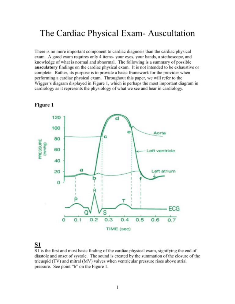
The Cardiac Physical Exam- Auscultation There is no more important component to cardiac diagnosis than the cardiac physical exam. A good exam requires only 4 items- your eyes, your hands, a stethoscope, and knowledge of what is normal and abnormal. The following is a summary of possible ausculatory findings on the cardiac physical exam. It is not intended to be exhaustive or complete. Rather, its purpose is to provide a basic framework for the provider when performing a cardiac physical exam. Throughout this paper, we will refer to the Wigger’s diagram displayed in Figure 1, which is perhaps the most important diagram in cardiology as it represents the physiology of what we see and hear in cardiology. Figure 1 S1 S1 is the first and most basic finding of the cardiac physical exam, signifying the end of diastole and onset of systole. The sound is created by the summation of the closure of the tricuspid (TV) and mitral (MV) valves when ventricular pressure rises above atrial pressure. See point “b” on the Figure 1. 1 S1 is best heard at the left lower sternal border (LLSB) and apex using the diaphragm of the stethoscope. S1 may be heard to “split” occasionally- this is usually a normal finding. In those instances when splitting is appreciated, it is a result of the tricuspid valve closing shortly after the mitral valve. Splitting of S1 can be easily appreciated in right bundle branch block. Of note, when “splitting” of S1 is heard at the base of the heart, you should suspect that you are actually hearing an S1 and then an ejection click, rather than a split S1. Abnormalities of S1 S1 is made louder by: 1) Short PR interval or tachycardia- this occurs due to the distance between the valve leaflets at the onset of systole. The further the leaflets are from each other before they are “slammed shut” by the rise in ventricular pressure, the louder the sound. With a short PR or in tachycardia, the onset of systole occurs immediately after the leaflets have been maximally opened by atrial systole, thus the S1 is loud. It is analogous to slamming a door shut when it is wide open compared to slamming it when it is mostly closed (as in a normal PR interval). 2) Mitral stenosis- calcified or thickened valve leaflets create a louder sound when they move against each other compared to pliable, noncalcified leaflets. Listen carefully for other evidence of mitral stenosis when you hear an unusually loud S1. 3) Hypertension- elevated systemic blood pressure leads to elevated ventricular pressure (especially LV Pressure). Higher ventricular pressure leads to greater force with which the atrioventricular valves are closed. This then generates a louder sound. S1 is made softer by: 1) Long PR interval- this mechanism is basically the opposite of the effect of a short PR. When ventricular systole is delayed after atrial systole, the valve leaflets have already moved closer to each other at the time when they forced to closure. The effect is a softer sound. 2) Aortic insufficiency- in patients with significant AI, the volume of blood returning to the ventricle during diastole raises LV pressure during diastole. The elevated pressure partially closes the mitral valve even prior to ventricular systole. The effect is a softer S1. 3) Insulation of the heart- air in COPD, fluid in pericardial effusions, and fat in obesity all serve to diminish sound propagation, thus limiting the listener’s ability to hear S1 (or many other heart sounds). 4) Mitral regurgitation- when the valve fails to fully close due an intrinsic defect, S1 may be softer. 5) Low cardiac output or low EF- in cases of severe systolic dysfunction, the LV cannot generate a sufficient force rapidly enough to create a “crisp” S1. The effect is a soft S1. 2 S2 S2 is the second, basic sound of the cardiac cycle and signifies the end of systole and onset of diastole. S2 is created by the closure of the aortic and pulmonic valves and is best heard at the left or right upper sternal borders with the diaphragm of the stethoscope. See point “e” on the Figure 1. It is important for any auscultator to understand the different forms of splitting of S2: Normal- normally the aortic component of S2 (S2a) closes prior to the pulmonic component of S2 (S2p). During expiration, S2a and S2p are so closely timed, no splitting can be appreciated. However, when one inspires, the RV takes longer to eject due to a decrease in the pulmonary artery pressure and an increase in RV preload due to enhanced venous return. Thus, S2p moves away from S2a and one appreciates a splitting of the sound. [WINE- widens inspiration, narrows expiration] Expiration S1 Inspiration S2a&p S1 S2a S2p Persistent splitting or Widely split- When the RV ejection is delayed abnormally, splitting can be heard in expiration and inspiration. Delays in RV ejection result from right bundle branch block, pulmonary embolism, pulmonary hypertension, and pulmonic stenosis. In addition, persistent splitting can be heard when the LV ejection is shortened due to a second low pressure outlet for LV ejection- most commonly, this would result from severe mitral regurgitation or a ventricular septal defect. Of note, when this type of splitting occurs, S2p still varies with respiration, but the S2a and S2p never “come together”. Expiration S1 S2a Inspiration S2p S1 S2a S2p Paradoxical- When the LV ejection is delayed, S2a occurs after S2p. Thus, inspiration moves S2p closer to S2a. The result is splitting during expiration which goes away with inspiration, hence “paradoxical”. The main causes include LBBB, paced ventricular rhythm, severe AS, severe systolic dysfunction. 3 Expiration S1 S2p Inspiration S2a S1 S2p&a Fixed splitting- in patients with an atrial septal defect, changes in respiration may not change RV preload. However, due to the left-to-right shunt, RV preload is always greater than LV preload, and hence RV systole is prolonged but never changes with inspiration or expiration. Hence, S2 splitting is audible and fixed. Expiration S1 S2a Inspiration S2p S1 S2a S2p Abnormalities of S2 S2 is made louder by: 1) S2a: systemic hypertension- the pressure closing the valve is higher 2) S2p: pulmonary hypertension- same as above S2 is made softer by: 1) S2a: aortic stenosis and systemic hypotension 2) S2p: pulmonic stenosis Sounds in Diastole Early: (occurring near point “f” in figure 1) 1) S3- a low pitched sound occurring 0.1-0.2 seconds after S2. It is best heard with the bell of the stethoscope with the patient in the left lateral decubitus position. S3 has numerous causes which can be divided into 3 separate states: a. Normal/athletic hearts- S3 occur in normal healthy individuals under the age of 40. A “normal” S3 is easily heard when a young patient is hyperdynamic- pregnancy, anemia, postexercise, or thyrotoxic. b. Diastolic overload states- when a large amount of blood moves from the atrium into the ventricle during early diastole, an S3 may be heard. Examples include severe mitral regurgitation 4 and lesions leading to L-R shunting with RV volume overload such as a ventricular septal defect or a patent ductus arteriosis. c. LV dysfunction- classically an S3 signifies global systolic dysfunction and usually an elevated BNP. 2) Opening Snap (OS) - the classic sound of mitral stenosis. It typically is associated with a loud S1 (see above) and is created by the opening of the calcified mitral cusps early in diastole. The timing of the OS can be a clue as to the severity of the stenosis: the earlier the OS (the shorter the S2-OS interval), the worse the stenosis. OS is usually heard best between the apex and the left sternal border with the patient in the left lateral decubitus position. It is higher pitched than an S3 and can be heard with the diaphragm or bell. 3) Tumor Plop- this early diastolic sound is created by the movement of a myxoma through the mitral valve. It is usually low pitched and heard best with the bell of the stethoscope. 4) Pericardial Knock- in patients with constrictive pericarditis, the initial movement of blood from the atrium into the ventricle in early diastole causes the ventricular walls to suddenly abut the stiff pericardium. The result is a medium-pitched sound which mimics an S3 or an opening snap. This sound is best heard with the diaphragm of the stethoscope at the apex with the patient in the left lateral decubitus position. 5) Intraaortic Balloon Pump Sound- patients who have had an intraaortic balloon pump inserted will have a early diastolic sound created by the inflation of the balloon pump after closure of the aortic valve. Late: (occurring near point “a” in figure 1) 1) S4- a low-pitched sound which follows atrial systole by 0.1-0.2s. The sound is created by the sudden movement of blood into a ventricle with a higher than normal diastolic pressures. The sudden, further increase in pressure created by the new influx of blood causes stiffening of the ventricle and tensing of the mitral apparatus, which then generates sound. An S4 is best heard with the bell of the stethoscope at the apex with the patient in the left lateral decubitus position. Causes of S4 include: a) Aging- as patients age, it is normal to lose some of the compliance of the left ventricle. Thus, many elderly patients have an S4. b) Ventricular hypertrophy- an increase in wall thickness leads to elevated intraventricular pressure as per LaPlace’s Law (wall tension= pressure x radius / wall thickness). Thus, any cause of LVH can lead to an S4: 5 i. HCM ii. Systemic hypertension iii. Aortic stenosis iv. Athletic heart (controversial) c) Ischemic heart disease- both chronic and acute ischemic disease lead to stiffening of the ventricle and increased intraventricular pressures. 2) Split S1- in some normal patients, splitting of S1 can be appreciated. The timing of the split may mimic an S4-S1; however, the components of S1 are higher pitched and heard best with the diaphragm of the stethoscope at the lower left sternal border. Sounds in Systole 1) Ejection click/sound (occurring near point “c” in figure 1) - this high pitched sound occurring soon after S1 and is heard best at the right or left upper sternal border. The interval between S1 and the ejection sound is essentially the time of isovolumic contraction. The ejection sound is a result of one of two possible etiologies: a. Valvular- an abnormal aortic or pulmonic valve can abruptly “dome” in early systole leading to an ejection sound. In adults, this abnormality is usually a bicuspid aortic valve. b. Arterial- the root of the aorta or pulmonary artery can abruptly tense in some patients leading to an ejection sound. 2) Mitral Valve Prolapse (occurring near “d” in figure 1) - a redundant mitral valve leaflet may tense in mid or late systole leading to a high-pitched snapping sound, termed a “click”. This sound is best heard at the lower left sternal border and apex. Any maneuver which decreases LV preload, such as the Valsalva, will move the click closer to S1. On the other hand, any maneuver which increases preload will move the click away from S1. Murmurs In general, murmurs can be characterized by 8 different qualities: 1) Timing (systole, diastole, continuous) 2) Intensity or grade I- heard with concentration only II- soft, but easily heard during auscultation III- moderate intensity IV- loud with a palpable thrill V- loud with the rim of the stethoscope barely on chest VI- loud with stethoscope off of the chest 3) Frequency (high, low) 4) Configuration (flat, crescendo-decrescendo, decrescendo, crescendo, holosystolic) 5) Quality (harsh, blowing, musical, rumble) 6 6) Duration (short, pansystolic) 7) Radiation (neck, axilla, back) 8) Maneuvers In this review, we will classify murmurs by timing and then discuss the murmurs heard best during those timing periods. The grade or intensity of each individual murmur depends upon the disease severity and thus will not be discussed further. 1) Systolic a. Aortic stenosis- the murmur of aortic stenosis arises from the pressure gradient between the high pressure left ventricular chamber and the low pressure aortic root due to obstruction to left ventricular outflow from the immobile aortic valve leaflets. As seen in figure 2, the LV pressure rises above aortic pressure; initially there is a small pressure difference which soon rises to its maximum and then decreases. This pattern of change in pressure gradient leads to the crescendo-decrescendo nature of the murmur. This murmur is heard best at the right upper sternal border with the diaphragm. It is important to note the location of the peak given that typically, worse stenosis peaks later in systole. Also important is the quality of S2, for reasons described above. i. Frequency- typically medium to high pitched ii. Configuration- crescendo-decrescendo for reasons described above iii. Quality- harsh or coarse iv. Duration- ends prior to or at S2 v. Radiation- the murmur of AS follows the direction of blood flow leaving the aortic valve. Thus, the murmur radiates into the upper chest and into the carotids. In most patients it can also be heard in the left subclavian artery. vi. Maneuvers- any maneuver which increases LV filling (and hence LV stroke volume) will tend to increase the murmur intensity. For example, a PVC will lead to a compensatory pause, which increases diastolic LV filling. Based on the Starling mechanism, the LV will then contract with a greater force, hence a greater pressure and stroke volume will be generated within the left ventricle. The aortic pressure will stay the same so the absolute LV-Ao gradient will increase. This will serve to increase the murmur. vii. NB- Occasionally the murmur of severe aortic stenosis can be heard as a musical murmur (not a harsh murmur) at the apex. This is referred to as the Gallaverdin’s murmur or phenomenon. 7 b. Mitral regurgitation- The murmur of MR derives from the movement of blood from the high pressure LV chamber to the low pressure left atrium. In contradistinction from aortic stenosis, the LV and LA never reach an equal pressure, so the murmur of MR will last throughout systole (“holosystolic”) and may even last past S2. This murmur is best heard at the apex with the diaphragm. i. Frequency- medium to high pitched ii. Configuration- usually flat (the intensity does not change during systole) iii. Quality- blowing iv. Duration- holosystolic v. Radiation- classically, the MR murmur radiates to the axilla. However, in patients with either a prolapsed leaflet, a flail leaflet, or a restricted leaflet, the murmur will radiate in the direction of the blood. For example, patients with a posterior leaflet which prolapses have an anterior directed jet. Their murmur then is directed towards the anterior chest wall and can mimic aortic stenosis. Posteriorly-directed jets radiate to the back and can even produce murmurs on the crown of the head through radiation up the spine. vi. Maneuvers- Any maneuver which increases afterload will increase the amount of blood moving into the LA because LV pressure correlates with aortic pressure. Thus, classically handgrip and squatting will increase the intensity of an MR murmur. c. TR- TR has the same characteristics as MR, but the murmur is best heard at the left sternal border or subdiaphragmatically. The murmur 8 of TR should increase with inspiration secondary to increase in systemic venous return. d. VSD- Most VSDs found incidentally in adults are small and restrictive. Thus, the murmur of a VSD is typically a loud, highpitched, holosystolic at the lower left sternal border. i. Frequency- usually high pitched in adults ii. Configuration- usually flat or decrescendo iii. Quality- blowing or musical iv. Duration- pansystolic v. Radiation- the murmur of a VSD can radiate throughout the precordium since it is such a loud murmur in most adults. Other VSDs radiate to the right sternal border. vi. Maneuvers -Any maneuver which increases LV pressure, such as hand grip, will increase the intensity of the murmur. e. Hypertrophic obstructive cardiomyopathy (HOCM)- Not all patients with hypertrophic cardiomyopathy have a murmur because not all hypertrophic cardiomypathies lead to outflow obstruction. However, when there is outflow obstruction from septal hypertrophy, a murmur is generated with its loudness dependent upon the degree of obstruction and height of the pressure gradient. Like aortic stenosis, the murmur of HOCM usually has a crescendo-decrescendo pattern, but the murmur of HOCM is usually best heard along the left sternal border, lower down the sternum that the murmur of aortic stenosis. The murmur also does not typically radiate to the carotids which helps distinguish it from aortic stenosis. i. Frequency- medium to high ii. Configuration- crescendo-decrescendo iii. Quality- harsh iv. Duration- usually begins after S1 and does not last throughout systole v. Radiation- as above, does not radiate to the neck vi. Maneuvers- any maneuver which leads to either a reduction in LV preload (e.g. Valsalva), decreases afterload (squat-stand), or increases LV contractile force (post-PVC) will increase the murmur of HOCM. The Valsalva maneuver helps distinguish HOCM from the murmur it most closely resembles, aortic stenosis. In HOCM, the decrease in preload and stoke volume created by the strain phase of the Valsalva will increase the murmur due to more obstruction from the hypertrophic ventricular septum. However, in aortic stenosis, the decrease in stroke volume created by the strain phase of Valsalva will lead to a decrease in the left ventricular-aorta gradient, and hence a reduction in the volume of the murmur. 9 2) Diastolic a. Aortic insufficiency- the murmur of aortic insufficiency is created by the regurgitation of blood from the aortic root into the left ventricle during diastole. The murmur thus begins early in diastole, as soon as left ventricular pressure falls below aortic pressure. It is best heard with the diaphragm of the stethoscope at the right or left upper sternal border. The length of the murmur depends upon the severity of the regurgitation and the compliance of the ventricle. For example, in acute severe aortic regurgitation, the noncompliant left ventricle equilibrates with aortic pressure very quickly; thus the murmur is brief. In chronic severe AI, pressure in the more compliant, dilated left ventricle rises more slowly. Thus the murmur may last throughout diastole. i. Frequency- variable, usually higher pitch indicates more severe regurgitation ii. Configuration- decrescendo (always) iii. Quality- blowing iv. Duration- depends on severity: very mild AI may be so short a murmur that it only makes S2 seem long. More severe AI is usually associated with longer, decrescendo murmur heard throughout diastole. N.B. however that some very severe, especially acute severe AI may lead to rapid equalization of aortic and LV pressure which shortens the murmur. v. Radiation- none vi. Maneuvers- any maneuver which increases afterload (handgrip) will increase the amount of regurgitation. b. MS- the murmur of mitral stenosis is one of the more challenging physical exam auscultation findings. It usually requires a quiet room with the patient in the left lateral decubitus position with the diaphragm or bell of the stethoscope at the apex. The murmur usually begins after an opening snap and then has a decrescendo pattern; in patients in normal sinus rhythm there may be a brief crescendo immediately before S1 (when the atrial kick pushes blood through the noncompliant mitral valve). i. Frequency- low ii. Configuration- decrescendo after S2, may crescendo prior to S1 (presystolic accentuation in patients in sinus rhythm) iii. Quality- rumbling iv. Duration- brief v. Radiation- none vi. Maneuvers- any maneuver which increases cardiac output will increase the murmur of MS. This is most easily accomplished by having the patient exercise briefly (sit-ups or jogging in place) and 10 then immediately examing the patient in the left lateral decubitus position. Pericardial Friction Rub The pericardial friction rub is created by movement of the heart within an inflamed pericardium. Classically, it has 3 components- two in diastole and one in systole. The diastolic components occur when the left ventricle is stretched during diastole, namely with early diastolic filling and later with the atrial kick. Each of these events, changes the left ventricular volume which makes it more likely that the visceral and parietal pericardial surfaces will contact each other. Ventricular systole causes motion of the ventricle which then creates the 3rd component. The pericardial friction rub may be transient and requires a prolonged auscultation. It is best heard with the diaphragm, usually with at the lower left sternal border. 11 12 Mitral Valve Prolapse Pulmonary Stenosis S1 normal S2 normal splitting Mid Systolic Click Earlier-Standing, Valsalva Later – Squatting, Handgrip Mid to Late Systolic Murmur Longer-Standing, Valsalva Shorter-Squatting, Handgrip S1 normal S2 Wide Splitting P2 Loud S3 Often Present Pansystolic Murmur Filling Rumble IHSS (HOCM) A2 normal Paradoxical Splitting S2 S4 Usually Present Systolic Ejection Murmur Valsalva, Standing, post PVC Handgrip, Squatting Bisferiens Carotid Pulse Tricuspid Insufficiency Mitral Insufficiency Atrial Septal Defect S1 normal S2 Paradoxical Splitting Pansystolic-Holosystolic Murmur With Inspiration, Squatting With Expiration, Standing Giant GV Waves S1 – Split S2 Wide Fixed Splitting Pulmonic SEM Tricuspid Mid Diastolic Filling Murmur Prominent JVP Diminished S1 Wide Splitting S2 S3 (moderate to severe) High Frequency Holosystolic Murmur Occasional Diastolic Filling Rumble Brisk-Low Amplitude Carotid Pulse Aortic Stenosis Ejection Sound (non-calc) Diminished A2 (Immobile) Paradoxical Splitting S2 S3 and S4 (severe) Late Peaking Systolic Ejection Murmur Delayed-Low Amplitude Carotid Pulse (Parvus Tardes) Delayed Brachial-Radial, ApicalCarotid Interval Mitral Stenosis Aortic Insufficiency Ventricular Septal Defect Loud S1 Narrow Splitting S2 Loud P2 Opening Snap Narrow A2-OS Interval (severe) Diastolic Rumble with Presystolic Accentuation Duration = Severity S1 Diminished Soft A2 and Narrow Splitting S2 S3 – Common Diastolic Decrescendo Murmur Early Peaking SEM S1 normal S2 Wide Splitting P2 Loud S3 Often Present Pansystolic Murmur Filling Rumble 13 14 15 16
