View/Open
advertisement
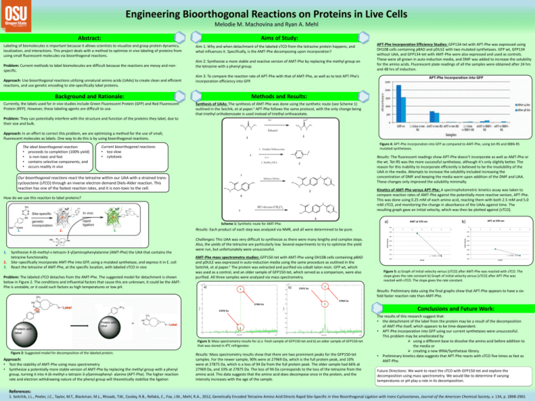
Engineering Bioorthogonal Reactions on Proteins in Live Cells Melodie M. Machovina and Ryan A. Mehl Aims of Study: Abstract: Labeling of biomolecules is important because it allows scientists to visualize and grasp protein dynamics, localization, and interactions. This project deals with a method to optimize in vivo labeling of proteins from using small fluorescent molecules via bioorthogonal reactions. Problem: Current methods to label biomolecules are difficult because the reactions are messy and nonspecific. Approach: Use bioorthogonal reactions utilizing unnatural amino acids (UAAs) to create clean and efficient reactions, and use genetic encoding to site-specifically label proteins. Aim 1: Why and when detachment of the labeled sTCO from the tetrazine protein happens, and what influences it. Specifically, is the AMT-Phe decomposing upon incorporation? Aim 2: Synthesize a more stable and reactive version of AMT-Phe by replacing the methyl group on the tetrazine with a phenyl group. Aim 3: To compare the reaction rate of APT-Phe with that of AMT-Phe, as well as to test APT-Phe’s incorporation efficiency into GFP. Background and Rationale: Currently, the labels used for in vivo studies include Green Fluorescent Protein (GFP) and Red Fluorescent Protein (RFP). However, these labeling agents are difficult to use. APT-Phe Incorporation Efficiency Studies: GFP134-tet with APT-Phe was expressed using DH10B cells containing pBAD and pDULE with two mutated synthetases. GFP wt, GFP134 without UAA, and GFP134-tet with AMT-Phe were also expressed and used as controls. These were all grown in auto-induction media, and DMF was added to increase the solubility for the amino acids. Fluorescent plate readings of all the samples were obtained after 24 hrs and 48 hrs of induction. APT-Phe Incorporation into GFP Methods and Results: Synthesis of UAAs: The synthesis of AMT-Phe was done using the synthetic route (see Scheme 1) outlined in the Seichik, et al paper.1 APT-Phe follows the same protocol, with the only change being that triethyl orthobenzoate is used instead of triethyl orthoacetate. Problem: They can potentially interfere with the structure and function of the proteins they label, due to their size and bulk. Approach: In an effort to correct this problem, we are optimizing a method for the use of small, fluorescent molecules as labels. One way to do this is by using bioorthogonal reactions. Current bioorthogonal reactions: • too slow • cytotoxic Results: The fluorescent readings show APT-Phe doesn’t incorporate as well as AMT-Phe or the wt. Tet-RS was the more successful synthetase, although it’s only slightly better. The reason for this inability to incorporate efficiently is believed to be the insolubility of the UAA in the media. Attempts to increase the solubility included increasing the concentration of DMF and keeping the media warm upon addition of the DMF and UAA. These changes only improved the solubility minimally. Our bioorthogonal reactions react the tetrazine within our UAA with a strained transcyclooctene (sTCO) through an inverse electron demand Diels-Alder reaction. This reaction has one of the fastest reaction rates, and it is non-toxic to the cell. Kinetics of AMT-Phe versus APT-Phe: A spectrophotometric kinetics assay was taken to compare reaction rates of AMT-Phe against the potentially more reactive version, APT-Phe. This was done using 0.25 mM of each amino acid, reacting them with both 2.5 mM and 5.0 mM sTCO, and monitoring the change in absorbance of the UAAs against time. The resulting graph gave an initial velocity, which was then be plotted against [sTCO]. a) Scheme 1: Synthetic route for AMT-Phe. 1. 1. 2. 3. 2. 3. Synthesize 4-(6-methyl-s-tetrazin-3-yl)aminophenylalanine (AMT-Phe) the UAA that contains the tetrazine functionality Site–specifically incorporate AMT-Phe into GFP, using a mutated synthetase, and express it in E. coli React the tetrazine of AMT-Phe, at the specific location, with labeled sTCO in vivo Problem: The labeled sTCO detaches from the AMT-Phe. The suggested model for detachment is shown below in Figure 2. The conditions and influential factors that cause this are unknown; it could be the AMTPhe is unstable, or it could such factors as high temperatures or low pH. 0 1 2 3 a) b) 27875 Da 4 5 0 6 -150 -100 -155 -200 -160 -165 -180 1 2 3 4 5 -300 -400 -500 -170 -175 AMT-Phe mass spectrometry studies: GFP150-tet with AMT-Phe using DH10B cells containing pBAD and pDULE was expressed in auto-induction media using the same procedure as outlined in the Seitchik, et al paper.1 The protein was extracted and purified via cobalt talon resin. GFP wt, which was used as a control, and an older sample of GFP150-tet, which served as a comparison, were also purified. All three samples were analyzed via mass spectrometry. APT at 370 nm 0 -145 Results: Each product of each step was analyzed via NMR, and all were determined to be pure. Challenges: This UAA was very difficult to synthesize as there were many lengthy and complex steps. Also, the yields of the tetrazine are particularly low. Several experiments to try to optimize the yield were run, but unfortunately were unsuccessful. b) AMT at 370 nm Initial Velocity How do we use this reaction to label proteins? Initial Velocity The ideal bioorthogonal reaction: • proceeds to completion (100% yield) • is non-toxic and fast • contains selective components, and • occurs readily in vivo Figure 4: APT-Phe incorporation into GFP as compared to AMT-Phe, using tet-RS and IBBN-RS mutated synthetases. y = -10.08x - 126 [sTCO] -600 -700 y = -59.04x - 327.6 [sTCO] Figure 5: a) Graph of initial velocity versus [sTCO] after AMT-Phe was reacted with sTCO. The slope gives the rate constant b) Graph of initial velocity versus [sTCO] after APT-Phe was reacted with sTCO. The slope gives the rate constant. Results: Preliminary data using the final graphs show that APT-Phe appears to have a sixfold faster reaction rate than AMT-Phe. 27969 Da 27969 Da 27878 Da Figure 3: Mass spectrometry results for a) a fresh sample of GFP150-tet and b) an older sample of GFP150-tet that was stored in 4⁰C refrigerator. Figure 2: Suggested model for decomposition of the labeled protein. Approach: • Test the stability of AMT-Phe using mass spectrometry • Synthesize a potentially more stable version of AMT-Phe by replacing the methyl group with a phenyl group, turning it into 4-(6-methyl-s-tetrazin-3-yl)aminophenyl- alanine (APT-Phe). The higher reaction rate and electron withdrawing nature of the phenyl group will theoretically stabilize the ligation Results: Mass spectrometry results show that there are two prominent peaks for the GFP150-tet samples. For the newer sample, 90% were at 27969 Da, which is the full protein peak, and 10% were at 27875 Da, which is a loss of 94 Da from the full protein peak. The older sample had 66% at 27969 Da, and 33% at 27875 Da. The loss of 94 Da corresponds to the loss of the tetrazine from the amino acid. This data suggests that the amino acid does decompose once in the protein, and the intensity increases with the age of the sample. Conclusions and Future Work: The results of this research suggest that: • the detachment of the label from the protein may be a result of the decomposition of AMT-Phe itself, which appears to be time-dependent. • APT-Phe incorporation into GFP using our current synthetases were unsuccessful. This problem may be ameliorated by using a different base to dissolve the amino acid before addition to the media or creating a new tRNA/Synthetase library. • Preliminary kinetics data suggests that APT-Phe reacts with sTCO five times as fast as AMT-Phe. Future Directions: We want to react the sTCO with GFP150-tet and explore the decomposition using mass spectrometry. We would like to determine if varying temperatures or pH play a role in its decomposition. References: 1. Seitchik, J.L., Peeler, J.C., Taylor, M.T., Blackman, M.L., Rhoads, T.W., Cooley, R.B., Refakis, C., Fox, J.M., Mehl, R.A., 2012, Genetically Encoded Tetrazine Amino Acid Directs Rapid Site-Specific in Vivo Bioorthogonal Ligation with trans-Cyclooctenes, Journal of the American Chemical Society, v. 134, p. 2898-2901 6


