Finger Deformities: Swan Neck, Mallet, Boutonniere
advertisement
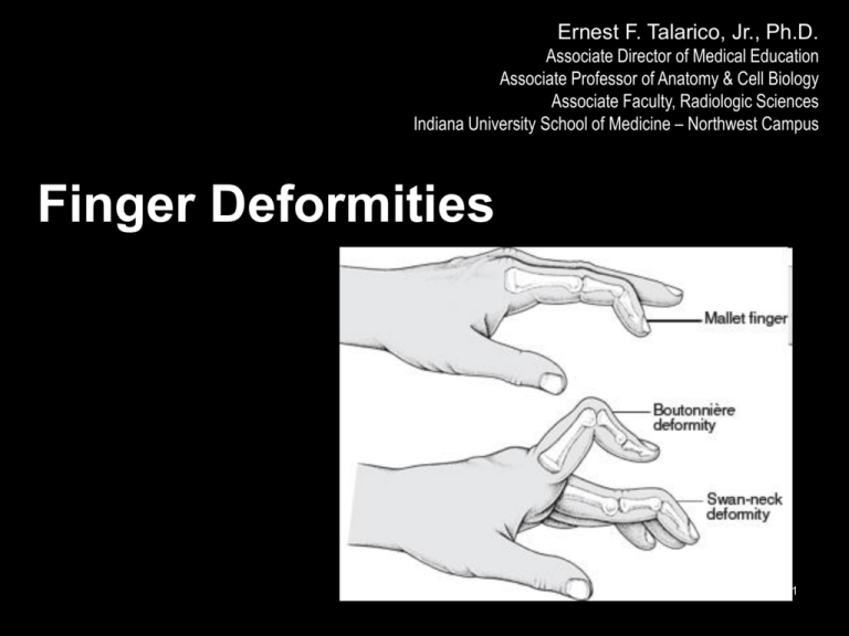
Ernest F. Talarico, Jr., Ph.D. Associate Director of Medical Education Associate Professor of Anatomy & Cell Biology Associate Faculty, Radiologic Sciences Indiana University School of Medicine – Northwest Campus Finger Deformities IUSM-NW F12 1 Swan Neck Deformity IUSM-NW F12 2 A swan neck deformity describes a finger with a hyperextended PIP joint and a flexed DIP joint. IUSM-NW F12 3 IUSM-NW F12 4 What parts of the finger are involved? The fingers are actually made up of three bones, called phalanges. The three phalanges in each finger are separated by two joints, called interphalangeal joints (IP joints). The joint near the end of the finger is called the distal IP joint (DIP joint). (Distal means further away.) The proximal IP joint (PIP joint) is the middle joint between the main knuckle and the DIP joint. (Proximal means closer in.) The IP joints of the fingers work like hinge joints when you bend and straighten your hand. The tendons that allow each finger joint to straighten are called the extensor tendons. The extensor tendons of the fingers begin as muscles that arise from the backside of the forearm bones. These muscles travel toward the hand, where they eventually connect to the extensor tendons before crossing over the back of the wrist joint. As they travel into the fingers, the extensor tendons become the extensor hood. The extensor hood flattens out to cover the top of the finger and sends out branches on each side that connect to the bones in the middle and end of the finger. When the extensor muscles contract, they tug on the extensor tendon and straighten the finger. Ligaments are tough bands of tissue that connect bones together. Several small ligaments connect the extensor hood with other tendons that travel into the finger to bend the finger. These connections help balance the motion of the finger so that all the joints of the finger work together, giving a smooth bending and straightening action. In the PIP joint (the middle joint between the main knuckle and the DIP joint), the strongest ligament is the Ivolar plate. This ligament connects the proximal phalanx to the middle phalanx on the palm side of the joint. The ligament tightens as the joint is straightened and keeps the PIP joint from bending back too far (hyperextending). Swan neck deformity can occur when the volar plate loosens from disease or injury. IUSM-NW F12 5 How does this condition occur? Conditions that loosen the PIP joint and allow it to hyperextend can produce a swan neck deformity of the finger. Rheumatoid arthritis (RA) is the most common disease affecting the PIP joint. Chronic inflammation of the PIP joint puts a stretch on the volar plate. (As mentioned earlier, the volar plate is a supportive ligament in front of the PIP joint that normally keeps the PIP joint from hyperextending.) As the volar plate becomes weakened and stretched, the PIP joint becomes loose and begins to easily bend back into hyperextension. The extensor tendon gets out of balance, which allows the DIP joint to get pulled downward into flexion. As the DIP joint flexes and the PIP joint hyperextends, the swan neck deformity occurs. Other conditions that weaken the volar plate can produce a swan neck deformity. The small (intrinsic) muscles of the hand and fingers can tighten up from hand trauma, RA, and various nerve disorders, such as cerebral palsy, Parkinson's disease, or stroke. The muscle imbalance tends to weaken the volar plate and pull the PIP joint into extension. Weakness in the volar plate can also occur from a finger injury that forces the PIP joint into hyperextension, stretching or rupturing the volar plate. As mentioned, looseness (laxity) in the volar plate can lead to a swan neck deformity. Clearly, PIP joint problems can produce a swan neck deformity. But so can problems that start in the DIP joint at the end of the finger. Injury or disease that disrupts the end of the extensor tendon can cause the DIP joint to droop (flex). An example from sports is a jammed finger that tears or ruptures the extensor tendon at the end of the finger (distal phalanx). Without treatment, the DIP joint droops and won't straighten out. This condition is called a mallet finger. The extensor tendon may become imbalanced and begin to pull the PIP joint into hyperextension, forming a swan neck deformity. Chronic inflammation from RA can also disrupt the very end of the extensor tendon. Inflammation and swelling in the DIP joint stretches and weakens the extensor tendon where it passes over the top of the DIP joint. A mallet deformity occurs in the DIP, followed by hyperextension of the PIP joint. Again, the result is a swan neck deformity. IUSM-NW F12 6 IUSM-NW F12 7 Mallet Finger IUSM-NW F12 8 IUSM-NW F12 9 How do these injuries of the DIP joint occur? A mallet finger results when the extensor tendon is cut or torn from the attachment on the bone. Sometimes, a small fragment of bone may be pulled, or avulsed, from the distal phalanx. The result is the same in both cases: the end of the finger droops down and cannot be straightened. IUSM-NW F12 10 IUSM-NW F12 11 IUSM-NW F12 12 IUSM-NW F12 13 Boutonniere Injury IUSM-NW F12 14 IUSM-NW F12 15 Boutonnière Deformity (Buttonhole Deformity) Boutonnière deformity (buttonhole deformity) is a deformity in which the middle finger joint is bent in a fixed position inward (toward the palm) and the outermost finger joint is bent excessively outward (away from the palm). This disorder most often results from rheumatoid arthritis but can also occur from injury (such as deep cuts, joint dislocation, or fractures) or osteoarthritis. People with rheumatoid arthritis can develop the disorder because they have long-standing inflammation of the middle joint of a finger. If the deformity is caused by an injury, the injury usually occurs at the base of a tendon (called the middle phalanx extensor tendon). As a result, the middle joint (called the proximal interphalangeal joint) becomes “buttonholed” between the outer bands of the tendon that runs to the end of the finger. The deformity can, but need not, interfere with hand function. The doctor makes the diagnosis by examining the finger. Treatment A boutonnière deformity caused by an extensor tendon injury can usually be corrected with a splint that keeps the middle joint fully extended for 6 weeks. When splinting is ineffective, or when boutonnière deformity is due to rheumatoid arthritis, surgery may be needed. IUSM-NW F12 16 IUSM-NW F12 17 IUSM-NW F12 18
