Pneumonia and pleural effusion
advertisement
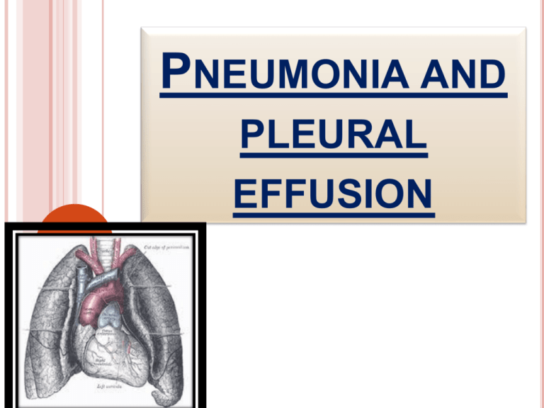
PNEUMONIA AND PLEURAL EFFUSION DEFINITION Is an acute inflammation of lung parenchyma caused by various micro organism Pneumonitis is a general term that describe an inflammatory process in the lung tissue that may predispose or place the at risk for microbial invasion. The discovery of sulfa drugs and penicillin was pivotal in treatment of it .Since that time , there has been remarkable progress in the development of antibiotics to treating pneumonia . However despite the new antimicrobial agents ,this is still common and is associated with significant morbidity and mortality ETIOLOGY Normally airway distal to larynx is sterile because of protective defense mechanism These includes Filtration of air ,macropha ges Epiglottis closure over trachea Warming , humidification inspired air Cough reflex , IgA FACTORS THAT PREDISPOSE When defense mechanism become incompetent or overwhelmed by virulence or quantity of inflammatory agents Pneumonia results decrease consciousness depresses cough and epiglottal reflex Aspiration CONT… Tracheal intubation interferes with normal cough reflex and muco ciliary escalator mechanism ; also bypasses upper airway Muco ciliary mechanism is interfered with air pollution , cigarette , viral URI , aging . In case of mal nutrition functions of lymphocytes and PMN leucocytes are altered Alcoholism , DM , Leukemia are associated with GNB in oropharynx Altered oropharyngeal flora Secondary to antibiotic therapy drugs Bed rest , prolonged immobility Feeding via NG tubes Head injury seizures , drug overdose Tracheal intubation Chronic dx ACQUISITION OF ORGANISMS Aspiration Inhalation Hematogenous CLASSIFICATION Typical Oppur tunistic Atypical Anaerobic VAP/HAP Opport unistic CATEGORY Aspiration CAP COMMUNITY ACQUIRED PNEUMONIA is defined as LRTI of lung parenchyma with onset in community / during first 2 days / 48 hrs after hospitalization CAUSATIVE AGENTS ARE : Strep.pneumoniae Myco . pneumoniae H.influenza Respiratory virus Clamydia pneumonia legionella pnemophila Oral anaerobes Nocardia M.tb , enteric GNB Staph.aureus , fungi STREPTOCOCCUS PNEUMONIAE Commonest in age < 60 yrs without co morbidity and > 60yrs with co morbidity Prevalent in winter and spring when URTI is frequent Gram positive , capsulated non motile coccus resides naturally in URT Organism colonizes URT and cause disseminated invasive infection , LRTI , URTI , otitis , sinusitis ,pneumonia Bacteremia – 15% - 25 % cases lobar Broncho pneumonia forms TREATMENT Cefotaxime / ceftriazone Antipseudomonal fluroquinolones Levofloxacin MYCOPLASMA PNEUMONIA Most common in older children and young adult is spread by infected respiratory droplets through person to person contact . Patient tested for Mycoplasma antibodies Inflammatory infiltrate is primarily interstial rather than alveolar Mortality = < 0.1% Spreads throughout entire tract including bronchioles , has characteristic of bronchopneumonia. CONT………. Sore throat , pleuritic pain Ear ache myalgia Nasal congestion , pharyngitis interstitial infiltrate = CXR Doxycyclin , macrolides fluroquinolones Aseptic meningitis Meningo encephalitis Comp lication Transverse myelitis Peri , myocardits Cranial nerve plasty HEMOPHILUS INFLUENZA Affects elderly and those with co morbid illness Mortality = 30% Associated with URTI = 2 – 6 wks before onset of illness Fever , chills , productive cough usually involves one or more lobes , sub acute bacteremia CXR = multilobar patchy bronchopneumonia / area of consolidation Cephalosporin , macrolides , quinolones LEGIONNAIRE DISEASE High in smokers / immunosuppressive therapy Epidemic / sporadic Lobar consolidatio n Broncho pneumonia flu Summerhigh TREATED WITH………….. • Erythromycin , Rifampin • clarithromycin • Macrolides • fluroquinolones CHLAMYDIAL PNEUMONIA Single infiltrate on chest x-ray Pleural effusion , upper respiratory tract infection Tetracyclin , erythromycin Complication include acute respiratory failure VIRAL PNEUMONIA Influenza A ,B , adeno virus , RSV Parainfluenza , CMV , Corono virus Winter months , epidemics occur 2 – 3yrs Patchy infiltrate on CXR with effusion , URTI , bronchitis , pleurisy TYPE A = AMANTIDINE , RIMANTIDINE TYPE A / B = ZANAMIVIR ,OSELTAMIVIR CONT.. Acute stage – within ciliated cells Infiltration of tracheo bronchial trees Extends into alveolar area – edema , exudation IDSA , ATS – 3 STEP APPROACH STEP 1 = assessment of ability to treat the patient at home STEP 2 = calculation of PORT PSI with recommendation , for home care and clinical judgment .this scale is produce by agency of health care research and quality based on multiple factors and scores indicates patient’s risk class STEP 3 = clinical judgment in final decision to treatment , either as OP / IP PNEUMONIA PATIENT OUTCOMES RESEARCH TEAM SEVERITY INDEX DRUG THERAPY HOSPITAL ACQUIRED , VENTILATOR ASSOCIATED , HEALTH CARE ASSOCIATED PNEUMONIA HAP occurring 48hrs / longer after hospital admission and not incubating at time of hospitalization. VAP refers to pneumonia that occurs 48 – 72 hrs after ETT intubation HCAP INCLUDES ANY PATIENT WITH NEW ONSET WHO hospitalized in acute care hospital for 2 or more days with in 90 days of infection resided in a long term care facility received recent IV antibiotic , chemo / wound care with in past 30 days of current infection CONT.. attended a hospital HAP OCCURS WHEN AT LEAST ONE OF 3 CONDITIONS OCCUR; host defenses are impaired an inoculums of organism reaches the LRT an overwhelms the host defenses highly virulent organism is present PREDISPOSING FACTORS Acute / chronic illness coma Co morbidities supine Hypo tension aspiration Prolonged hospitalization malnutrition INTERVENTION RELATED FACTORS Agents R/T CNS depression Impaired secretions removal Thoraco Respiratory therapy abdominal devices , procedure equipments COMMON ORGANISMS ARE….. Entero bacter species E- coli H.Influenza Proteus Serratia P.Aeruginosa MRSA , S.pneumonia P.AERUGINOSA High in pre existing lung disease / cancer / homograft transplants , burns , tracheostomy , suctioning Diffuse consolidation = chest x-ray Toxic appearance , fever , productive cough , relative bradycardia , leucocytosis Amino glycosides and Antipseudomonal agents – ticarcillin , piperacillin Lung cavitations STAP.AUREUS Severe hypoxemia , cyanosis , bacteremia necrotizing infection As a complication of epidemic influenza Accounts for 10 – 30% of HAP Mortality rate – 25 - 60% Complications include effusion , pneumothorax , lung abscess , emphyema Nafcillin , oxacillin , clindamycin , linezolid KLEBSIELLA Greater in elder / alcoholics Mortality – 40 – 50% Tissue necrosis , bronchopneumonia , lung abscess , lobar consolidation Cephalosporin , amino glycosides ,, Antipseudomonal penicillin , monobactum , quinolones ASPIRATION PNEUMONIA Refers to sequlae occurring abnormal entry of secretion or substances into lower airway . it usually follows aspiration of material from mouth or stomach into trachea and subsequently to lungs history of LOC , depressed gag or cough reflex , RT feeds dependent portion of lung – superior segments of lower lobe , posterior segments of upper lobe ASPIRATION Inert substance – barium Mechanical airway obstruction Toxics – gastric juices – chemical injury 48 – 72 hrs – chemical pneumonitis Bacterial infection Food , water , vomitus , toxics OPPURTUNISTIC INFECTION Severe PEM Immuno deficiency Immuno Suppression Chemo therapy Radiation PNEUMOCYSTITIS JIROVECI Fungal infection pulmonary diffuse bilateral alveolar pattern of infiltration. in wide spread infection lungs are massively consolidated fever , tachycardia , tachypnea , hypoxemia , non productive cough TMP – SMZ , dapsone to those intolerant to bacterim , aerosolized pentamident , primaquine ,clindamycin CYTO MEGALO VIRUS Particularly in transplant recipient , gives rise to latent infection . Reactivation with shedding of infectious virus Ganciclovir is recommended PATHOPHYSIOLOGY Congestion Red hepatisation Resolution Grey hepatisation CLINICAL FEATURES Chills – sudden onset Rapidly raising fever Pleuritic chest pain – aggravated by deep breathing and coughing Tachypnea – 45b/m , respiratory distress Use of accessory muscles for respiration Relative bradycardia Purulent sputum , poor appetite Rusty blood tinged sputum Diaphoresis , myalgia , pharyngitis Preferred to be in propped up / sitting position leaning forward Mucoid or mucopurulent sputum Hypoxemia , orthopnea Central cyanosis Physical examination reveals ……………….. increased tactile fremitus crackles ego phony whispered pectoriloquy dullness on percussion bronchial breath sounds DIAGNOSTIC STUDIES History collection , physical examination Chest X-Ray lab Microbiology serology ABG COLLABERATIVE CARE Amantidine , Rimantidine Neuroaminase Inhibitors – Zanamivir , Oseltamivir Inactivated Influenza Vaccine , Live Attenuated Virus Vaccine LAIV– Flumist – Intranasal – 5- 49yrs Inactivated - . 6 mths Pneumococcal Vaccine COMPLICATIONS Lung abscess Pleural effusion Atelectasis Respiratory failure ,shock Peri , myocarditis RESTRICTIVE DISORDERS These are characterized by restriction in lung volume either caused by decreased compliance of lungs or chest wall as opposed to obstructive disorders are characterized by increased resistance to airflow PLEURAL EFFUSION Collection of fluid in a pleural space , rarely a primary disease , usually secondary to other disease It is a sign of serious disease Normally it contains 5 – 15 ml IT MAY BE COMPLICATION OF….. heart failure TB Pneumonia pulmonary infection / embolus bronchogenic carcinoma nephrotic syndrome NORMAL PHYSIOLOGY Fluid enters pleural space from capillary in parietal pleura Removed by lymph situated in parietal pleura Fluid also enter pleural space from interstial space of lung Via visceral pleura / peritoneum via small holes in diaph Lymph can absorb 20 times more fluid than is normally formed So , either excess formation / impaired lymph absorption EFFUSIONS CAN OCCUR DUE TO heart failure pulmonary embolisation malignancy , TB mesothelioma hepatic hydrothorax , viral infection parapneumonic effusion , AIDS CHYLOTHORAX Occurs when the thoracic duct is disrupted and chlye accumulates in pleural space Most common cause – trauma Dyspnea , large effusion Milky fluid , TGL – exceeds 1.2mmol/l Chest tube with octreotide Pleuroperitoneal shunt HEMOTHORAX When diagnostic thoracentesis – bloody pleural effusion , a HCT – on fluid If more than half of that in periperal blood – hemothorax Trauma , tumor , rupture of vessels thoracostomy T YPES TRANSUDATE Primarily non inflammatory conditions and is an accumulation of protein poor , cell poor fluid EXUDATE Accumulation of fluid in the area of inflammation Results from increased capillary permeability Occurs secondary to Hydro thoraces caused by Pulmonary malignancy increased hydrostatic pressure Pulmonary infection , embolus decreased oncotic pressure High protein content Clear and pale yellow Pancreatic disease Dark amber / yellow EMPYEMA Is a pleural effusion which contain pus , caused by TB pneumonia lung abscess infections of chest FIBROTHORAX Complication of emphyema , in which there is a fibrous fusion of visceral and parietal pleura EFFUSION MAY BE………. clear bloody purulent CLINICAL FEATURES Progressive dyspnea Decreased movement of chest wall on affected side Pleuritic pain Dullness on percussion Absent or decreased breath sounds ASSESSMENT THORA CENTESIS - CHEMICAL PLEURODESIS SURGICAL TREATMENT Pleurx catheter pleurectomy Pleuro peritoneal shunt EMPHYEMA Accumulation of thick , purulent fluid within pleural space often with fibrin development and a loculated area where infection is located PATHOPHYSIOLOGY Fluid is thin with low leukocyte count Fibro purulent stage Loculated emphyema MANAGEMENT Needle aspiration Tube thoracotomy Open chest drainage via thoracotomy decortications NURSING MANAGEMENT
