2.3 Page 1
AS Unit BY2: Biodiversity and Physiology of Body Systems
Name:
Topic 2.3 Transport (Animals) – Page 1
Date:
Prior to AS level you probably only looked at the structure of the heart as well as the basic structure and function of arteries, veins and capillaries.
1. Read about open and closed circulatory systems p 4–6 and answer the questions within the text.
Completed
2. Be clear what is meant by a single and double circulatory system
Read p7
3. Label the heart on page 8 and answer the questions
4. Compare the structures of arteries, veins and capillaries.
Complete the questions on p9-11
6. Read Toole and Toole Understanding Biology Textbook p429-433
2.3 Animal Transport – Page 1 S. Preston 1
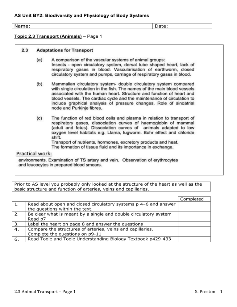
End of topic checklist for 2.3 Transport
Tick as appropriate:
RED: I do not know about this
AMBER: I have heard about this but have not learned this yet. I am unsure on this.
GREEN: I have heard about this and I have learned this. I am confident about this.
Topic
1.
Multicellular animals have a transport system.
2.
Insects have an open circulatory system, with a dorsal tube shaped heart, and a fluid filled cavity (haemocoel).
3.
The earthworm has a closed circulatory system, with blood pressure.
Organs are not in direct contact with the blood. Respiratory gases are transported in the blood.
4.
Mammals have a circulatory system comprising closed, double circulation and a heart with two atria and ventricles.
5.
The major blood vessels of the heart include: aorta, vena cava, pulmonary veins, pulmonary arteries and coronary arteries.
6.
The heart is a specialised organ having cardiac muscle, own blood supply, variation in the thickness of its walls and valves.
7.
Large vessels have 3 main layers in the walls: tough collagen, elastic muscular layer to sustain pressure and endothelium to which is smooth to reduce friction; capillary walls are one cell thick.
8.
Veins have a thinner muscle layer than arteries and along their length are semi-lunar valves to ensure flow in one direction.
9.
Arteries have thick walls to resist pressure.
10.
Arterioles adjust diameter to adjust blood pressure.
11.
Capillaries have a small diameter and friction with the walls slows the blood flow. Although the diameter is small, there are many capillaries in the capillary bed, providing a large cross sectional area, which further reduces blood flow. The low velocity in very thin walled vessels enhances their ability to exchange materials with the surrounding tissue fluid.
12.
Venules/veins have larger diameters and thinner walls than arterioles/arteries and the pressure is reduced; valves prevent back flow.
13.
The cardiac cycle refers to a sequence of events that takes place during the beating of the heart. The sinoatrial node is spontaneously active and its excitation spreads out across the atria, causing them to contract, but is prevented from spreading to the ventricles by a thin layer of connective tissue. Excitation spreads via the atrioventricular node, through the Bundle of His to the apex of the ventricle. The bundle branches into Purkinje fibres in the ventricle walls, which carry the wave of excitation upwards through the ventricle muscle. The contraction of the ventricles is therefore delayed after the atria. The pressure changes in the atria, ventricles and aorta during the cardiac cycle can be analysed graphically. These pressure changes are responsible for the opening and closing of the valves.
14.
When blood leaves the heart the highest pressures are found in the aorta and main arteries which show a rhythmic rise and fall which corresponds to ventricular contraction.
15.
Friction with vessel walls causes progressive pressure drop. Arterioles have a large total surface area and relatively narrow bore causing substantial reduction from aortic pressure. Their pressure depends on whether they are dilated or contracted.
16.
There is an even greater resistance in the capillaries with a larger crosssectional area.
17.
The velocity of blood of blood flow is directly related to pressure. In the capillary beds the pressure drops further due to leakage from the capillaries into tissues.
18.
Return flow to the heart is non-rhythmic and the pressure in the veins is low but can be increased by the massaging effect of the muscles.
19.
Heart rate can be modified by hormones or the nervous system.
20.
Blood components carry gases – haemoglobin carries oxygen as oxyhaemoglobin.
21.
The functioning of different types of haemoglobin is demonstrated by plotting oxygen dissociation curves for normal mammalian haemoglobin
2.3 Animal Transport – Page 1
RED AMBER GREEN
S. Preston 2
compared to foetal haemoglobin.
22.
Oxygen dissociation curves for llama haemoglobin and lugworm demonstrate a physiological adaptation for life in oxygen-depleted conditions.
23.
The release of oxygen involved the Bohr effect where the lowered pH due to dissolving carbon dioxide reducing the oxygen affinity for haemoglobin, causing it to release oxygen where it is most required.
24.
Some carbon dioxide is transported in red blood cells, but most is converted in the red blood cells to bicarbonate, which is then dissolved in the plasma. The chloride shift refers to the influx of chloride ions into the red blood cells to preserve electrical neutrality as the bicarbonate leaves.
25.
The blood transports other substances in the blood plasma: digested food products, hormones, proteins, albumin, fibrinogen, antibodies and ions and it also distributes heat.
26.
Water and small solutes pass through the capillary endothelium at the beginning of the capillary beds. The hydrostatic pressure here (forcing liquid out) is greater than the osmotic pressure (drawing water in). At the end of the capillary bed the hydrostatic pressure has dropped to a low value and the water potential gradient causes an inward flow about 99% of the fluid that leaves the blood at the arterial end of the capillary bed returns at the venous end.
27.
The rest of the tissue fluid is returned via the lymphatic system.
Transport in Animals
In larger animals, diffusion is simply too slow for all the cells to exchange materials quickly enough or in sufficient quantities to stay active and healthy.
2.3 Animal Transport – Page 1 S. Preston 3
An effective transport system will include:
A fluid or medium to carry nutrients and (gases) around the body
A pump to create pressure that will push the fluid around the body
Exchange surfaces that enable (gases) and nutrients to enter the transport fluid and then leave the transport fluid where they are needed
Can you think of a reason why I put (gases) into brackets in the above statements?
More efficient and well-developed transport systems will also have:
Tubes or vessels to carry the transport (usually blood).
Two circuits, one to puck up gases from the gas exchange surface and a second to then deliver it to the places it is needed in the body.
Open and Closed Circulatory Systems
Circulatory systems can be classified as:
Open systems, e.g. insects
Closed systems e.g. fish and mammals
Open Circulatory System
In an open circulatory system the artery, which leaves the heart, branches into short arteries which themselves open into large, blood filled spaces collectively called haemocoel. Blood from these spaces gradually returns to the heart through a few open-ended veins. The blood seeps around the organs which are literally bathed in blood.
Label where y ou think the ‘short arteries’, ‘haemocoel’ and ‘open-ended veins’ would be on the diagram below on the left.
Closed Circulatory System
In a closed circulatory system blood is enclosed within blood vessels and is distinct from the tissue fluid.
2.3 Animal Transport – Page 1 S. Preston 4
Blood is moved from a muscular pump to the tissues of the body and back again entirely through blood vessels. High pressure in the capillaries forces water and other small molecules through the thin capillary walls to form a tissue fluid that bathes the cells. Label the heart and capillaries below.
Which system do you think will have the highest blood pressure and why?
Insect Circulatory System – An example of an Open Circulatory System
In an insect there is a muscular pumping organ much like a heart. This is a long tube that lies under the dorsal (upper) surface of the insect. The heart pumps the blood towards the head via peristalsis. At the forward end of the heart nearest the head, the blood simply pours out into the spaces of the body cavity called haemocoel. (the blood is referred to as haemolymph in the diagram below and the sinuses refer to the body cavities called haemocoel).
The blood bathes the tissues directly; exchange of materials takes place with the cells. Blood slowly returns to the heart. Valves and muscular contractions move the blood towards the heart where it enters back into the heart through pores called ostia.
Do you think that the blood will contain a respiratory pigment, explain your answer:
This open circulatory system is under low pressure and so it seen as being not very efficient.
Why do you think it works for insects?
Closed Circulatory Systems (shown by annelids, fish and mammals)
2.3 Animal Transport – Page 1 S. Preston 5
Annelids
An annelid has a closed circulatory system, blood never comes into direct contact with the tissues and body cavities of the body, it is always confined to blood vessels.
A simple organism, the earthworm (an example of an annelid) has a closed circulation system. It has dorsal and ventral vessels running the length of its body and these are connected via 5 pairs of
‘pseudohearts’. Blood is pushed through the vessels by the pumping action of the ‘pseudohearts’.
Summary of Open and Closed Circulatory Systems
Single and Double Circulation
All chordates possess a closed circulatory system. There are two types.
2.3 Animal Transport – Page 1 S. Preston 6
Single Circulatory Systems e.g. fish
Double Circulatory Systems e.g. mammals
Fish – Single Circulatory System
In fish, deoxygenated blood is pumped by the heart to the gills, the oxygenated blood then flows from the gills around the body and then returns to the heart. The blood flows through the heart only once for every complete circuit of the body. This is why it is called a single circulatory system.
Problems
When the blood flows to the gills it has to go through the capillary networks of the lamellae before travelling around the rest of the body and reaching more capillary networks.
Capillaries offer resistance to blood flow, this leads to a large drop in blood pressure. This means that the flow of blood back from the tissues to the heart is extremely slow and sluggish.
Single Circulation Double Circulation
Mammals - Double Circulatory System
The problems of a drop in blood pressure are overcome with a double circulatory system in which deoxygenated blood is pumped from the heart to the lungs, after which the oxygenated blood is returned to the heart and is then pumped around the rest of the body. So the blood flows twice for each complete circuit of the body. Mammalian hearts are being divided into completely separate right and left sides separates the deoxygenated and oxygenated blood.
What do the terms pulmonary and systemic mean?
The Heart
2.3 Animal Transport – Page 1 S. Preston 7
Highlight the aorta, vena cava, pulmonary veins, pulmonary arteries and valves.
Explain the importance of the blood vessels that run over the surface of the cardiac muscle , the coronary arteries :
Explain why the left ventricle has a thicker more muscular wall than the right ventricle.
What is the purpose of the valves?
What is special about cardiac muscle?
Arteries
2.3 Animal Transport – Page 1 S. Preston 8
Large blood vessels contain three main layers in their walls tough collagen, elastic muscular layer and endothelium as the inner layer.
Describe the how the blood is flowing in the arteries as it leaves the heart:
Describe how the structure of the artery adapts it to cope with the blood leaving the heart:
Arteries branch into smaller arterioles that carry blood to the capillary beds. They can adjust their diameter.
How do they do this and why would it be useful?
Veins
2.3 Animal Transport – Page 1 S. Preston 9
Blood enters venules from the capillary beds and the venules converge into veins.
Describe the blood in the veins.
Describe how the structure and location of veins adapts them to cope with returning blood to the heart:
Contrast the structure and function of an artery with a vein.
Capillaries
2.3 Animal Transport – Page 1 S. Preston 10
The diameter of capillaries is small and friction with the walls will slow down blood flow.
This slow velocity will enhance their function of exchange.
Compare the structures and functions of arteries, veins and capillaries.
Draw a table for comparison purposes: e.g.
Feature
Description of blood in vessel
Function
Artery
Pulsating and at high pressure
To transport blood away from the heart
Vein
Slow Moving, low pressure.
To transport blood back to the heart
Capillary
Slow moving.
Exchange of materials, form tissue fluid
