Practical 5 Plant Structure
advertisement
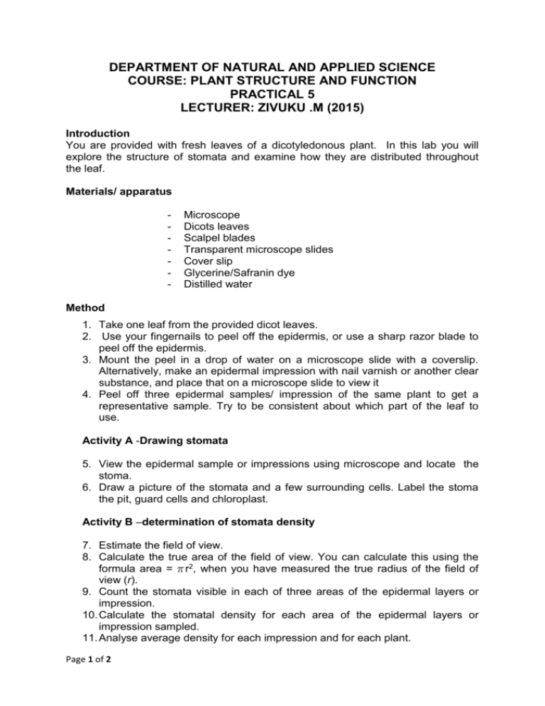
DEPARTMENT OF NATURAL AND APPLIED SCIENCE COURSE: PLANT STRUCTURE AND FUNCTION PRACTICAL 5 LECTURER: ZIVUKU .M (2015) Introduction You are provided with fresh leaves of a dicotyledonous plant. In this lab you will explore the structure of stomata and examine how they are distributed throughout the leaf. Materials/ apparatus - Microscope Dicots leaves Scalpel blades Transparent microscope slides Cover slip Glycerine/Safranin dye Distilled water Method 1. Take one leaf from the provided dicot leaves. 2. Use your fingernails to peel off the epidermis, or use a sharp razor blade to peel off the epidermis. 3. Mount the peel in a drop of water on a microscope slide with a coverslip. Alternatively, make an epidermal impression with nail varnish or another clear substance, and place that on a microscope slide to view it 4. Peel off three epidermal samples/ impression of the same plant to get a representative sample. Try to be consistent about which part of the leaf to use. Activity A -Drawing stomata 5. View the epidermal sample or impressions using microscope and locate the stoma. 6. Draw a picture of the stomata and a few surrounding cells. Label the stoma the pit, guard cells and chloroplast. Activity B –determination of stomata density 7. Estimate the field of view. 8. Calculate the true area of the field of view. You can calculate this using the formula area = r2, when you have measured the true radius of the field of view (r). 9. Count the stomata visible in each of three areas of the epidermal layers or impression. 10. Calculate the stomatal density for each area of the epidermal layers or impression sampled. 11. Analyse average density for each impression and for each plant. Page 1 of 2 12. Repeat 1,2, 3, 4, 7, 8, 9, 10 and 11 for different types of leaves and record your results in the form a table. Your table for stomata density in leaf from plant A; Plant Number and leaf True type radius Plant A Leaf 1 Leaf 2 Leaf 3 Average Page 2 of 2 Area view of Number of Density Stomata of stomata

