17. Female Reproductive System - PEER
advertisement
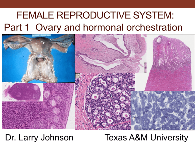
FEMALE REPRODUCTIVE SYSTEM: Part 1 Ovary and hormonal orchestration Dr. Larry Johnson Texas A&M University Objectives: female reproductive system Part 1 Ovary and hormonal orchestration • Identify the tissues of the ovary, and distinguish between primordial, primary, secondary, tertiary, and atretic follicles. • Hormonal orchestration Part 2 Uterine tube and uterus • Describe the three layers of the uterine tube and compare the structure in the different regions along its length. • Describe the three layers of the uterus. Understand the morphological changes in the endometrium and myometrium during the normal menstrual cycle and during pregnancy. Part 3 Cervix, mammary gland, and placenta • Compare the structures and functions of the mammary glands at different stages. • Identify the three layers of the cervix and the vagina. • Identify the villi and intervillous space in the placenta. Relate these structures with placental function. FUNCTIONS OF THE FEMALE REPRODUCTIVE SYSTEM • OVA PRODUCTION • OVA and SPERM TRANSPORTATION • MICROENVIRONMENTS FOR FERTILIZATION • IMPLANTATION AND FETAL-PLACENTAL GROWTH • NOURISHMENT AND SUPPORT OF OFFSPRING • POSTNATAL REPETITION OVARY GENERAL STRUCTURE • • • • GERMINAL EPITHELIUM TUNICA ALBUGINEA MEDULLA CORTEX FUNCTIONAL OVERVIEW Slide 79: Ovary Medullary blood vessels Tunica albuginea Cortex Medulla Cortex Germinal epithelium Ovary, monkey 1 2 Ovarian Cortex: 3 1. Germinal epithelium 2. Tunica albunginea 3. Primordial and primary follicles Follicle development and changes within ovary Copyright McGraw-Hill Companies FOLLICLE MATURATION PRIMORDIAL FOLLICLES • • OOCYTE FOLLICULAR (GRANULOSA) CELLS Ovary: 1. Primordial follicle 2. Granulosa cell Ovarian Cortex: 1. Germinal epithelium 2. Tunica albunginea 3. Primary follicle FOLLICLE MATURATION PRIMARY FOLLICLE • • • • ZONA PELLUCIDA STRATUM GRANULOSUM THECAL FOLLICULI CALL-EXNER BODIES FOLLICLE MATURATION SECONDARY (ANTRAL) FOLLICLE FOLLICULAR FLUID MEMBRANA GRANULOSA CUMULUS OOPHORUS CORONA RADIATA THECA INTERNA THECA EXTERNA FOLLICLES FOLLICLE MATURATION GRAAFIAN FOLLICLE GRAAFIAN FOLLICLE egg Slide 79: Ovary Secondary follicle Primordial follicle Primary follicle Theca externa & interna Zona pellucida Granulosa cells Antrum with liquor folliculi Corona radiata Cumulus oophorus Membrana granulosa EM 50: Ovary EM 25: early primary follicle 1. Cytoplasm of primary oocyte 2. Zona pellucida 3. Follicular cell Ovary Ovary Ovary, guinea pig Ovary, guinea pig – primary and secondary follicles; follicle with atresia and its final form as a glassy membrane OVULATION Ovary, corpus luteum 1. Granulosa lutein 2. Theca lutein 3. Central clot Corpus luteum of ovary 268 Slide 80: Ovary with corpus luteum Corpus luteum Corpus hemorrhagicum remnants Theca lutein cells Granulosa lutein cells Corpus luteum of Ovary, mouse (toluidine blue) 268 EM 24: corpus luteum 1. Capillary 2. Nucleus of granulosa lutein cell 3. erythrocyte Corpus luteum and corpus albicans of ovary 170 Slide 81: Ovary Vasculature Corpus albican Corpus luteum OOGENESIS - FORMATION AND DEVELOPMENT OF OVA MITOSIS (OOCYTOGENESIS) – OOGONIA • PRENATAL DEVELOPMENT (RUMINANTS, RODENT, SWINE, HUMAN) • POSTNATAL DEVELOPMENT (CARNIVORES) OOGENESIS - FORMATION AND DEVELOPMENT OF OVA MEIOSIS – OOCYTES EARLY DEVELOPMENT MATURATION ARREST (DICTYATE STEP OF MEIOTIC PROPHASE) LATER DEVELOPMENT SYNCHRONIZED WITH DEVELOPMENT AND MATURATION OF FOLLICLES DIVISION • FIRST MEIOTIC DIVISION – REDUCTION DIVISION – FIRST POLAR BODY • SECOND MEIOTIC DIVISION – • EQUATIONAL DIVISION – SECOND POLAR BODY Oogenesis Copyright McGraw-Hill Companies The number of chromosomes are present in the human secondary oocyte is 23 chromosomes, the haploid number. MEIOSIS Zygote MEIOSIS (ONLY IN SPERMATOGENESIS AND OOGENESIS) EXCHANGE OF GENETIC MATERIAL IN HOMOLOGOUS CHROMOSOMES (LEPTOTENE, ZYGOTENE, PACHYTENE, AND DIPLOTENE STEPS OF DEVELOPMENT) PRODUCES HAPLOID CONDITION OF GAMETES MEIOSIS (ONLY IN SPERMATOGENESIS AND OOGENESIS) PRODUCES HAPLOID CONDITION OF GAMETES SPECIES DIFFERENCE IN TIME OF POLAR BODY FORMATION POLAR BODY EXTRUSION FIRST SECOND MANY MAMMALS PREOVULATION HORSE & DOG POSTOVULATION ZONA PENETRATION OF SPERM ZONA PENETRATION OF SPERM FIRST POLAR BODY birth FOLLICULAR DEVELOPMENT SYNCHRONIZED WITH GAMETE DEVELOPMENT TYPE PRIMORDIAL PRIMARY SECONDARY DESCRIPTION FLATTENED FOLLICULAR CELLS GERM CELL TYPE PRIMARY OOCYTE LOW CUBOIDAL FOLLICULAR CELLS PRIMARY OOCYTE YOLK FORMATION PRIMARY OOCYTE ZONA PELLUCIDA THECA INTERNA THECA FOLLICULI EXTERNA FOLLICULAR DEVELOPMENT - SYNCHRONIZED WITH GAMETE DEVELOPMENT (cont’d) TYPE DESCRIPTION GERM CELLTYPE VESICULAR FOLLICLE- CALL-EXNER BODIES PRIMARY OOCYTE (TERTIARY FOLLICLE) (PRECURSORS OF LIQUOR FOLLICULI) - FORMATION OF FOLLICULAR ANTRUM - CUMULUS OOPHORUS- MOUND OF CELLS - CORONA RADIATA- ADJACENT TO OOCYTE NUTRIENTS - STRATUM GRANULOSUM MATURE FOLLICLE - PREOVULATORY (GRAAFIAN FOLLICLE) LARGEST, EXTEND FROM CORTEX TO SURFACE OF OVARY PRIMARY/ SECONDARY OOCYTE HORMONES ORCHESTRATE THE PROCESS OVARIAN HORMONES ANDROGENS • LH STIMULATION CAUSES THECA INTERNA CELLS TO PRODUCE ANDROGENS (ANDOSTENEDIONE AND TESTOSTERONE) ESTROGENS • FSH STIMULATION CAUSES MEMBRANA GRANULOSA CELLS TO PRODUCE ESTROGENS (ESTRODIOL 17B) • GRANULOSA CELLS AROMATIZE ANDROGENS FROM THECA INTERNA CELLS TO PRODUCE ESTROGENS • ESTROGENS - INDUCE FURTHER PROLIFERATION OF GRANULOSAL CELLS OVARIAN HORMONES INHIBITION OF FSH • FOLLICULOSTATIN - INHIBITORY SUBSTANCE HAS NEGATIVE FEEDBACK ON FSH • STEROIDSTHEMSELVES WORK THROUGH NEGATIVE FEEDBACK PROGESTERONE - GRANULOSA LUTEIN CELLS OF CORPUS LUTEUM • UTERINE GLAND DEVELOPMENT • MAINTENANCE OF PREGNANCY RELAXIN - PRODUCED BY CORPUS LUTEUM • CAUSES RELAXATION OF LIGAMENTS ASSOCIATED WITH PUBIC SYMPHYSIS BEFORE PARTARITION Hormonal regulation of ovarian function Copyright McGraw-Hill Companies Female reproductive system cycles Copyright McGraw-Hill Companies Many illustrations in these VIBS Histology YouTube videos were modified from the following books and sources: Many thanks to original sources! • • Bruce Alberts, et al. 1983. Molecular Biology of the Cell. Garland Publishing, Inc., New York, NY. Bruce Alberts, et al. 1994. Molecular Biology of the Cell. Garland Publishing, Inc., New York, NY. • William J. Banks, 1981. Applied Veterinary Histology. Williams and Wilkins, Los Angeles, CA. • Hans Elias, et al. 1978. Histology and Human Microanatomy. John Wiley and Sons, New York, NY. • • Don W. Fawcett. 1986. Bloom and Fawcett. A textbook of histology. W. B. Saunders Company, Philadelphia, PA. Don W. Fawcett. 1994. Bloom and Fawcett. A textbook of histology. Chapman and Hall, New York, NY. • Arthur W. Ham and David H. Cormack. 1979. Histology. J. S. Lippincott Company, Philadelphia, PA. • • Luis C. Junqueira, et al. 1983. Basic Histology. Lange Medical Publications, Los Altos, CA. L. Carlos Junqueira, et al. 1995. Basic Histology. Appleton and Lange, Norwalk, CT. • L.L. Langley, et al. 1974. Dynamic Anatomy and Physiology. McGraw-Hill Book Company, New York, NY. • W.W. Tuttle and Byron A. Schottelius. 1969. Textbook of Physiology. The C. V. Mosby Company, St. Louis, MO. • • Leon Weiss. 1977. Histology Cell and Tissue Biology. Elsevier Biomedical, New York, NY. Leon Weiss and Roy O. Greep. 1977. Histology. McGraw-Hill Book Company, New York, NY. • • • Nature (http://www.nature.com), Vol. 414:88,2001. Arthur C. Guyton,1971.Textbook of Medical Physiology W.B. Saunders company, Philadelphia, PA WW Tuttle and BA Schottelius 1969 Textbook of Physiology C.V. Mosby Co. • A.L. Mescher 2013 Junqueira’s Basis Histology text and atlas, 13th ed. McGraw • Douglas P. Dohrman and TAMHSC Faculty 2012 Structure and Function of Human Organ Systems, Histology Laboratory Manual - Slide selections were largely based on this manual for first year medical students at TAMHSC End of Part 1 Ovary and hormonal orchestration FEMALE REPRODUCTIVE SYSTEM: Part 2 Uterine tube and uterus Dr. Larry Johnson Texas A&M University Objectives: female reproductive system Part 1 Ovary and hormonal orchestration • Identify the tissues of the ovary, and distinguish between primordial, primary, secondary, tertiary, and atretic follicles. • Hormonal orchestration Part 2 Uterine tube and uterus • Describe the three layers of the uterine tube and compare the structure in the different regions along its length. • Describe the three layers of the uterus. Understand the morphological changes in the endometrium and myometrium during the normal menstrual cycle and during pregnancy. Part 3 Cervix, mammary gland, and placenta • Compare the structures and functions of the mammary glands at different stages. • Identify the three layers of the cervix and the vagina. • Identify the villi and intervillous space in the placenta. Relate these structures with placental function. Uterine tubes and uterus Copyright McGraw-Hill Companies image 179 Copyright McGraw-Hill Companies Fallopian tube = uterine tube Uterine tube - infundibulum frimbriae lymphatic vessels help give rigidity to the projections Uterine tube: infundibulum frimbriae - ciliated epithelium Slide 82: Oviduct (ampulla) Serosa Muscularis externa Blood vessels Ciliated simple columnar epithelium Lamina propria Uterine tube: isthmus Copyright McGraw-Hill Companies Uterine tube Copyright McGraw-Hill Companies 472 Uterine tube - ciliated epithelium Slide 145, uterus 1. Endometrium 2. Myometrium 3. Perimetrium 1 3 2 GENERAL STRUCTURE PERIMETRIUM MYOMETRIUM ENDOMETRIUM 177 ZONA BASALIS ZONA FUNCTIONALIS The stratum basalis remains relatively unchanged during the menstrual cycles. ZONA FUNCTIONALIS SPIRAL ARTERIES 177 Uterus, early proliferative endometrium Uterus, early proliferative endometrium Slide 83: Uterus (early proliferative [follicular] stage) Perimetrium serosa lined by mesothelium Myometrium Endometrium Uterine lumen lined by simple columnar epithelium Slide 83: Uterus (early proliferative [follicular] stage) Arterial supply to endometrium Basal layer Functionalis layer Copyright McGraw-Hill Companies Straight uterine glands Uterus, late proliferative endometrium Uterus, late proliferative endometrium Uterus, early secretory endometrium 177 Uterus, early secretory endometrium Uterus, early secretory endometrium Uterus, early secretory endometrium and myometrium Slide 84: Uterus (secretory [luteal] stage Endometrium Functionalis layer Basal layer Hypertrophied and tortuous uterine glands Myometrium Uterus, late secretory endometrium spiraling arteries SPIRAL ARTERIES Uterus, late secretory endometrium smooth muscle Uterus, late secretory endometrium The phase of the uterine cycle when the glands produce the most glycogen is secretory phase. HORMONES ORCHESTRATE THE PROCESS Uterus, late secretory 1. Decidual cells 2. Spiral arteries ~ 10-day implantation 10 day old implant human #272 Many illustrations in these VIBS Histology YouTube videos were modified from the following books and sources: Many thanks to original sources! • • Bruce Alberts, et al. 1983. Molecular Biology of the Cell. Garland Publishing, Inc., New York, NY. Bruce Alberts, et al. 1994. Molecular Biology of the Cell. Garland Publishing, Inc., New York, NY. • William J. Banks, 1981. Applied Veterinary Histology. Williams and Wilkins, Los Angeles, CA. • Hans Elias, et al. 1978. Histology and Human Microanatomy. John Wiley and Sons, New York, NY. • • Don W. Fawcett. 1986. Bloom and Fawcett. A textbook of histology. W. B. Saunders Company, Philadelphia, PA. Don W. Fawcett. 1994. Bloom and Fawcett. A textbook of histology. Chapman and Hall, New York, NY. • Arthur W. Ham and David H. Cormack. 1979. Histology. J. S. Lippincott Company, Philadelphia, PA. • • Luis C. Junqueira, et al. 1983. Basic Histology. Lange Medical Publications, Los Altos, CA. L. Carlos Junqueira, et al. 1995. Basic Histology. Appleton and Lange, Norwalk, CT. • L.L. Langley, et al. 1974. Dynamic Anatomy and Physiology. McGraw-Hill Book Company, New York, NY. • W.W. Tuttle and Byron A. Schottelius. 1969. Textbook of Physiology. The C. V. Mosby Company, St. Louis, MO. • • Leon Weiss. 1977. Histology Cell and Tissue Biology. Elsevier Biomedical, New York, NY. Leon Weiss and Roy O. Greep. 1977. Histology. McGraw-Hill Book Company, New York, NY. • • • Nature (http://www.nature.com), Vol. 414:88,2001. Arthur C. Guyton,1971.Textbook of Medical Physiology W.B. Saunders company, Philadelphia, PA WW Tuttle and BA Schottelius 1969 Textbook of Physiology C.V. Mosby Co. • A.L. Mescher 2013 Junqueira’s Basis Histology text and atlas, 13th ed. McGraw • Douglas P. Dohrman and TAMHSC Faculty 2012 Structure and Function of Human Organ Systems, Histology Laboratory Manual - Slide selections were largely based on this manual for first year medical students at TAMHSC End of Part 2: Uterine tube and uterus FEMALE REPRODUCTIVE SYSTEM: Part 3 Cervix, mammary gland, and placenta Dr. Larry Johnson Texas A&M University Objectives: female reproductive system Part 1 Ovary and hormonal orchestration • Identify the tissues of the ovary, and distinguish between primordial, primary, secondary, tertiary, and atretic follicles. • Hormonal orchestration Part 2 Uterine tube and uterus • Describe the three layers of the uterine tube and compare the structure in the different regions along its length. • Describe the three layers of the uterus. Understand the morphological changes in the endometrium and myometrium during the normal menstrual cycle and during pregnancy. Part 3 Cervix, mammary gland, and placenta • Compare the structures and functions of the mammary glands at different stages. • Identify the three layers of the cervix and the vagina. • Identify the villi and intervillous space in the placenta. Relate these structures with placental function. Slide 184, cervix 1. Lumen 2. Cervical glands 3. Lamina propria 4. Smooth muscle Cervix Cervix Cervix Cervix Cervix, 1. junction of endocervix and ectocervix 2. Vagina cervix ENDOCERVIX ends in the Vagina which has no MUCUS glands Vagina Vagina epithelium 1. Stem cells 2. Prickle cell layer 3. Nonkeratinized squamous epithial cells The wall of the vagina lacks glands. Any mucus in the vagina is produced by cervical glands and other liberation comes from blood plasma. Vagina Slide 85: Cervix / vagina Mucous glands Endocervix Simple columnar epithelium Nonkeratinized stratified squamous epithelium lining the portio vaginalis Slide 86: Vagina 2 1 Nonkeratinized stratified squamous epithelium 1. Lamina propria 2. Muscularis externa Slide 89: Mammary gland (inactive) Interlobular CT Adipocytes Lobule Copyright McGraw-Hill Companies Alveolar duct Intralobular CT NOURISHMENT AND PROTECTION OF OFFSPRING NOURISHMENT and PROTECTION of OFFSPRING NOURISHMENT AND PROTECTION OF OFFSPRING OTHER GLANDS OF EPIDERMAL ORIGIN – mammary gland Gland for which our class,, Mammalia, was named. Mammals are characterized by hair on skin, special ear bones, and in females, milk-producing mammary glands for nourishment of young. Nipple Breast inactive 482 Breast, pregnancy 482 Breast, early pregnancy 182 Breast during late pregnancy Slide 90: Mammary gland (early proliferative) Intralobular CT Lobule Interlobular CT Alveolar duct Adipocytes Slide 91: Mammary gland (active) Lobule Large duct with colostrum Interlobular CT Alveoli with colostrum Intralobular CT Myoepithelial cell In summary Mammary gland Late pregnancy Copyright McGraw-Hill Companies Histo 091 IMPLANTATION AND FETAL-PLACENTAL GROWTH Slide 272 IMPLANTATION AND FETAL-PLACENTAL GROWTH Slide 87: Umbilical cord http://homepages.cae.wisc.edu/~bme300/umbilical_f07/ Wharton’s jelly Umbilical vein Umbilical arteries The umbilical vein carries oxygenated blood to the fetus. Copyright McGraw-Hill Companies Placenta Blood flow from the mother to the fetus. Maternal artery > placenta > umbilical vein > liver OR ductus venosus (bypasses liver) > R atrium of heart > foramen ovale OR R ventricle Foramen ovale (bypass lungs) > L atrium > L ventricle > aorta > brain and body R ventricle > pulmonary artery > ductus arteriosis (bypass lungs) > aorta > body Body > 2 umbilical arteries > placenta > maternal vein Slide 88: Placenta Chorionic villi Intervillous space Placenta Cytotrophoblast cell Fetal capillaries Mesenchyme Synctiotrophoblast cells Decidua basalis Decidual cells EM 26; trophoblast, 20,000x 1. Nucleus 2. Microvilli 3. Tubular cristae hemochorial placenta = human placenta 273 A hemochorial placenta type has the maternal blood in direct contact with the chorionic trophoblast. However, there are three layers of cells 273 Mesenchyme Cytotrophoblast cell Synctiotrophoblast cells Fetal capillaries 129 CONNECTIVE TISSUE CLASSIFICATION Clinical Correlation Stimulation of receptors in the nipple during breast-feeding results in release of oxytocin from the posterior pituitary. Where are the cell bodies of the cells that release oxytocin located? • The cell bodies of the cells that release oxytocin are located in the hypothalamus. Oxytocin is responsible for the milk-ejection reflex. Which cells in the breast are stimulated oxytocin? • Oxytocin stimulates contraction of the myoepithelial cells around the alveoli, thereby ejecting milk. http://animalsciences.missouri.edu/reprod/notes/endocrin/oxytocin.jpg Clinical Correlation The structure of the endometrium changes dramatically during the menstrual cycle. Menstrual phase Proliferative phase Secretory phase All images Copyright McGraw-Hill Companies Many illustrations in these VIBS Histology YouTube videos were modified from the following books and sources: Many thanks to original sources! • • Bruce Alberts, et al. 1983. Molecular Biology of the Cell. Garland Publishing, Inc., New York, NY. Bruce Alberts, et al. 1994. Molecular Biology of the Cell. Garland Publishing, Inc., New York, NY. • William J. Banks, 1981. Applied Veterinary Histology. Williams and Wilkins, Los Angeles, CA. • Hans Elias, et al. 1978. Histology and Human Microanatomy. John Wiley and Sons, New York, NY. • • Don W. Fawcett. 1986. Bloom and Fawcett. A textbook of histology. W. B. Saunders Company, Philadelphia, PA. Don W. Fawcett. 1994. Bloom and Fawcett. A textbook of histology. Chapman and Hall, New York, NY. • Arthur W. Ham and David H. Cormack. 1979. Histology. J. S. Lippincott Company, Philadelphia, PA. • • Luis C. Junqueira, et al. 1983. Basic Histology. Lange Medical Publications, Los Altos, CA. L. Carlos Junqueira, et al. 1995. Basic Histology. Appleton and Lange, Norwalk, CT. • L.L. Langley, et al. 1974. Dynamic Anatomy and Physiology. McGraw-Hill Book Company, New York, NY. • W.W. Tuttle and Byron A. Schottelius. 1969. Textbook of Physiology. The C. V. Mosby Company, St. Louis, MO. • • Leon Weiss. 1977. Histology Cell and Tissue Biology. Elsevier Biomedical, New York, NY. Leon Weiss and Roy O. Greep. 1977. Histology. McGraw-Hill Book Company, New York, NY. • • • Nature (http://www.nature.com), Vol. 414:88,2001. Arthur C. Guyton,1971.Textbook of Medical Physiology W.B. Saunders company, Philadelphia, PA WW Tuttle and BA Schottelius 1969 Textbook of Physiology C.V. Mosby Co. • A.L. Mescher 2013 Junqueira’s Basis Histology text and atlas, 13th ed. McGraw • Douglas P. Dohrman and TAMHSC Faculty 2012 Structure and Function of Human Organ Systems, Histology Laboratory Manual - Slide selections were largely based on this manual for first year medical students at TAMHSC End of Part 3 Cervix, mammary gland, and placenta The End!
