sheep_brain_dissection - IB-Biology
advertisement
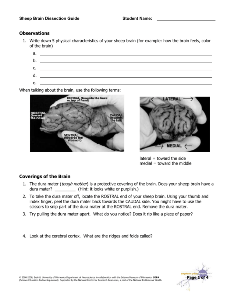
Sheep Brain Dissection Guide Student Name: Observations 1. Write down 5 physical characteristics of your sheep brain (for example: how the brain feels, color of the brain) a. b. c. d. e. When talking about the brain, use the following terms: lateral = toward the side medial = toward the middle Coverings of the Brain 1. The dura mater (tough mother) is a protective covering of the brain. Does your sheep brain have a dura mater? _________ (Hint: it looks white or purplish.) 2. To take the dura mater off, locate the ROSTRAL end of your sheep brain. Using your thumb and index finger, peel the dura mater back towards the CAUDAL side. You might have to use the scissors to snip part of the dura mater at the ROSTRAL end. Remove the dura mater. 3. Try pulling the dura mater apart. What do you notice? Does it rip like a piece of paper? 4. Look at the cerebral cortex. What are the ridges and folds called? © 2000-2008, BrainU, University of Minnesota Department of Neuroscience in collaboration with the Science Museum of Minnesota. SEPA (Science Education Partnership Award) Supported by the National Center for Research Resources, a part of the National Institutes of Health. Page 1 of 4 Sheep Brain Dissection Guide Student Name: 5. Draw the LATERAL (outer side) view of your sheep brain; include the cerebellum and brainstem. You can peel the covering halfway back. 6. Flip the brain over and examine the underside of the brain. Locate on your brain the structures labeled in the diagram below: a. What does the optic chiasm lead to? b. What do the olfactory bulbs lead to? Midline/Medial Anatomy 7. Cut through the middle of the brain up and down. Gently force the two cerebral hemispheres apart. Draw the MEDIAL (inner side) view of the sheep brain. Label 3-5 brain parts that you see. © 2000-2008, BrainU, University of Minnesota Department of Neuroscience in collaboration with the Science Museum of Minnesota. SEPA (Science Education Partnership Award) Supported by the National Center for Research Resources, a part of the National Institutes of Health. Page 2 of 4 Sheep Brain Dissection Guide Student Name: 8. Using the diagram below, locate the thalamus, hypothalamus, and pituitary in your sheep brain. Exploring the Neuron and its Parts 9. Look closely at the inside of the cerebellum. You should see a branching "tree" of lighter tissue surrounded by darker tissue. The branches are _______ matter, which is made up of nerve axons. The darker tissue is ______ matter, which is a collection of nerve cell bodies. 10. Place your sheep brain with the cut side (MEDIAL) down. Using the plastic knife, cut the brain about 1 inch from the rostral end (where the dent is located). Look at the inside surface view of the piece of brain cut off. a. What do you notice about the cut surface? b. Using the probe stick, poke the dark area. How does it feel? c. Using the probe stick, poke the light area. How does it feel? d. What do you think the dark and light areas are? © 2000-2008, BrainU, University of Minnesota Department of Neuroscience in collaboration with the Science Museum of Minnesota. SEPA (Science Education Partnership Award) Supported by the National Center for Research Resources, a part of the National Institutes of Health. Page 3 of 4 Sheep Brain Dissection Guide Student Name: Comparing Sheep Brains to Human Brains MEDIAL View of the Human Brain MEDIAL View of the Sheep Brain 1. Label the brain parts in both the human and sheep brain pictures. 2. How are these brains similar? 3. Why might both brains be similar? 4. How are they different? 5. Why would they be different? © 2000-2008, BrainU, University of Minnesota Department of Neuroscience in collaboration with the Science Museum of Minnesota. SEPA (Science Education Partnership Award) Supported by the National Center for Research Resources, a part of the National Institutes of Health. Page 4 of 4
