World Journal of Engineering PREPARATION OF Fe3O4/CaP
advertisement
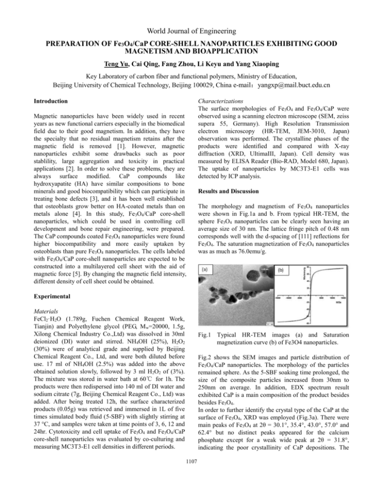
World Journal of Engineering PREPARATION OF Fe3O4/CaP CORE-SHELL NANOPARTICLES EXHIBITING GOOD MAGNETISM AND BIOAPPLICATION Teng Yu, Cai Qing, Fang Zhou, Li Keyu and Yang Xiaoping Key Laboratory of carbon fiber and functional polymers, Ministry of Education, Beijing University of Chemical Technology, Beijing 100029, China e-mail:yangxp@mail.buct.edu.cn Introduction Magnetic nanoparticles have been widely used in recent years as new functional carriers especially in the biomedical field due to their good magnetism. In addition, they have the specialty that no residual magnetism retains after the magnetic field is removed [1]. However, magnetic nanoparticles exhibit some drawbacks such as poor stablility, large aggregation and toxicity in practical applications [2]. In order to solve these problems, they are always surface modified. CaP compounds like hydroxyapatite (HA) have similar compositions to bone minerals and good biocompatibility which can participate in treating bone defects [3], and it has been well established that osteoblasts grow better on HA-coated metals than on metals alone [4]. In this study, Fe3O4/CaP core-shell nanoparticles, which could be used in controlling cell development and bone repair engineering, were prepared. The CaP compounds coated Fe3O4 nanoparticles were found higher biocompatibility and more easily uptaken by osteoblasts than pure Fe3O4 nanoparticles. The cells labeled with Fe3O4/CaP core-shell nanoparticles are expected to be constructed into a multilayered cell sheet with the aid of magnetic force [5]. By changing the magnetic field intensity, different density of cell sheet could be obtained. Characterizations The surface morphologies of Fe3O4 and Fe3O4/CaP were observed using a scanning electron microscope (SEM, zeiss supera 55, Germany). High Resolution Transmission electron microscopy (HR-TEM, JEM-3010, Japan) observation was performed. The crystalline phases of the products were identified and compared with X-ray diffraction (XRD, UItimaIII, Japan). Cell density was measured by ELISA Reader (Bio-RAD, Model 680, Japan). The uptake of nanoparticles by MC3T3-E1 cells was detected by ICP analysis. Results and Discussion The morphology and magnetism of Fe3O4 nanoparticles were shown in Fig.1a and b. From typical HR-TEM, the sphere Fe3O4 nanoparticles can be clearly seen having an average size of 30 nm. The lattice fringe pitch of 0.48 nm corresponds well with the d-spacing of [111] reflections for Fe3O4. The saturation magnetization of Fe3O4 nanoparticles was as much as 76.0emu/g. Experimental Materials FeCl2·H2O (1.789g, Fuchen Chemical Reagent Work, Tianjin) and Polyethylene glycol (PEG, Mw=20000, 1.5g, Xilong Chemical Industry Co.,Ltd) was dissolved in 30ml deionized (DI) water and stirred. NH4OH (25%), H2O2 (30%) were of analytical grade and supplied by Beijing Chemical Reagent Co., Ltd, and were both diluted before use. 17 ml of NH4OH (2.5%) was added into the above obtained solution slowly, followed by 3 ml H2O2 of (3%). The mixture was stored in water bath at 60℃ for 1h. The products were then redispersed into 140 ml of DI water and sodium citrate (7g, Beijing Chemical Reagent Co., Ltd) was added. After being treated 12h, the surface characterized products (0.05g) was retrieved and immersed in 1L of five times simulated body fluid (5-SBF) with slightly stirring at 37 °C, and samples were taken at time points of 3, 6, 12 and 24hr. Cytotoxicity and cell uptake of Fe3O4 and Fe3O4/CaP core-shell nanoparticles was evaluated by co-culturing and measuring MC3T3-E1 cell densities in different periods. Fig.1 Typical HR-TEM images (a) and Saturation magnetization curve (b) of Fe3O4 nanoparticles. Fig.2 shows the SEM images and particle distribution of Fe3O4/CaP nanoparticles. The morphology of the particles remained sphere. As the 5-SBF soaking time prolonged, the size of the composite particles increased from 30nm to 250nm on average. In addition, EDX spectrum result exhibited CaP is a main composition of the product besides besides Fe3O4. In order to further identify the crystal type of the CaP at the surface of Fe3O4, XRD was employed (Fig.3a). There were main peaks of Fe3O4 at 2θ = 30.1°, 35.4°, 43.0°, 57.0° and 62.4° but no distinct peaks appeared for the calcium phosphate except for a weak wide peak at 2θ = 31.8°, indicating the poor crystallinity of CaP depositions. The 1107 World Journal of Engineering saturation magnetization of Fe3O4/CaP nanoparticles dropped greatly comparing with pure Fe3O4 nanoparticles (Fig.3b) because CaP surrounded the Fe3O4 cores which would weaken the magnetism. Besides, as the biomineralization time increased, Ms of composite particles was lower. reason is unclear. It might be related to the higher uptake ability of Fe3O4/CaP nanoparticles than pure Fe3O4 by osteoblasts owing to the increased affinity of CaP component to cells (Fig. 4b). Fig.4 Cytotoxicities of Fe3O4 and Fe3O4/CaP nanoparticles with concentration of 100μg/ml after 1, 3 and 5 days of culture (a), Amount of iron up taken by MC3T3-E1 cells after 2, 4 and 24 h incubation with Fe3O4 and Fe3O4/CaP nanoparticles with concentration of 100μg/ml (b). Conclusion Fe3O4/CaP magnetic nanoparticles were synthesized by hydrothermal and biomineralization methods. The pure Fe3O4 nanoparticles had an average size of 30nm and high magnetism. With the deposition of CaP compounds on surface, the particle size increased, while the magnetism dropped greatly. But with increased biocompatibility and affinity to cells, the Fe3O4/CaP core-shell nanoparticles were more easily uptaken by osteoblasts than pure Fe3O4 nanoparticles. These primary results indicate that Fe3O4/CaP core-shell nanoparticles have potential use in bone defects repairing and further studies are going on. . Fig.2 SEM images and particle distribution of Fe3O4/CaP nanoparticles obtained at different biominerlization time: (a) 3h, (b) 6h, (c) 12h, (d) 24h. EDX spectrum of Fe3O4/CaP nanoparticles-3h (e). References 1.Tsang, S. C., Yu, C. H., Gao, X. and Tam, K. Metal nanoparticle encapsulated in oxide. J. Phys. Chem. B., 110 (2006) 16914-16922. 2.Yang, Z. P., Gong, X. Y. and Zhang, C. J. Recyclable Fe3O4/hydroxyapatite composite nanoparticles for photocatalytic applications. Chem. Eng. J., 165 (2010) 117-121. 3. Nhiem, T. and Webster, T. J. Increased osteblast functions in the presence of hydroxyapatite-coated iron oxide nanoparticles. Acta Biomater., 7 (2011) 1298-1306. 4.Webster, T. J., Siegel, R. W. and Bizios, R. Osteoblast adhesion on nanophase ceramics. Biomaterial. 20 (1999) 1221-1127. 5.Ito, A., Jitsunobu, H., Kawabe, Y. and Kamihira, M. Construction of Heterotypic cell sheets by magnetic force-based 3-D coculture of HepG2 and NIH3T3 cells. J. Fig.3 XRD patterns (a) and saturation magnetization curve (b) of Fe3O4/CaP nanoparticles at different biomineralization times. The results of cytotoxic tests showed that osteoblast densities were similar in the presence of Fe3O4 and Fe3O4/CaP nanoparticles at day 1 and 3, and both were comparable with the control. However, cells cultured associating with Fe3O4/CaP nanoparticles showed much higher cell densities than the others at day 5 (Fig.4a). The 1108 World Journal of Engineering Biosci. Bioeng., 5 (2007) 371-378. 1109
