European Heart Journal Advance Access published May 26, 2011
CARDIOVASCULAR FLASHLIGHT
doi:10.1093/eurheartj/ehr135
.............................................................................................................................................................................
Living on an atrial kick – an unusual case of a stuck mitral valve
Yoram Neuman*, David Pereg, and Morris Mosseri
Division of Cardiology, Meir Medical Center, Kfar-Saba, Israel
* Corresponding author. Echocardiography Laboratory, Meir Medical Center, 59 Tchernichovsky St, Kfar saba 44281, Israel. Tel: +972 97472587; fax: +972 97401704,
Email: yoram.neuman@clalit.org.il
Supplementary material
Supplementary material is available at European Heart Journal online.
Published on behalf of the European Society of Cardiology. All rights reserved. & The Author 2011. For permissions please email: journals.permissions@oup.com.
Downloaded from http://eurheartj.oxfordjournals.org/ by guest on March 6, 2016
A 53-year-old woman was admitted for dyspnoea. Her past history included mitral valve
replacement (mechanical, tilting disc) 20
years earlier. The patient was well coumadinized both on admission and during the
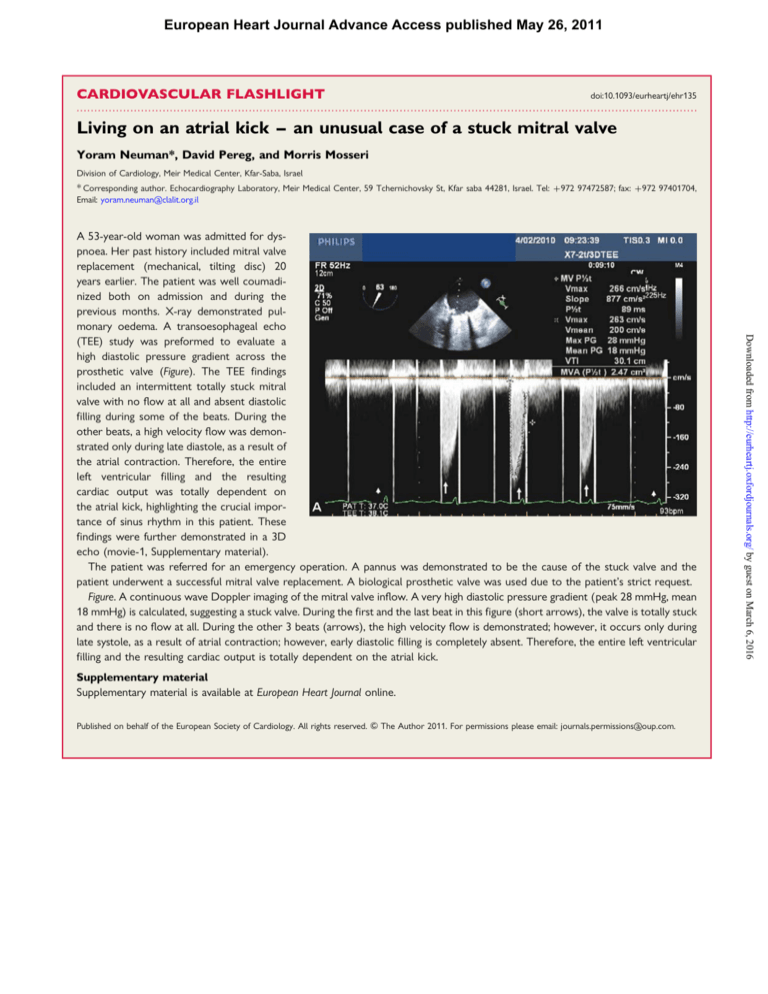
previous months. X-ray demonstrated pulmonary oedema. A transoesophageal echo
(TEE) study was preformed to evaluate a
high diastolic pressure gradient across the
prosthetic valve (Figure). The TEE findings
included an intermittent totally stuck mitral
valve with no flow at all and absent diastolic
filling during some of the beats. During the
other beats, a high velocity flow was demonstrated only during late diastole, as a result of
the atrial contraction. Therefore, the entire
left ventricular filling and the resulting
cardiac output was totally dependent on
the atrial kick, highlighting the crucial importance of sinus rhythm in this patient. These
findings were further demonstrated in a 3D
echo (movie-1, Supplementary material).
The patient was referred for an emergency operation. A pannus was demonstrated to be the cause of the stuck valve and the
patient underwent a successful mitral valve replacement. A biological prosthetic valve was used due to the patient’s strict request.
Figure. A continuous wave Doppler imaging of the mitral valve inflow. A very high diastolic pressure gradient (peak 28 mmHg, mean
18 mmHg) is calculated, suggesting a stuck valve. During the first and the last beat in this figure (short arrows), the valve is totally stuck
and there is no flow at all. During the other 3 beats (arrows), the high velocity flow is demonstrated; however, it occurs only during
late systole, as a result of atrial contraction; however, early diastolic filling is completely absent. Therefore, the entire left ventricular
filling and the resulting cardiac output is totally dependent on the atrial kick.

