Soft Tissue Wound Healing Review
advertisement
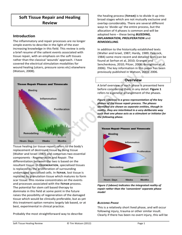
Soft Tissue Repair and Healing Review Introduction The inflammatory and repair processes are no longer simple events to describe in the light of the ever increasing knowledge in this field. This review is only a brief resume of the salient events associated with tissue repair, with an emphasis on the soft tissues rather than the classical 'wounds' approach. I have covered the electrical stimulation modalities for wound healing (ulcers, pressure sores etc) elsewhere (Watson, 2008). the healing process (REPAIR) is to divide it up into broad stages which are not mutually exclusive and overlap considerably. There are several different ways to ‘divide up’ the entire process, but the allocation of 4 phases is common and will be adopted here – these being BLEEDING, INFLAMMATION, PROLIFERATION and REMODELLING. In addition to the historically established texts (Walter and Israel, 1987; Hardy, 1989; Peacock, 1984) some more recent and detailed texts can be found at Serhan et al, 2010; Granger and Senchenkova, 2010; Pitzer, 2006; Broughton et al, 2006). The key information in this paper has been previously published in Watson, 2003; 2006. Overview A brief overview of each phase is presented here before considering them in any detail. Figure 1 refers to a general arrangement of the phases. Figure 1(below) is a gross representation of the key phases of the tissue repair process. The phases identified are shown as separate entities, though in reality, they are interlinked in a very deliberate way such that one phase acts as a stimulant or initiator for the following phase. Tissue healing (or tissue repair) refers to the body's replacement of destroyed tissue by living tissue (Walter and Israel 1987) and comprises two essential components - Regeneration and Repair. The differentiation between the two is based on the resultant tissue. In REGENERATION, specialised tissues is replaced by the proliferation of surrounding undamaged specialised cells. In REPAIR, lost tissue is replaced by granulation tissue which matures to form scar tissue. This review concentrates on the events and processes associated with the REPAIR process. The potential for stem cell based therapy to dominate in this field at some point in the future raises the possibility of regeneration of the damaged tissue which would be clinically preferable, but as yet this treatment option remains largely lab based, or at best, experimental in clinical practice. Probably the most straightforward way to describe Soft Tissue Healing Review © Tim Watson (2012) Figure 2 (above) indicates the integrated reality of repair rather than the ‘convenient’ separate phase model BLEEDING PHASE This is a relatively short lived phase, and will occur following injury, trauma or other similar insult. Clearly if there has been no overt injury, this will be Page 1 of little or no importance, but following soft tissue injury, there will have been some bleeding. The normal time for bleeding to stop will vary with the nature of the injury and the nature of the tissue in question. The more vascular tissues (e.g. muscle) will bleed for longer and there will be a greater escape of blood into the tissues. Other tissues (e.g. ligament) will bleed less (both in terms of duration and volume). It is normally cited that the interval between injury and end of bleeding is a matter of a few hours (4-6 hours is often quoted) though this of course is the average duration after the average injury in the average patient. Some tissues may continue to bleed for a significantly longer period, albeit at a significantly reduced rate. INFLAMMATORY P HASE : OVERVIEW The inflammatory phase is an essential component of the tissue repair process and is best regarded in this way rather than as an 'inappropriate reaction' to injury. There are, of course, numerous other initiators of the inflammatory process (e.g. repetitive minor trauma, mechanical irritation), though for the purpose of this paper, the injury model will be adopted. The inflammatory phase has a rapid onset (few hours at most) and swiftly increases in magnitude to its maximal reaction (1-3 days) before gradually resolving (over the next couple of weeks). It can result in several outcomes (see below) but in terms of tissue repair, it is normal and essential. The onset and resolution are swifter in more vascular tissues and slower in the relatively poorly vascularised tissues. The alternative initiators of the inflammatory events include mechanical irritation, repeated minor trauma, excessive heating and cooling plus others that may be less significant in therapy such as infection and a wide range of autoimmune disorders. The inflammatory events are essentially the same whichever 'route' is relevant for the initiation. PROLIFERATION PHASE : OVERVIEW The proliferative phase essentially involves the generation of the repair material, which for the majority of musculoskeletal injuries, involves the production of scar (collagen) material. The proliferative phase has a rapid onset (24-48 hours) but takes considerably longer to reach its peak reactivity, which is usually between 2-3 weeks post injury (the more vascular the tissue, the shorter the time taken to reach peak proliferative production). This peak in activity does not represent the time at which scar production (repair) is complete, but the Soft Tissue Healing Review © Tim Watson (2012) time phase during which the bulk of the scar material is formed. The production of a final product (a high quality and functional scar) is not achieved until later in the overall repair process. In general terms it is usually considered that proliferation runs from the first day or two post injury through to its peak at 2-3 weeks and decreases thereafter through to a matter of several months (typically 4-6) post trauma. REMODELLING PHASE : OVERVIEW The remodelling phase is an often overlooked phase of repair in terms of its importance, especially in the context of therapy and rehabilitation. It is neither swift nor highly reactive, but does result in an organised, quality and functional scar which is capable of behaving in a similar way to the parent tissue (that which it is repairing). The remodelling phase has be widely quoted as starting at around the same time as the peak of the proliferative phase (2-3 weeks post injury), but more recent evidence would support the proposal that the remodelling phase actually starts rather earlier than this, and it would be reasonable to consider the start point to be in the first week. The final outcome of these combines events is that the damaged tissue will be repaired with a scar which is not a ‘like for like’ replacement of the original, but does provide a functional, long term ‘mend’ which is capable of enabling quality recovery from injury. For most patients, this is a process that will occur without the need for drugs, therapy or other intervention. It is designed to happen, and for those patients in whom problems are realised, or in whom that magnitude of the damage is sufficient, some ‘help’ may be required in order to facilitate the process. It would be difficult to argue that therapy is ‘essential’ in some sense. The body has an intricately complex and balanced mechanism through which these events are controlled. It is possible however, that in cases of inhibited response, delayed reactions or repeated trauma, therapeutic intervention is of value. It would also be difficult to argue that there was any need to change the process of tissue repair. If there is an efficient (usually) system through which tissue repair is initiated and controlled, why would there be any reason to change it? The more logical approach would be to facilitate or promote the normality of tissue repair, and thereby enhance the sequence of events that take the tissues from their injured to their ‘normal’ state. This is the argument Page 2 that will be followed in this paper – the promotion of normality, rather than trying to achieve a better normality. The best of the available evidence would also support this approach. If the tissue repair process is slowed, stalled or in some way delayed, encouraging the 'normal' sequence is the best evidenced way forward. This can be achieved with the same essential techniques as those used for a 'normally' progressing repair sequence, though it may take a 'stronger' or more 'intense' therapy to initiate a tissue response. In therapy practice, our view of tissue repair is somewhat skewed by the patients that are seen. The majority of patients whose tissues are repairing 'on track' do not need therapy help in order to achieve a quality result. The majority of the patients that arrive in the clinical environment are those for whom the normal repair sequence has been disturbed, has not happened or is in some way delayed. Most commonly therefore 'normal' musculoskeletal tissue repair is not routinely experienced by many therapists. The mechanism through which therapy can be effective throughout the repair sequence is becoming better understood, though as a general comment, these effects appear to be achieved by 'stimulating' rather than 'changing' the events. Inflammatory Events Inflammation is a normal and necessary prerequisite to healing (Aller et al, 2006; Hardy 1989; Serhan et al 2010 with Medzhitov (2008) providing an insightful analysis). Following the tissue bleeding which clearly will vary in extent depending on the nature of the damage, a number of substances will remain in the tissues which make a contribution to the later phases. Fibrin and fibronectin form a substratum which is hospitable to the adhesion of various cells. The complex chemically mediated amplification cascade that is responsible for both the initiation and control of the inflammatory response can be started by numerous events, one of which is trauma. Mechanical irritation, thermal or chemical insult, and a wide variety of immune responses are some of the alternative initiators, and for a wide range of patients experiencing an inflammatory response in the musculoskeletal tissues, these are more readily identified causes. For the purposes of this review, only the traumatic route will be pursued though the key events and control systems involved in an Soft Tissue Healing Review © Tim Watson (2012) inflammatory response subsequent to mechanical irritation etc are all but identical. There are two essential elements to the inflammatory events, namely the vascular and cellular cascades. Importantly, these occur in parallel and are significantly interlinked. Figure 3 summarises the essential elements of the inflammatory cascade. The chemical mediators that make an active contribution to this process are myriad. They are usefully summarised in several reviews including Jiminez and Jiminez, (2004) and Singer and Clark (1999). Whilst these are clearly not the newest reviews, they do provide a useful background to the topic. Smith et al (2008) provide a useful review of the mediators associated with muscle injury, whilst Molloy et al (2003) have reviewed the role of these mediators in relation to ligament and tendon injury. Rutkowski et al (2010) review the role of the complement cascade in relation to growth and regeneration. A more detailed account can be found in Serhan et al (2010). In recent years, the identification of numerous cytokines and 'growth factors' had led to several important discoveries and potential new treatment lines (e.g. Wagner et al 2003; Leung et al 2006). The effect of various therapies on the cytokine cascades is becoming more obvious with the increasing volume of research in this field (further reference support in the latter part of this paper). VASCULAR EVENTS In addition to the vascular changes associated with bleeding, there are also marked changes in the state of the intact vessels. There are changes in the calibre of the blood vessels, changes in the vessel wall and in the flow of blood through the vessels. Vasodilation follows an initial but brief vasoconstriction and persists for the duration of the inflammatory response. Flow increases through the main channels and additionally, previously dormant capillaries are opened to increase the volume through the capillary bed. The cause of this dilation is primarily by chemical means (histamine, prostaglandins and complement cascade components C3 and C5 and many others) whilst the axon reflex and autonomic system may exert additional influences. There is an initial increase in velocity of the blood followed by a prolonged slowing of the stream. The white cells Page 3 marginate, platelets adhere to the vessel walls and the endothelial cells swell. leukotreines together with a potentiating effect from the prostaglandins. In addition to the vasodilation response, there is an increase in the vasopermeability of the local vessels (also mediated by numerous of the chemical mediators), and thus the combination of the vasodilation and vasopermeability response is that there is an increased flow through vessels which are more ‘leaky’, resulting in an increased exudate production. The effect of the exudate is to dilute any irritant substances in the damaged area and due to the high fibrinogen content of the fluid, a fibrin clot can also form, providing an initial union between the surrounding intact tissues and a meshwork which can trap foreign particles and debris. The meshwork also serves as an aid to phagocytic activity (see below). Mast cells in the damaged region release hyaluronic acid and other proteoglycans which bind with the exudate fluid and create a gel which limits local fluid flow, and further traps various particles and debris (Hardy 1989). INFLAMMATION Tissue Damage / Insult / Injury Mast Cells, Platelets Chemical Mediators, Cytokines VASCULAR RESPONSE Cellular Response Chemical Mediators, Cytokines Vasodilation Vasopermeability Attraction of Phagocytes (Neutrophils, PMN’s) Increase Flow Volume Increase Exudate Increased Phagocytic Activity Tissue Oedema Debris Clearance Macrophages Release of Proliferative mediators Figure 3 : Key Inflammatory elements The flow and pressure changes in the vessels allows fluid and the smaller solutes to pass into the tissue spaces. This can occur both at the arterial and venous ends of the capillary network as the increased hydrostatic pressure is sufficient to overcome the osmotic pressure of the plasma proteins. The vessels show a marked increase in permeability to plasma proteins. There are several phases to the permeability changes but essentially, there is a separation of the endothelial cells, particularly in the venules, and an increased escape of protein rich plasma to the interstitial tissue spaces. The chemical mediators responsible for the permeability changes include histamine, serotonin (5-HT), bradykinin and Soft Tissue Healing Review © Tim Watson (2012) CELLULAR EVENTS The cellular components of the inflammatory response include the early emigration (within minutes) of the phagocytes (neutrophils; polymorphonucleocytes or PMN's) from the vessels. This is followed by several other species leaving the main flow, including monocytes, lymphocytes, eosinophils, basophils (Lorena et al 2002) and smaller numbers of red cells (though these leave the vessel passively rather than the active emigration of the white cells). Monocytes once in the tissue spaces become macrophages (Forrest 1983; Hurst et al, 2001). The main groups of chemical mediators responsible for chemotaxis are some components of the complement cascade, lymphokines, factors released for the PMN's and peptides released from the mast cells in the damaged tissue (Rankin, 2004; Egozi et al 2003; Luster, 1998; Vernon-Roberts 1988). Butterfield et al (2006) usefully consider the beneficial and the potentially detrimental effects of neutrophila and macrophages in inflammation. The PMN escapees act as early debriders of the wound. Numerous chemical mediators have been identified as having a chemotactic role, for example, PDGF (platelet derived growth factor) released from damaged platelets in the area. Components of the complement cascade (C3a and C5a), leukotreines (released from a variety of white cells, macrophages and mast cells) and lymphokines (released from polymorphs) have been identified (see Walter and Israel 1987; Vernon-Roberts 1988; Dierich et al 1987; Smith et al 2008) These cells exhibit a strong phagocytic activity and are responsible for the essential tissue debridement role. Dead and dying cells, fibrin mesh and clot Page 4 reside all need to be removed. As a ‘bonus’, one of the chemicals released as an end product of phagocytosis is lactic acid which is one of the stimulants of proliferation – the next sequence of events in the repair process. The inflammatory response therefore results in a vascular response, a cellular and fluid exudate, with resulting oedema and phagocytic activation. The complex interaction of the chemical mediators not only stimulates various components of the inflammatory phase, but also stimulates the proliferative phase. The course of the inflammatory response will depend upon the number of cells destroyed, the original causation of the process and the tissue condition at the time of insult. Chronic inflammation does not necessarily imply inflammation of long duration, and may follow a transient or prolonged acute inflammatory stage (Vernon-Roberts 1988). Essentially there are two forms of chronic inflammation : either the chronic reaction supervenes on the acute reaction or may in fact develop slowly with no initial acute phase (ab initio) (Hurley 1985). Chronic inflammation ab initio can have many causes including local irritants, poor circulation, some micro-organisms or immune disturbances. Chronic inflammation is usually more productive than exudative - it produces more fibrous material than inflammatory exudate. Frequently there is some tissue destruction, inflammation and attempted healing occurring simultaneously (Serhan et al, 2010; Metz et al, 2007;Hurly, 1985; Walters and Israel 1987). INFLAMMATORY OUTCOMES Resolution is a possible outcome at this stage on condition that less than a critical number of cells have been destroyed. For most patients that come Figure 4 : Inflammatory Outcomes to our attention, this is an unlikely scenario unless tissue irritation rather than overt damage is the initiator. There is some considerable debate with regard 'micro injury' or 'micro trauma' and whether it leads to a repair event or a resolution. It is possible that they should result in a micro repair, and if the tissues fail to respond in this way, the microdamaged tissue fails to mount a repair response, thus resulting in accumulative damage and possible longer term issues. This debate continues with interesting evidence e.g. Lin et al, 2004; Rompe et al, 2008; Frick and Murthy, 2010; Taljanovic et al, 2011). Suppuration, in the presence of infective microorganisms will result in pus formation. Pus consists of dead cell debris, living, dead and dying polymorphs suspended in the inflammatory exudate. Clearly the presence of an infection will delay the healing of a wound (Zederfelt 1979). Clearly in some areas of clinical practice, infection in the tissues is a key issue. Whilst not ignoring its importance, it will not be considered further in this context. Soft Tissue Healing Review © Tim Watson (2012) Healing/ Repair by fibrosis will most likely be taking place in the tissue repair scenario considered here. The fibrin deposits from the inflammatory stage will be partly removed by the fibrinolytic enzymes (from the plasma and PMN's) and will be gradually replaced by granulation tissue which becomes organised to form the scar tissue. Macrophages are largely responsible for the removal of the fibrin, allowing capillary budding and fibroblastic activity to proceed (proliferation). The greater the volume of damaged tissue, the greater the extent of, and the greater the density of the resulting scar tissue. Chronic inflammation is usually accompanied by some fibrosis even in the absence of significant tissue destruction (e.g. Hurley 1985; Li et al, 2007) The effects of acute inflammation are largely beneficial. The fluid exudate dilutes the toxins and escaped blood products include antibodies (and systemic drugs). The fibrinogen forms fibrin clots providing a mechanical barrier to the spread of micro-organisms (if present) and additionally assists phagocytosis. The gel like consistency of the inflammatory exudate also makes a positive contribution by preventing the spread of the inflammatroy mediators to surrounding, intact tissues. Transportation of invading bacteria (if present) to the lymphatic system stimulates an immune response whilst the increased blood flow contributes to the increased cell metabolism necessary for the proliferative stage by increasing local oxygen content, supply of necessary nutrients and removal of waste products. The leucocytes provide a mechanism for the phagocytosis of foreign material, bacteria, dead cells, with the Page 5 neutrophils (PMN's) and monocytes (becoming macrophages) making the greatest contribution. There are several detrimental aspects of inflammation which deserve mention. Firstly the increased local hydrostatic pressure from the oedema can restrict blood flow if the injured tissue space is limited, produce pain and therefore limit function and additionally reduce local oxygen levels. There have been suggestions that free radicals produced as a result of acute inflammatory responses may have detrimental effects on cell membrane processes as may overproduction of lysosomal enzymes from PMN activity. There are many aspects of the inflammatory events that can be influenced by therapeutic intervention, ranging from the mechanical to the biochemical. There is a growing body of evidence to support the effects of manual and exercise therapy on the ‘soup’ of chemical mediators, cytokines and growth factors. Various therapy modalities can also exert influence when applied at appropriate doses e.g. (there are hundreds of these papers - this is a mini selection): EXERCISE AND MECHANICAL STRESS Caltrioni et al (2008) – link between exercise and plasma glycosaminoglycan levels Fujiwara et al (2005) - mechanical stress and bFGF Handschin and Spiegelman (2008) - exercise and PGC1 Kahn and Scott (2009) - mechanical stress and IGF Kido et al (2009) - mechanical stress and IL-11 expression Li et al (2004) Mechanical stretching and fibroblast behaviour Ostrowski et al (2000) – link between exercise and Interleukin-6 (IL-6) production Takao et al (2011) - mechanical stress and COX-2, interleukin-1β, PGE2 ULTRASOUND (LIPUS AND TRADITIONAL ) Khanna et al (2009) - LIPUS and a range of cytokine actions reviewed Leung et al (2006) –ultrasound and TGF-β in knee ligament healing Li et al (2003) - LIPUS and various cytokines (TNF and TGF-β1 and IL-6) McBrier et al (2007) - US and Mechano Growth Factor (MGF) Nussbaum and Locke (2007) - US and Heat Shock Proteins Rego et al (2010) - US and PGE2 synthesis Soft Tissue Healing Review © Tim Watson (2012) Sugita et al (2008) - US and nitric oxide (NO) LASER Bjordal et al (2006) - laser therapy and altered prostaglandin levels in the tissue (Achilles tendon) Mesquita Ferrari et al (2011) - laser therapy, TNF- and TGF-β Safavi et al (2008) - laser and a range of inflammatory cytokines Sawasaki et al (2009) - laser and mast cell degranulation Saygun et al (2008) - laser therapy and bFGF and IGF-1 OTHER THERAPIES Zhang et al (2004) – demonstrated link between electroacupuncture and peripheral inflammatory responses Sakurai et al (2008) - magnetic fields and prostaglandin E2 secretion In addition to the ‘classic’ modalities in this regard, it remains possible that small (endogenous) electric currents can exert an influence (e.g. Watson, 2008). The application of microcurrent based therapies is thought to enhance this component of the inflammatory/repair sequence (reviewed in Poltawski and Watson, 2009) and whilst most electrical stimulation modalities do not have a direct influence on the tissue repair sequence, microcurrent based therapies do appear to be increasingly supported by the research evidence in this regard. Proliferative Events The repair process restores tissue continuity by the deposition of repair (scar) tissue. This is initially granulation tissue which matures to form scar tissue. Repair tissue is a connective tissue distinct right from the onset in several ways from the connective tissue native to the site (Forrest 1983). Interesting recent developments have identified that in muscle there is a degree of regenerative activity post trauma, linked to the activation of a mechanosensitive growth factor and subsequent activation of muscle satellite (stem) cells (Hill et al 2003). A range of growth factors have been identified as being active in the processes of proliferation, leading again to some new potential treatments (e.g. Hildebrand et al 1998). The source of the majority of these cytokines is the Page 6 inflammatory phase, thus 'turning off' or limiting the inflammatory events also reduces the signal strength stimulating these proliferative events (e.g. Boursinos et al, 2009; Beck et al, 2005; Dimmen et al, 2009; Radi et al, 2005). Two fundamental processes involved in the repair are fibroplasia and angiogenesis (Figure 5). The function of the fibroblast is to repair the connective tissue (Vanable 1989). PROLIFERATION There is growing evidence that various therapies are able to (positively) influence these proliferative and angiogenic events include : Cytokine Based Drivers from Inflammatory Events Fibroblasts products. Oxygen is critical for many of the reparative processes, but especially for collagen production (Vanables 1989, Niinikoski 1980). A wide range of growth factors and chemical mediators have been identified which exert influences on the developing capillaries. These include macrophage derived factors, PDGF, lactic acid and fibroblast growth factor (Vernon-Roberts 1988). Some of these mediators are produced during the inflammatory phase, thus making an essential link between the inflammatory and proliferative phases. Endothelial Cells Proliferate and Increase Activity Levels Collagen Based Scar Tissue Myofibroblasts Angiogenesis (Neovascularisation) enhanced local circulatory activity Wound Contraction and Enhanced repair tissue Strength Figure 5 : Key Proliferative elements Fibroblasts appear to migrate to the area from surrounding tissue. Fibroblastic activation appears to be chemically mediated, particularly by chemicals released from the macrophages during the inflammatory stage. Fibroblasts migrate into the damaged area and proliferate within the first few days after the tissue damage. Macrophage Derived Growth Factors (MGDF's) are a complex group of mediators responsible, at least in part for the activation of fibroblasts. Alongside the fibroblastic activation, capillaries in the region of the tissue damage bud and grow towards the repair zone. Loops and arcades are formed together with anastamoses which re-establish a blood flow through the region, providing oxygen and nutrients whilst removing metabolic and repair waste Soft Tissue Healing Review © Tim Watson (2012) Azuma et al (2001) demonstrate that LIPUS influences angiogenesis in relation to fracture healing. Reher et al (2002) demonstrate influence of ultrasound in relation to NO and PGE2 production. Zhao et al (2004) demonstrate link between electrical stimulation and angiogenic enhancement by means of VEGF mediated response. Fitzsimmons et al (2008) demonstrate link between pulsed electric fields, chondrocyte activity and nitric oxide pathways . Chao et al (2008) demonstrate links between shockwave therapy, TGF beta and nitric oxide pathways. Rego et al (2010) ultrasound stimulation of prostaglandin (PGE2) synthesis. Cheung et al (2011) ultrasound (LIPUS) and angiogenesis in osteoporotic fractures Kuo et al (2009) shockwave therapy increases several cytokines including VEGF in a wound healing model Bossini et al (2009) demonstrate the influence of laser therapy on the angiogenic events in wound repair Lu et al (2008) ultrasound (LIPUS) and VEGF regulation in fracture healing Granulation tissue invasion follows the 'demolition' phase (when autolytic enzymes are released from PMN's and dead cells) (Walter and Israel 1987). The activation of fibroblasts and capillary budding would normally occur by about the third day after the tissue insult. The combination of capillary budding and collagen production results in a more vascular than usual repair site. The fibroblasts initially produce predominantly type III collagen Page 7 which will become type I collagen as the repair matures – during remodelling (Walter and Israel 1987). Fibroblasts also produce fibronectins and proteoglycans which are essential components of the ground substance (Figure 5) (Walter and Israel 1987, Forrest 1983, Hardy 1989). PHAGOCYTES PROCOLLAGEN Type I collagen with more cross links and greater tensile strength (Vanables 1989, Forrest 1983). Collagen synthesis and lysis both occur at a greater rate in a normal wound compared with non wounded tissue as old fibrous tissue is removed and new scar tissue is laid down. The maturing scar is therefore a dynamic system rather than a static one. REMODELLING LACTIC ACID PROCOLLAGEN PROCOLLAGEN COLLAGEN FIBRIL PROCOLLAGEN Collagen Fibre FIBROBLAST ORIENTATION OF COLLAGEN FIBRES Collagen Bundle Glycosaminoglycans (GAG’s) E.G. LUBRICATION + SPACING BETWEEN Water Binding Properties MDGF FIBRILS Proteoglycans MACROPHAGES REABSORPTION OF EARLY, TYPE III COLLAGEN PREDOMINANTLY REPRESENTATION OF FIBROBLAST / COLLAGEN PRODUCTION PATHWAY REPLACEMENT WITH TYPE I COLLAGEN PREDOMINANTLY Figure 6 : Fibroblast activity during proliferation Myofibroblasts are derived from fibroblasts activated by a variety of chemical mediators, and are responsible for wound contraction and the early strength of the repair. They draw the edges of the wound together, thus reducing the size of the final scar (Gabbiani 2003; Lorena et al 2002; Peacock 1984; Hardy 1989; Wipff et al, 2009; McAnulty, 2007). Granulation tissue matures with lymphatic development (in much the same way as capillary development), nerve fibre ingrowth and mast cell invasion. Collagen fibres are oriented in response to local stress thus providing tensile strength in the required directions (see Forrest 1983 and Hardy 1989 for useful collagen reviews). As the granulation tissue matures, there is a process of devascularisation with obliteration of the lumen of the vessels. Remodelling Events The remodelling phase primarily involves the refinement of the collagen and its associated extracellular matrix. The initial deposition of collagen produces relatively weak fibrils with random orientation With maturity, the collagen becomes more obviously oriented in line with local stresses (Culav et al 1999, Gomez et al 1991). A proportion of the original fine (Type III) collagen is reabsorbed (due to the action of collagenases) and is replaced with Soft Tissue Healing Review © Tim Watson (2012) Structure of Final Scar Tissue Resembles Parent Tissue as Closely as Possible – provides enhanced QUALITY and FUNCTIONAL CAPACITY Figure 7 : Principal events of remodelling There are several influential factors during this long phase, including physical stress. This remodelling process is initiated whilst the proliferative stage proceeds, therefore providing a considerable overlap between the phases. Final remodelling will continue for months, and typically at least a year from the initial damage. See Hardy (1989) for a comprehensive consideration of collagen behaviour in remodelling and Culav et al (1999) for an excellent review of collagen and its roles). The potential mechanism by which physical stress can influence cell and tissue behaviour is usefully considered by Ingber (2003, 2008). Kahn and Scott (2009) and Killian et al (2012) provide more recent papers linking mechanical stress and tissue repair, as do Bring et al (2007) and Cyr and Ross (1998) whilst Mackey et al (2008) also provide a valuable review. It is suggested that the strength of the final repair, whilst impressive, will not match that of the preinjury strength, as illustrated in Figure 8 (after Lin et al, 2004) Page 8 Figure 8 : Representation of regained tissue strength (after Lin et al, 2004) FACTORS KNOWN TO DELAY HEALING are divided into general and local: General: Age, Protein deficiency, Low Vitamin C levels, Steroids & NSAID's (inhibitory effect), Temperature (lower rate when colder) Local: Poor blood supply / ischeamia, Adhesion to bone or other underlying tissue, Prolonged inflammation, Drying of the wound, Excessive movement or mechanical stress (restarts inflammation) THERAPY INFLUENCES : Clearly the effects of the whole range of therapies can not be considered in any significant detail here but in principle a therapy which is beneficial to the repair events is a therapy which stimulates rather than ‘changes’ the natural sequence. Promoting or stimulating the inflammatory events is not intended to achieve a ‘bigger’ inflammatory response, but to maximise its efficiency. Similarly, if delivering therapy during the proliferative phase, there would be no benefit in simply creating a bigger volume of scar tissue. The advantage of appropriate intervention is that it stimulates a maximally efficient response, and therefore the required repair material is generated with best quality and minimal time. In the remodelling phase, the refinement of the scar tissue is the aim and the use of therapy can have a significant effect, especially given the growing body of evidence relating the effects of mechanical stress and collagen behaviour. Inappropriate therapy at any stage is perfectly capable of inhibiting these events and therefore results in a less good repair – therapy is not guaranteed to be beneficial – one has to be mindful of the events needed and be selective of the most appropriate (evidenced) therapy at each stage. supports the idea that existing therapies have an effect on the chemical environment of the repairing tissue (Watson, 2011). Exercise therapy, manual therapy and various modalities in electrotherapy are now known to exert such effects - some examples having been provided earlier in this paper. This need not ‘replace’ the current explanations for the mode of action of therapy, but do offer an extended effects model in which there are mechanical, neurological, gross physiological, chemical and bioelectric effects of therapy. The mode of action of those therapies, historically employed, is actually a lot more complex than was originally conceived and hitherto understood. Conclusion : Tissue healing is a complex and dynamic system which enables effective repair of damaged tissue. The repair control system and links between its various components are complex, and there is an ever increasing volume of literature which continues to identify new mediators, cytokines and variants. Whilst this knowledge base continues to expand, the links between the effects of therapy and these chemical control systems is also growing. There is little doubt that appropriate therapy has the capacity to influence the process in a positive way and the most logical and best evidenced approach to intervention is to stimulate or promote the ‘normal’ events rather than trying to change them to something better. If repair is underway, then keep it moving. If it is delayed, then stimulate it in order to help get it back on track. Whilst there are myriad approaches, those that are most effective appear to follow this philosophy. REFERENCES : Aller, M. A., J. L. Arias, et al. (2006). "The inflammatory response: an efficient way of life." Med Sci Monit 12(10): RA225-234. Azuma, Y., M. Ito, et al. (2001). "Low-intensity pulsed ultrasound accelerates rat femoral fracture healing by acting on the various cellular reactions in the fracture callus." J Bone Miner Res 16(4): 671-680. Beck, A., K. Salem, et al. (2005). "Nonsteroidal antiinflammatory drugs (NSAIDs) in the perioperative phase in traumatology and orthopedics effects on bone healing." Oper Orthop Traumatol 17(6): 569-578. The other interesting recent development is that there is an increasing body of knowledge which Soft Tissue Healing Review © Tim Watson (2012) Page 9 Bjordal, J. M., R. A. Lopes-Martins, et al. (2006). "A randomised, placebo controlled trial of low level laser therapy for activated Achilles tendinitis with microdialysis measurement of peritendinous prostaglandin E2 concentrations." Br J Sports Med 40(1): 76-80; discussion 76-80. Bossini, P. S., R. Fangel, et al. (2009). "Low-level laser therapy (670 nm) on viability of random skin flap in rats." Lasers Med Sci 24(2): 209-213. Boursinos, L. A., T. Karachalios, et al. (2009). "Do steroids, conventional non-steroidal anti-inflammatory drugs and selective Cox-2 inhibitors adversely affect fracture healing?" J Musculoskelet Neuronal Interact 9(1): 44-52. Bring, D. K., A. Kreicbergs, et al. (2007). "Physical activity modulates nerve plasticity and stimulates repair after Achilles tendon rupture." J Orthop Res 25(2): 164-172. Broughton, G., 2nd, J. E. Janis, et al. (2006). "The basic science of wound healing." Plast Reconstr Surg 117(7 Suppl): 12S-34S. Butterfield, T. A., T. M. Best, et al. (2006). "The dual roles of neutrophils and macrophages in inflammation: a critical balance between tissue damage and repair." J Athl Train 41(4): 457-465. Calatroni, A., A. Avenoso, et al. (2008). "Transient increase with strenuous exercise of plasma levels of glycosaminoglycans in humans and horses." Connect Tissue Res 49(6): 416-425. Chao, Y. H., Y. H. Tsuang, et al. (2008). "Effects of shock waves on tenocyte proliferation and extracellular matrix metabolism." Ultrasound Med Biol 34(5): 841-852. Cheung, W. H., W. C. Chin, et al. (2011). "Low intensity pulsed ultrasound enhances fracture healing in both ovariectomy-induced osteoporotic and age-matched normal bones." J Orthop Res. Culav, E. M., C. H. Clark, et al. (1999). "Connective tissues : Matrix composition and its relevance to physical therapy." Physical Therapy 79(3): 308-319. Cyr, L. M. and R. G. Ross (1998). "How controlled stress affects healing tissues." Journal of Hand Therapy 11(2): 125-130. Dierich, M. P., O. Forster, et al. (1987). "Inflammation and phagocytosis." J Clin Chem Clin Biochem 25: 785-793. Dimmen, S., L. Nordsletten, et al. (2009). "The effect of parecoxib and indometacin on tendon-to-bone healing in a bone tunnel: an experimental study in rats." J Bone Joint Surg Br 91(2): 259-263. Soft Tissue Healing Review © Tim Watson (2012) Egozi, E. I., A. M. Ferreira, et al. (2003). "Mast cells modulate the inflammatory but not the proliferative response in healing wounds." Wound Repair Regen 11(1): 46-54. Fitzsimmons, R. J., S. L. Gordon, et al. (2008). "A pulsing electric field (PEF) increases human chondrocyte proliferation through a transduction pathway involving nitric oxide signaling." J Orthop Res 26(6): 854-859. Forrest, L. (1983). "Current concepts in soft connective tissue wound healing." Br J Surgery 70: 133-140. Frick, M. A. and N. S. Murthy (2010). "Imaging of the elbow: muscle and tendon injuries." Semin Musculoskelet Radiol 14(4): 430-437. Fujiwara, Y., M. Uesugi, et al. (2005). "Down-regulation of basic fibroblast growth factor production from cartilage by excessive mechanical stress." J Orthop Sci 10(6): 608-613. Gabbiani, G. (2003). "The myofibroblast in wound healing and fibrocontractive diseases." J Pathol 200(4): 500-503. Gomez, M. A., S. L. Woo, et al. (1991). "The effects of increased tension on healing medical collateral ligaments." Am J Sports Med 19(4): 347-354. Granger, D. N. and E. Senchenkova (2010). Inflammation and the Microcirculation. San Francisco, Morgan and Claypool. Handschin, C. and B. M. Spiegelman (2008). "The role of exercise and PGC1[alpha] in inflammation and chronic disease." Nature 454(7203): 463-469. Hardy, M. A. (1989). "The biology of scar formation." Physical Therapy 69(12): 1014-1024. Hildebrand, K. A., S. L. Woo, et al. (1998). "The effects of platelet-derived growth factor-BB on healing of the rabbit medial collateral ligament. An in vivo study." Am J Sports Med 26(4): 549-554. Hill, M., A. Wernig, et al. (2003). "Muscle satellite (stem) cell actiation during local tissue injury and repair." J Anat 203: 89-99. Hurley, J. V. (1985). Inflammation. Muir's Textbook of Pathology. J.R.Anderson. Hurst, S. M., T. S. Wilkinson, et al. (2001). "IL-6 and Its Soluble Receptor Orchestrate a Temporal Switch in the Pattern of Leukocyte Recruitment Seen during Acute Inflammation." Immunity 14(6): 705-714. Ingber, D. E. (2003). "Mechanobiology and diseases of mechanotransduction." Annals of Medicine 35: 564-577. Page 10 Ingber, D. E. (2008). "Tensegrity and mechanotransduction." J Bodyw Mov Ther 12(3): 198-200. Jimenez, P. A. and S. E. Jimenez (2004). "Tissue and cellular approaches to wound repair." The American Journal of Surgery 187: 56s-64s. Khan, K. M. and A. Scott (2009). "Mechanotherapy: how physical therapists' prescription of exercise promotes tissue repair." Br J Sports Med 43(4): 247-252. Khanna, A., R. T. Nelmes, et al. (2009). "The effects of LIPUS on soft-tissue healing: a review of literature." Br Med Bull 89: 169-182. Kido, S., R. Kuriwaka-Kido, et al. (2009). "Mechanical stress induces Interleukin-11 expression to stimulate osteoblast differentiation." Bone 45(6): 1125-1132. Killian, M. L., L. Cavinatto, et al. (2012). "The role of mechanobiology in tendon healing." J Shoulder Elbow Surg 21(2): 228-237. Kuo, Y. R., C. T. Wang, et al. (2009). "Extracorporeal shockwave therapy enhanced wound healing via increasing topical blood perfusion and tissue regeneration in a rat model of STZ-induced diabetes." Wound Repair Regen 17(4): 522-530. Luster, A. D. (1998). "Chemokines — Chemotactic Cytokines That Mediate Inflammation." New England Journal of Medicine 338(7): 436-445. Mackey, A. L., K. M. Heinemeier, et al. (2008). "Dynamic adaptation of tendon and muscle connective tissue to mechanical loading." Connect Tissue Res 49(3): 165-168. McAnulty, R. J. (2007). "Fibroblasts and myofibroblasts: their source, function and role in disease." Int J Biochem Cell Biol 39(4): 666-671. McBrier, N. M., J. M. Lekan, et al. (2007). "Therapeutic ultrasound decreases mechano-growth factor messenger ribonucleic acid expression after muscle contusion injury." Arch Phys Med Rehabil 88(7): 936-940. Medzhitov, R. (2008). "Origin and physiological roles of inflammation." Nature 454(7203): 428-435. Mesquita-Ferrari, R. A., M. D. Martins, et al. (2011). "Effects of low-level laser therapy on expression of TNFalpha and TGF-beta in skeletal muscle during the repair process." Lasers Med Sci 26(3): 335-340. Metz, M., M. A. Grimbaldeston, et al. (2007). "Mast cells in the promotion and limitation of chronic inflammation." Immunol Rev 217: 304-328. Leung, M. C., G. Y. Ng, et al. (2006). "Therapeutic ultrasound enhances medial collateral ligament repair in rats." Ultrasound Med Biol 32(3): 449-452. Metz, M. and M. Maurer (2007). "Mast cells--key effector cells in immune responses." Trends Immunol 28(5): 234241. Li, J., J. Chen, et al. (2007). "Pathophysiology of acute wound healing." Clin Dermatol 25(1): 9-18. Molloy, T., Y. Wang, et al. (2003). "The roles of growth factors in tendon and ligament healing." Sports-Med. 33(5): 381-394. Li, J. K., W. H. Chang, et al. (2003). "Cytokine release from osteoblasts in response to ultrasound stimulation." Biomaterials 24(13): 2379-2385. Li, Z., G. Yang, et al. (2004). "Inflammatory response of human tendon fibroblasts to cyclic mechanical stretching." Am J Sports Med 32(2): 435-440. Lin, T. W., L. Cardenas, et al. (2004). "Biomechanics of tendon injury and repair." Journal of Biomechanics 37(6): 865-877. Lorena, D., K. Uchio, et al. (2002). "Nornal scarring : importance of myofibroblasts." Wound Repair Regen 10(2): 86-92. Lu, H., L. Qin, et al. (2008). "Low-intensity pulsed ultrasound accelerated bone-tendon junction healing through regulation of vascular endothelial growth factor expression and cartilage formation." Ultrasound Med Biol 34(8): 1248-1260. Soft Tissue Healing Review © Tim Watson (2012) Niinikoski, J. (1979). Current concepts in wound nutrition. Symposium on Wound Healing, Helsinki, Finland, A Lindgren & Soner. Nussbaum, E. L. and M. Locke (2007). "Heat shock protein expression in rat skeletal muscle after repeated applications of pulsed and continuous ultrasound." Arch Phys Med Rehabil 88(6): 785-790. Ostrowski, K., P. Schjerling, et al. (2000). "Physical activity and plasma interleukin-6 in humans--effect of intensity of exercise." Eur J Appl Physiol 83(6): 512-515. Peacock, E. E. (1984). Wound Repair, W B Saunders. Pitzer, J. A. (2006). Progress in Inflammation Research, Nova Science Pub Inc. Poltawski, L. and T. Watson (2009). "Bioelectricity and microcurrent therapy for tissue healing - a narrative review." Physical Therapy Reviews 14(2): 104-114. Page 11 Radi, Z. A. and N. K. Khan (2005). "Effects of cyclooxygenase inhibition on bone, tendon, and ligament healing." Inflamm Res 54(9): 358-366. Rankin, J. A. (2004). "Biological mediators of acute inflammation." AACN Clinical Issues: Advanced Practice in Acute and Critical Care 15(1): 3-17. Rego, E. B., T. Inubushi, et al. (2010). "Ultrasound stimulation induces PGE(2) synthesis promoting cementoblastic differentiation through EP2/EP4 receptor pathway." Ultrasound Med Biol 36(6): 907-915. Reher, P., M. Harris, et al. (2002). "Ultrasound stimulates nitric oxide and prostaglandin E2 production by human osteoblasts." Bone 31(1): 236-241. Rompe, J. D., J. P. Furia, et al. (2008). "Mid-portion Achilles tendinopathy--current options for treatment." Disabil Rehabil 30(20-22): 1666-1676. Rutkowski, M. J., M. E. Sughrue, et al. (2010). "The complement cascade as a mediator of tissue growth and regeneration." Inflamm Res 59(11): 897-905. Safavi, S. M., B. Kazemi, et al. (2008). "Effects of low-level He-Ne laser irradiation on the gene expression of IL-1beta, TNF-alpha, IFN-gamma, TGF-beta, bFGF, and PDGF in rat's gingiva." Lasers Med Sci 23(3): 331-335. Sakurai, T., S. Terashima, et al. (2008). "Enhanced secretion of prostaglandin E2 from osteoblasts by exposure to a strong static magnetic field." Bioelectromagnetics 29(4): 277-283. Sawasaki, I., V. R. Geraldo-Martins, et al. (2009). "Effect of low-intensity laser therapy on mast cell degranulation in human oral mucosa." Lasers Med Sci 24(1): 113-116. Saygun, I., S. Karacay, et al. (2008). "Effects of laser irradiation on the release of basic fibroblast growth factor (bFGF), insulin like growth factor-1 (IGF-1), and receptor of IGF-1 (IGFBP3) from gingival fibroblasts." Lasers Med Sci 23(2): 211-215. Serhan, C. N., P. A. Ward, et al. (2010). Fundamentals of inflammation. Cambridge ; New York, Cambridge University Press. Singer, A. J. and R. A. Clark (1999). "Cutaneous wound healing." N Engl J Med 341(10): 738-746. Smith, C., M. J. Kruger, et al. (2008). "The inflammatory response to skeletal muscle injury: illuminating complexities." Sports Med 38(11): 947-969. Sugita, Y., S. Mizuno, et al. (2008). "Nitric oxide generation directly responds to ultrasound exposure." Ultrasound Med Biol 34(3): 487-493. Soft Tissue Healing Review © Tim Watson (2012) Takao, M., T. Okinaga, et al. (2011). "Role of heme oxygenase-1 in inflammatory response induced by mechanical stretch in synovial cells." Inflamm Res 60(9): 861-867. Taljanovic, M. S., J. K. Nisbet, et al. (2011). "Humeral avulsion of the inferior glenohumeral ligament in college female volleyball players caused by repetitive microtrauma." Am J Sports Med 39(5): 1067-1076. Vanable, J. (1989). Integumentary potentials and wound healing. Electric Fields in Vertebrate Repair. R. Borgens. New York, Alan Liss Inc: 171-224. Vernon Roberts, B. (1988). "Inflammation 1987; An overview." Agents Actions Suppl 24: 1-18. Wagner, S., S. Coerper, et al. (2003). "Comparison of inflammatory and systemic sources of growth factors in acute and chronic human wounds." Wound Repair and Regeneration 11(4): 253-260. Walter, J. B. and M. S. Israel (1987). General Pathology, Churchill Livingstone. Watson, T. (2003). "Soft Tissue Healing." In Touch 104: 29. Watson, T. (2006). "Tissue repair: The current state of the art." Sportex-Medicine. 28: 8-12. Watson, T. (2008). Electrical Properties of Tissues. Electrotherapy : Evidence Based Practice. T. Watson. Edinburgh, Churchill Livingstone / Elsevier: 37-52. Watson, T., Ed. (2008). Electrotherapy : Evidence Based Practice. Edinburgh, Churchill Livingstone - Elsevier. Watson, T. (2011). An Extended Model of Physical Therapy Modes of Action. 16th International WCPT Congress. Amsterdam, Physiotherapy (2011)(). 97: A-2100032-02387. Wipff, P. J. and B. Hinz (2009). "Myofibroblasts work best under stress." J Bodyw Mov Ther 13(2): 121-127. Zederfeldt, B. (1979). Factors influencing wound healing. Symposium on Wound Healing, Helsinki, Finland, A Lindgren & Soner. Zhang, S. P., J. S. Zhang, et al. (2004). "Non-opioiddependent anti-inflammatory effects of low frequency electroacupuncture." Brain Res Bull 62(4): 327-334. Zhao, M., H. Bai, et al. (2004). "Electrical stimulation directly induces pre-angiogenic responses in vascular endothelial cells by signaling through VEGF receptors." J Cell Sci 117(Pt 3): 397-405. Page 12
