tutorial
advertisement
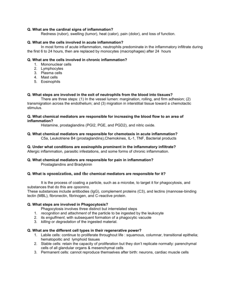
Q. What are the cardinal signs of inflammation? Redness (rubor), swelling (tumor), heat (calor), pain (dolor), and loss of function. Q. What are the cells involved in acute inflammation? In most forms of acute inflammation, neutrophils predominate in the inflammatory infiltrate during the first 6 to 24 hours, then are replaced by monocytes (macrophages) after 24 hours Q. What are the cells involved in chronic inflammation? 1. Mononuclear cells 2. Lymphocytes 3. Plasma cells 4. Mast cells 5. Eosinophils Q. What steps are involved in the exit of neutrophils from the blood into tissues? There are three steps: (1) In the vessel lumen: margination, rolling, and firm adhesion; (2) transmigration across the endothelium; and (3) migration in interstitial tissue toward a chemotactic stimulus. Q. What chemical mediators are responsible for increasing the blood flow to an area of inflammation? Histamine, prostaglandins (PGI2, PGE, and PGD2), and nitric oxide. Q. What chemical mediators are responsible for chemotaxis in acute inflammation? C5a, Leukotriene B4 (prostaglandins),Chemokines, IL-1, TNF, Bacterial products Q. Under what conditions are eosinophils prominent in the inflammatory infiltrate? Allergic inflammation, parasitic infestations, and some forms of chronic inflammation. Q. What chemical mediators are responsible for pain in inflammation? Prostaglandins and Bradykinin Q. What is opsonization, and the chemical mediators are responsible for it? It is the process of coating a particle, such as a microbe, to target it for phagocytosis, and substances that do this are opsonins. These substances include antibodies (IgG), complement proteins (C3), and lectins (mannose-binding lectin (MBL), fibronectin, fibrinogen, and C-reactive protein. Q. What steps are involved in Phagocytosis? Phagocytosis involves three distinct but interrelated steps 1. recognition and attachment of the particle to be ingested by the leukocyte 2. its engulfment, with subsequent formation of a phagocytic vacuole 3. killing or degradation of the ingested material. Q. What are the different cell types in their regenerative power? 1. Labile cells: continue to proliferate throughout life : squamous, columnar, transitional epithelia; hematopoitic and lymphoid tissues 2. Stable cells: retain the capacity of proliferation but they don’t replicate normally: parenchymal cells of all glandular organs & mesenchymal cells 3. Permanent cells: cannot reproduce themselves after birth: neurons, cardiac muscle cells Q. What steps are involved in wound healing? 24 hr.: hematoma & neutrophils, mitotic activity of basal layer, thin epithelial layer at the surface Day2 & 3: macrophages, granulation tissue day 5: collagen bridges the incision, epidermis thickens 2nd week: continued collagen and fibroblasts, blanching End of 1st month: scar Q. What is the difference between primary intention and secondary intention? The basic process of healing is the same in all wounds. In contrast to healing by primary intention, wounds healing by secondary intention Require more time to close because the edges are far apart Show a more prominent inflammatory reaction in and around the wound Contain more copious granulation tissue inside the tissue defect wound contraction (5 to 10%), ?myofibroblasts Q. What are the Factors that influence healing? 1. Infection 2. Poor blood supply 3. Foreign bodies in the wound 4. Mechanical factors 5. Nutritional deficiencies 6. Excess corticosteroids Q. What is Granulomatous inflammation? is a distinctive pattern of chronic inflammatory reaction characterized by aggregation (focal accumulations) of activated macrophages (epithelioid cells)

