Operative Report: Appendectomy for Perforated Appendicitis
advertisement
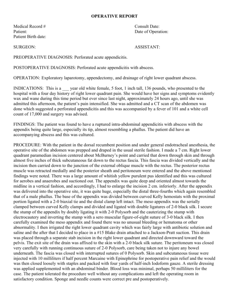
OPERATIVE REPORT Medical Record # Patient: Patient Birth date: Consult Date: Date of Operation: SURGEON: ASSISTANT: PREOPERATIVE DIAGNOSIS: Perforated acute appendicitis. POSTOPERATIVE DIAGNOSIS: Perforated acute appendicitis with abscess. OPERATION: Exploratory laparotomy, appendectomy, and drainage of right lower quadrant abscess. INDICATIONS: This is a ___ year old white female, 5 foot, 1 inch tall, 136 pounds, who presented to the hospital with a four day history of right lower quadrant pain. She would have her signs and symptoms evidently wax and wane during this time period but ever since last night, approximately 24 hours ago, until she was admitted this afternoon, the patient’s pain intensified. She was admitted and a CT scan of the abdomen was done which suggested a perforated appendicitis and this was accompanied by a fever of 101 and a white cell count of 17,000 and surgery was advised. FINDINGS: The patient was found to have a ruptured intra-abdominal appendicitis with abscess with the appendix being quite large, especially its tip, almost resembling a phallus. The patient did have an accompanying abscess and this was cultured. PROCEDURE: With the patient in the dorsal recumbent position and under general endotracheal anesthesia, the operative site of the abdomen was prepped and draped in the usual sterile fashion. I made a 7 cm. Right lower quadrant paramedian incision centered about McBurney’s point and carried that down through skin and through almost five inches of thick subcutaneous fat down to the rectus fascia. This fascia was divided vertically and the incision then carried down to the junction of the external oblique muscle with the rectus. The posterior rectus muscle was retracted medially and the posterior sheath and peritoneum were entered and the above mentioned findings were noted. There was a large amount of whitish yellow purulent pus identified and this was cultured for aerobes and anaerobes and suctioned out. The appendix was quite deep and oriented almost towards the midline in a vertical fashion, and accordingly, I had to enlarge the incision 2 cm. inferiorly. After the appendix was delivered into the operative site, it was quite huge, especially the distal three-fourths which again resembled that of a male phallus. The base of the appendix was divided between curved Kelly hemostats with the proximal portion ligated with a 2-0 biaxial tie and the distal clamp left intact. The meso appendix was the serially clamped between curved Kelly clamps and divided and ligated with double ligatures of 2-0 black silk. I secure the stump of the appendix by doubly ligating it with 2-0 Polysorb and the cauterizing the stump with electrocautery and inverting the stump with a sero muscular figure-of-eight suture of 3-0 black silk. I then carefully examined the meso appendix and found there was no unusual bleeding or hematoma or other abnormality. I then irrigated the right lower quadrant cavity which was fairly large with antibiotic solution and saline and the after that I decided to place in a #15 Blake drain attached to a Jackson-Pratt suction. This drain was placed through a separate stab incision in the right lower quadrant and directed downward toward the pelvis. The exit site of the drain was affixed to the skin with a 2-0 black silk suture. The peritoneum was closed very carefully with running continuous suture of 2-0 Polysorb, care being taken not to injure any bowel underneath. The fascia was closed with interrupted sutures of 0 Polysorb. Skin and subcutaneous tissue were injected with 10 milliliters if half percent Marcaine with Epinephrine for postoperative pain relief and the would was then closed loosely with staples and packed with four yards of half-inch Iodoform gauze. A sterile dressing was applied supplemented with an abdominal binder. Blood loss was minimal, perhaps 50 milliliters for the case. The patient tolerated the procedure well without any complications and left the operating room in satisfactory condition. Sponge and needle counts were correct pre and postoperatively. _________________________________ M.D. Signature D: T: CC:
