The Application of a Two-Dimensional Paper
advertisement
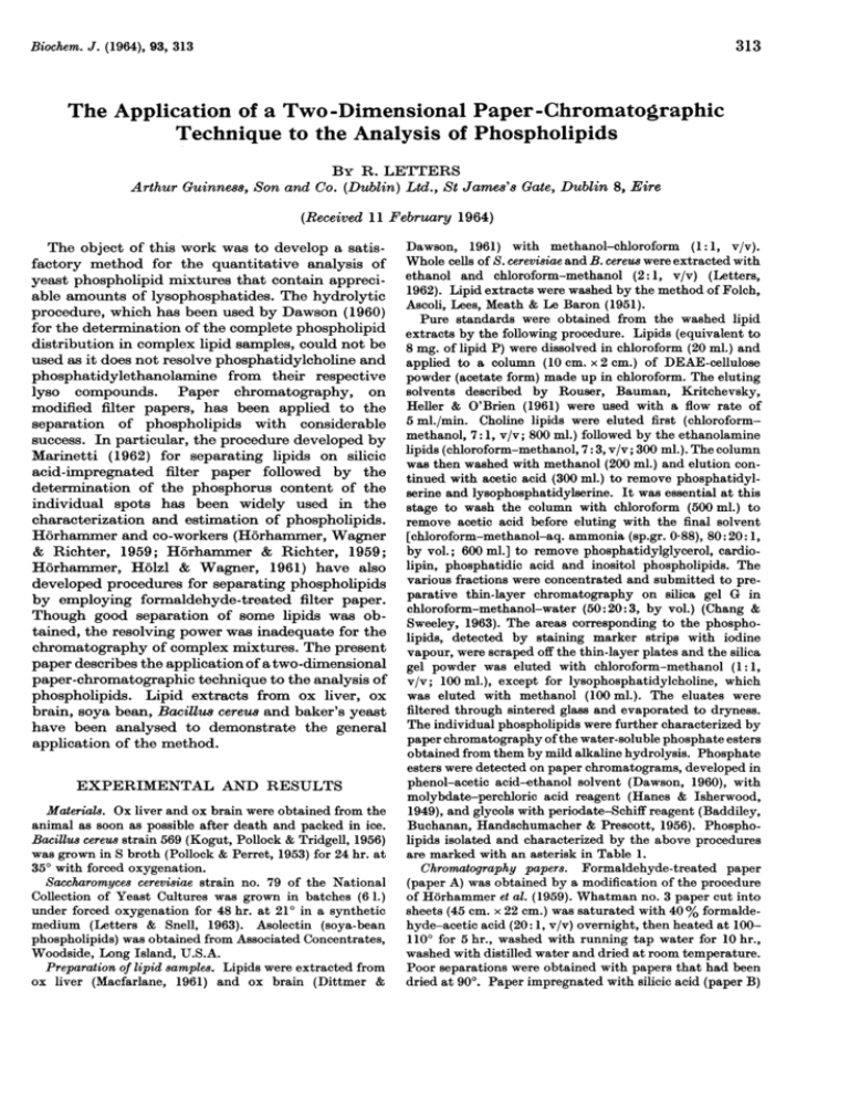
Biochem. J. (1964), 93, 313 313 The Application of a Two-Dimensional Paper-Chromatographic Technique to the Analysis of Phospholipids By R. LETTERS Arthur Guinness, Son and Co. (Dublin) Ltd., St James's Gate, Dublin 8, Eire (Received 11 February 1964) The object of this work was to develop a satisfactory method for the quantitative analysis of yeast phospholipid mixtures that contain appreciable amounts of lysophosphatides. The hydrolytic procedure, which has been used by Dawson (1960) for the determination of the complete phospholipid distribution in complex lipid samples, could not be used as it does not resolve phosphatidylcholine and phosphatidylethanolamine from their respective lyso compounds. Paper chromatography, on modified filter papers, has been applied to the separation of phospholipids with considerable success. In particular, the procedure developed by Marinetti (1962) for separating lipids on silicic acid-impregnated filter paper followed by the determination of the phosphorus content of the individual spots has been widely used in the characterization and estimation of phospholipids. Horhammer and co-workers (Horhammer, Wagner & Richter, 1959; Horhammer & Richter, 1959; Horhammer, Holzl & Wagner, 1961) have also developed procedures for separating phospholipids by employing formaldehyde-treated filter paper. Though good separation of some lipids was obtained, the resolving power was inadequate for the chromatography of complex mixtures. The present paper describes the applicationof atwo-dimensional paper-chromatographic technique to the analysis of phospholipids. Lipid extracts from ox liver, ox brain, soya bean, Bacillus cereus and baker's yeast have been analysed to demonstrate the general application of the method. EXPERIMENTAL AND RESULTS Material8. Ox liver and ox brain were obtained from the animal as soon as possible after death and packed in ice. Bacillus cereus strain 569 (Kogut, Pollock & Tridgell, 1956) was grown in S broth (Pollock & Perret, 1953) for 24 hr. at 350 with forced oxygenation. Saccharomnyces cerevisiae strain no. 79 of the National Collection of Yeast Cultures was grown in batches (6 1.) under forced oxygenation for 48 hr. at 210 in a synthetic medium (Letters & Snell, 1963). Asolectin (soya-bean phospholipids) was obtained from Associated Concentrates, Woodside, Long Island, U.S.A. Preparation of lipid samples. Lipids were extracted from ox liver (Macfarlane, 1961) and ox brain (Dittmer & Dawson, 1961) with methanol-chloroform (1: 1, v/v). Whole cells of S. cerevisiae and B. cereus were extracted with ethanol and chloroform-methanol (2: 1, v/v) (Letters, 1962). Lipid extracts were washed by the method of Folch, Ascoli, Lees, Meath & Le Baron (1951). Pure standards were obtained from the washed lipid extracts by the following procedure. Lipids (equivalent to 8 mg. of lipid P) were dissolved in chloroform (20 ml.) and applied to a column (10 cm. x 2 cm.) of DEAE-cellulose powder (acetate form) made up in chloroform. The eluting solvents described by Rouser, Bauman, Kritchevsky, Heller & O'Brien (1961) were used with a flow rate of 5 ml./min. Choline lipids were eluted first (chloroformmethanol, 7: 1, v/v; 800 ml.) followed by the ethanolamine lipids (chloroform-methanol, 7:3, v/v; 300 ml.). The column was then washed with methanol (200 ml.) and elution continued with acetic acid (300 ml.) to remove phosphatidylserine and lysophosphatidylserine. It was essential at this stage to wash the column with chloroform (500 ml.) to remove acetic acid before eluting with the final solvent [chloroform-methanol-aq. ammonia (sp.gr. 0.88), 80:20: 1, by vol.; 600 ml.] to remove phosphatidylglycerol, cardiolipin, phosphatidic acid and inositol phospholipids. The various fractions were concentrated and submitted to preparative thin-layer chromatography on silica gel G in chloroform-methanol-water (50:20:3, by vol.) (Chang & Sweeley, 1963). The areas corresponding to the phospholipids, detected by staining marker strips with iodine vapour, were scraped off the thin-layer plates and the silica gel powder was eluted with chloroform-methanol (1:1, v/v; 100 ml.), except for lysophosphatidylcholine, which was eluted with methanol (100 ml.). The eluates were filtered through sintered glass and evaporated to dryness. The individual phospholipids were further characterized by paper chromatography of the water-soluble phosphate esters obtained from them by mild alkaline hydrolysis. Phosphate esters were detected on paper chromatograms, developed in phenol-acetic acid-ethanol solvent (Dawson, 1960), with molybdate-perchloric acid reagent (Hanes & Isherwood, 1949), and glycols with periodate-Schiff reagent (Baddiley, Buchanan, Handschumacher & Prescott, 1956). Phospholipids isolated and characterized by the above procedures are marked with an asterisk in Table 1. Chromatography papers. Formaldehyde-treated paper (paper A) was obtained by a modification of the procedure of Horhammer et al. (1959). Whatman no. 3 paper cut into sheets (45 cm. x 22 cm.) was saturated with 40 % formaldehyde-acetic acid (20: 1, v/v) overnight, then heated at 1001100 for 5 hr., washed with running tap water for 10 hr., washed with distilled water and dried at room temperature. Poor separations were obtained with papers that had been dried at 900. Paper impregnated with silicic acid (paper B) 314 R. LETTERS was obtained from Schleicher and Schull (Dassel/Kr. Einbeck, paper no. 289). This paper is very fragile and gives less satisfactory resolution than the paper described by Marinetti (1962), but its properties have proved very suitable for the present work. Chambers and solvent systems. Papers A were chromatographed by the ascending technique in battery jars (29 cm. x 20 cm. x 47 cm. deep) with the upper phase of butan-l-ol-acetic acid-water (4:1:5, by vol.) equilibrated with 0-125 vol. of diethyl ether. Papers B were developed in a similar manner in battery jars (37 cm. x 22 cm. x 47 cm. deep) with di-isobutyl ketone-acetic acid-water (80:50:7, by vol.) (Marinetti, 1962). Two-dimensional technique for chromatography of phospholipids. The lipid sample (25-100l,g. of lipid P) was applied to paper A as a band (2 cm.) and after development the chromatogram was dried. A strip (29 cm. x 6 cm.) was cut from this chromatogram in such a way that the separated lipids were near one of the edges. This strip was then placed on top of a sheet (29 cm. x 35 cm.) of paper B overlapping the edge by about 3 cm. The papers were pressed together at the join between strips of glass, held in position with rubber bands, so that the leading edge of paper A was completely covered. Development in the second direction was then effected by immersing the lower edge of paper A in the required solvent, thus enabling the lipids to be eluted from paper A on to paper B. Detection of spots. The silicic acid-impregnated paper chromatograms were air-dried at room temperature for 1 hr. and then dipped in Rhodamine 6 G solution to locate lipid spots. The chromatograms were washed with distilled water and air-dried at room temperature. Other detection methods were employed to confirm the identity of spots: (a) ninhydrin for phospholipids containing free amino groups; (b) the modified Dragendorff reagent (Wagner, Horhammer & Wolff, 1961) for choline; (c) osmium tetroxide vapour (Hack, 1953) to detect compounds containing unsaturated acids or aldehydes. Fig. 1 shows a map of the phospholipids that have been detected in extracts from B. cereus, ox brain, ox liver, soya bean and baker's yeast. 1964 Estimation of phosphorus in spots. Each spot was cut out and chopped into a graduated centrifuge tube (15 ml.). Blank paper was added where necessary to ensure that all tubes contained the same amount of paper. Ammonium molybdate (5%, w/v; 1 drop) and perchloric acid (70%, w/w; 1-2 ml.) were added, and the tubes were capped with loose-fitting glass covers. The tubes were then placed in a convector air oven (Gallenkamp, heavy duty) at 165°. This temperature was raised to 2000 and the tubes were removed after a total heating time of 15 min. Perchloric acid (70 %, w/w) to 1-2 ml., water (5 ml.), 5 % (w/v) ammonium molybdate (0-5 ml.) and 0-4 ml. of the reducing agent (Fiske & Subbarow, 1925) were added and the volume of each tube was made up to 10 ml. with water. The tubes were shaken, 12 ~ B -2 4,-- CZ10S, 1 0-2 0-3 0-4 0-5 0-6 C) 16 CD---=) 1) A 0-7 0-8 0-9 RF A Fig. 1. Tracing of a two-dimensional chromatogram of yeastphospholipids ( ) superimposed with spots ( ---- -) detected on two-dimensional chromatograms of asolectin, ox liver and B. cereus. Ascending chromatography in direction A was carried out on formaldehyde-treated paper with the butan-l-ol-acetic acid-water solvent system, and in direction B on silicic acid-impregnated paper with diisobutyl ketone-acetic acid-water by using a strip-transfer technique. Identification of the spots is given in Table 1. Table 1. Distribution of individual phospholipids in various extracts Results express the P of the phospholipid as a percentage of the total lipid P recovered. Experimental details are given in the text. No. on map Ox liver Ox brain Compound B. cereus Asolectin 1 Unidentified 5-3 2 Unidentified 3-3 3 Lysophosphatidylinositol 3-6* 4 Unidentified 2-7 5 Lysophosphatidylserine 6 5-7 Phosphatidylinositol 5-2 14-4* 7 Lysophosphatidylcholine 8.8* 8 Lysophosphatidylethanolamine 2-8 3-7* 9 Phosphatidylserine 2-8* 2-9* 16.4* 10 Phosphatidylglycerol 27-0* 3.0* 11 Phosphatidylethanolamine 19.1* 24-4* 51-0 25-6 12 Phosphatidic acid 6-2* 13 Sphingomyelin 12-5* 7-6* 14 54-6* Phosphatidylcholine 37-1* 2-1 28-0* Unidentified 15 7-0 16 Cardiolipin 3-2* 1-6 5-7* 6-7* * Denotes compounds isolated by chromatography on DEAE-cellulose and on thin-layers of silica gel G. Vol. 93 ANALYSIS OF PHOSPHOLIPIDS 315 Table 2. Recovery of 8tandard pho8pholipid8 from 8ilicic acid-impregnated paper after 8eparation by two-dimen8ional 8trip-transfer chromatography Experimental details are given in the text. The values for average recovery are given as means ± S.D., with the numbers of experiments in parentheses. Average recovery of P (%) Amount applied Applied as mixture Applied singly Compounds (,ug. of P) 100-6±1-4 (4) 12-64 98-2±0-93 (6) Phosphatidylcholine 100-7±5-4 (4) 5-72 100-7±2-9 (6) Lysophosphatidylcholine 10-61 102-4±3-0 (6) 99-3±3-6 (4) Phosphatidylinositol Table 3. Quantitative analyist and recovery of yeast pho8pholipid8, exprea8ed as percentages of total pho8phoru8 A total of 54-6 ,g. of P was applied. Results are given as means ± S.D. of four replicate experiments. All compounds isolated and characterized as described in the text. For identification of spots see Table 1. Average recovery of P No. on map (%) 5 3-5±0-16 14-7 ±0-73 6 7 19-7±0-57 8 8-55+0-35 9 6-6±0-34 11 13-85±0-43 14 31-7 ±0-48 16 0-87±0-16 Total recovery 98-8±2-7 were then placed in a boiling-water bath for 7 min. and centrifuged. The supernatants were decanted into 15 ml. tubes and centrifuged again before measuring the blue colour at 830 m,u. Two paper blanks were determined for each chromatogram; such blanks were similar (E 0-06-0-09) to those obtained with ordinary filter paper. The calibration, which was obtained with a standard sample of lysophosphatidylcholine, was linear in the range 0-5-25Jg. of P. The distribution of phospholipids in a number of biological samples is shown in Table 1. The individual spots were identified on the basis of the position of the standard compounds and by the detection methods listed above. Phosphatidylinositol, phosphatidylcholine and lysophosphatidylcholine were used for recovery studies. Satisfactory recovery was obtained for all three phospholipids applied individually or as a mixture (Table 2). The recovery and quantitative analysis of yeast phospholipids is given in Table 3. DISCUSSION Normally in two-dimensional paper chromatography a sheet of filter-paper is developed in one direction with the first solvent system, dried and then developed in a direction at right angles to the first in another solvent system. By using a striptransfer method it has been possible to extend this technique so that phospholipids can be separated in the first direction on formaldehyde-treated paper and then on silicic acid-impregnated paper in the second direction. The two-dimensional chromatograms obtained in this way have shown that the method will prove useful in the characterization of phospholipids. The preparation of formaldehyde-treated papers has been simplified so that heating under pressure is not necessary. To obtain good resolution of mixtures of cardiolipin and phosphatidic acid on these papers I have used the upper phase of the butan-l-olacetic acid-water solvent system equilibrated with half of the quantity of ether used by previous workers (Horhammer et al. 1959). The striptransfer method for eluting lipids from formaldehyde-treated paper on to silicic acid-impregnated paper is very similar to the methods that have been described for the chromatography of radioactive phosphorus compounds (Schlogl & Siegel, 1953; Borst-Pauwels & de Mots, 1963). However, I have found that the minor modifications introduced in the present work have given more compact spots, but this may also be due in part to the properties of the solvent used for development in the second direction. For example, when the water content of the di-isobutyl ketone-acetic acid-water solvent was increased by 1 % the spots were flattened and tended to overlap, whereas a decrease of water content by 1 % resulted in elongation of spots with streaking in some cases. These observations support the recommendation of Marinetti (1962) that chromatography on silicic acid-impregnated paper should be carried out under conditions of controlled humidity. Previously the determination of the phosphorus content of a spot cut from a silicic acid-impregnatedpaper chromatogram has involved a double extraction of paper spots with methanolic hydrochloric acid (Marinetti, 1962). The errors and difficulties (for instance the chromatograms should be extracted on the same day if they are to be used for quantitative work) inherent in this procedure have been overcome by employing a simple perchloric acid-digestion procedure. The phosphorus content of the digests was then determined by a colorimetric method. The silicic acid present in the digests did not interfere with the determination provided that rigorously controlled digestion con- R. LETTERS 316 ditions were adhered to. To satisfy this requirement it was necessary to carry out digestions in a convector air oven, thus ensuring uniform heating of samples. Explosions during digestions with perchloric acid, which probably arise through overheating (Dawson, Hemington & Davenport, 1962), have not been observed in the present investigations. The analyses obtained by the present method of the phospholipids in ox liver and ox brain are in good agreement with those reported by Dawson et al. (1962). The minor spots (nos. 1 and 2 in Fig. 2) detected on two-dimensional chromatograms of B. cereus may correspond to the unidentified components observed by Houtsmuller & van Deenen (1963). However, the present analysis of phospholipids from B. cereus is in more reasonable agreement with the work of Kates, Kushner & James (1962). The presence of lysophosphatides in yeast and soya-bean extracts has been reported previously (Wagner et al. 1961; Letters & Snell, 1963), and their identification on paper chromatograms has been facilitated by their interaction with Rhodamine 6G, which results in white spots when the chromatograms are dried at room temperature. The soya-bean extract appears to be a useful source of phosphatidic acid and cardiolipin. The preliminary fractionation of phospholipid mixtures by chromatography on DEAE-cellulose followed by further purification by preparative thin-layer chromatography has proved valuable in confirming the presence of individual phospholipids in the various extracts examined. Phospholipids isolated by column chromatography on silicic acid were unsuitable for the recovery experiments as they were partly degraded during the time required for elution, whereas compounds isolated from thinlayer chromatograms could be eluted quickly and showed no sign of degradation. SUMMARY 1. A two dimensional paper-chromatographic technique for the separation of phospholipid mixtures on formaldehyde-treated paper and on silicic acid-impregnated paper is described. 1964 2. Phosphorus was determined, in spots located by staining, by direct digestion of the silicic acidimpregnated paper. Silicic acid did not interfere with the determination. 3. Analyses of the phospholipids of ox liver, ox brain, Bacillu8 cereu8, baker's yeast and soya bean are given. Thanks are due to Dr A. K. Mills for his encouragement. REFERENCES Baddiley, J., Buchanan, J. G., Handschumacher, R. E. & Prescott, J. F. (1956). J. chem. Soc. p. 2818. Borst-Pauwels, G. W. F. H. & de Mots, A. (1963). Experientia, 19, 51. Chang, T. L. & Sweeley, C. C. (1963). Biochemi8try, 2, 592. Dawson, R. M. C. (1960). Biochem. J. 75, 45. Dawson, R. M. C., Hemington, N. & Davenport, J. B. (1962). Biochem. J. 84, 497. Dittmer, J. C. & Dawson, R. M. C. (1961). Biochem. J. 81, 535. Fiske, C. H. & Subbarow, Y. (1925). J. biol. Chem. 66, 375. Folch, J., Ascoli, I., Lees, M., Meath, J. A. & Le Baron, N. F. (1951). J. biol. Chem. 191, 833. Hack, M. H. (1953). Biochem. J. 54, 602. Hanes, C. S. & Isherwood, F. A. (1949). Nature, Lond., 164, 1107. Horhammer, L., Holzl, J. & Wagner, H. (1961). Naturwi8sen8chaften, 48, 103. Horhammer, L. & Richter, G. (1959). Biochem. Z. 332, 186. Horhammer, L., Wagner, H. & Richter, G. (1959). Biochem. Z. 331, 155. Houtsmuller, U. M. T. & van Deenen, L. L. M. (1963). Biochem. J. 88, 43P. Kates, M., Kushner, D. J. & James, A. T. (1962). Canad. J. Biochem. Phy8iol. 40, 83. Kogut, M., Pollock, M. R. & Tridgell, E. J. (1956). Biochem. J. 62, 391. Letters, R. (1962). J. In8t. Brew. 68, 318. Letters, R. & Snell, B. K. (1963). J. chem. Soc. p. 5127. Macfarlane, M. G. (1961). Biochem. J. 78, 44. Marinetti, G. V. (1962). J. Lipid Re8. 3, 1. Pollock, M. R. & Perret, C. J. (1953). J. gen. Microbiol. 8, 186. Rouser, G., Bauman, A. J., Kritchevsky, G., Heller, D. & O'Brien, J. (1961). J. Amer. Oil Chem. Soc. 38, 544. Schl6gl, K. & Siegel, A. (1953). Hoppe-Seyl. Z. 292, 263. Wagner, H., Horhammer, L. & Wolff, P. (1961). Biochem. Z. 334, 175.
