Evidence Based Diagnosis of Placenta Increta/Percreta
advertisement
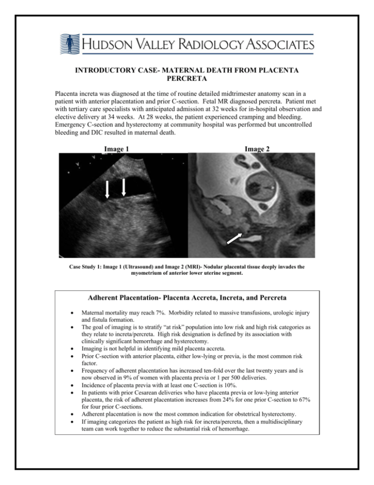
INTRODUCTORY CASE- MATERNAL DEATH FROM PLACENTA PERCRETA Placenta increta was diagnosed at the time of routine detailed midtrimester anatomy scan in a patient with anterior placentation and prior C-section. Fetal MR diagnosed percreta. Patient met with tertiary care specialists with anticipated admission at 32 weeks for in-hospital observation and elective delivery at 34 weeks. At 28 weeks, the patient experienced cramping and bleeding. Emergency C-section and hysterectomy at community hospital was performed but uncontrolled bleeding and DIC resulted in maternal death. Image 1 Image 2 Case Study 1: Image 1 (Ultrasound) and Image 2 (MRI)- Nodular placental tissue deeply invades the myometrium of anterior lower uterine segment. Adherent Placentation- Placenta Accreta, Increta, and Percreta Maternal mortality may reach 7%. Morbidity related to massive transfusions, urologic injury and fistula formation. The goal of imaging is to stratify “at risk” population into low risk and high risk categories as they relate to increta/percreta. High risk designation is defined by its association with clinically significant hemorrhage and hysterectomy. Imaging is not helpful in identifying mild placenta accreta. Prior C-section with anterior placenta, either low-lying or previa, is the most common risk factor. Frequency of adherent placentation has increased ten-fold over the last twenty years and is now observed in 9% of women with placenta previa or 1 per 500 deliveries. Incidence of placenta previa with at least one C-section is 10%. In patients with prior Cesarean deliveries who have placenta previa or low-lying anterior placenta, the risk of adherent placentation increases from 24% for one prior C-section to 67% for four prior C-sections. Adherent placentation is now the most common indication for obstetrical hysterectomy. If imaging categorizes the patient as high risk for increta/percreta, then a multidisciplinary team can work together to reduce the substantial risk of hemorrhage. Imaging Assessment of Adherent Placentation- 2D US, Color Doppler US, MR Identification of clinical risk factors: AMA, prior CS, placenta previa, prior uterine surgery, multiple D&C, unexplained elevated MSAFP. Placenta lacunae- number, morphology, doppler flow patterns. Placenta/myometrial interface- 2D and MR Bizarre vascularity- color flow mapping Imaging of Adherent Placentation Increta/Percreta- Multi-Modality Approach Meta Analysis of the Literature Suggests… “Low risk” designation- 93% true negative “High risk” – 10 to 15% false positive rate HVRA’s Imaging Census for Placenta Increta/Percreta from January 2008 through September 2011. 75 patients studied. True positive: True negative: False positive: False negative: Sensitivity: Specifity: 25 41 5 4 86% 84% Positive Predictive Value: Negative Predictive Value: 86% 91% CASE STUDY 2: PATIENT WITH PRIOR POSTERIOR FUNDAL HYSTEROTOMY (prior pregnancy with complete placenta previa) WHOSE OUTSIDE OBSTETRICAL MR SUGGESTED POSTERIOR PLACENTA PERCRETA WITH ADHERENCE TO AORTA. HVRA’s second opinion MR also suggested placental tissue abutting the aorta. Ultrasound by MD sonologist demonstrated an intact tissue plane separating placental tissue from aorta. Imaging identifies a distinct exophytic placental bulge with focal thinning of myometrium in the absence of placenta lacunae. These findings were of significant concern for placenta increta. The absence of lacunae is atypical and unusual in increta/percreta but not exclusionary. At 24-weeks, patient experienced sudden pain and uterine rupture requiring hysterectomy. Pathology demonstrated a deep increta with microscopic percreta. At the time of hysterectomy there was no adherence to the aorta or inferior vena cava. Image 3 Image 4 Case Study 2: At 24 weeks- sudden pain, uterine rupture requiring hysterectomy. Path- Deep increta with microscopic percreta. Image 3 (MRI)= placenta. Image 4 (ultrasound)- Thick white arrow denotes intact tissue plane separating placenta from aorta. Note focal exophytic bulge of placenta = increta. CASE STUDY 3: PLACENTA INCRETA REQURING HYSTERECTOMY. ONLY UTERINE INTERVENTION WAS ONE PRIOR DILATATION AND CURRETTAGE Serial ultrasound imaging demonstrated increasing lacunae in an unusual “balled-up” appearing left cornual placenta accompanied by premature dystrophic calcifications. Placenta increta/percreta can occur in the absence of prior C-section. Knowledge, experience and vigilance allowed us to alert the obstetrician to its possibility. The obstetrician was prepared for a possible hysterectomy which was ultimately required. Case Study 3: Arrows= lacunae. Note increasing number between 19 weeks and 23 weeks. Lacunae are irregular and ovoid. Placenta increta can occur in the absence of prior CS. Experience and vigilance allowed us to alert the OB to its possibility in this case. OB was prepared for possible hysterectomy, which was ultimately required. Daniel J. Cohen, MD Director of Maternal Fetal Imaging, Hudson Valley Radiology Associates (914) 391-0109. danjcohen@optonline.net
