1 AP Biology Name: Period: Date: Gel Electrophoresis Lab Results 1
advertisement
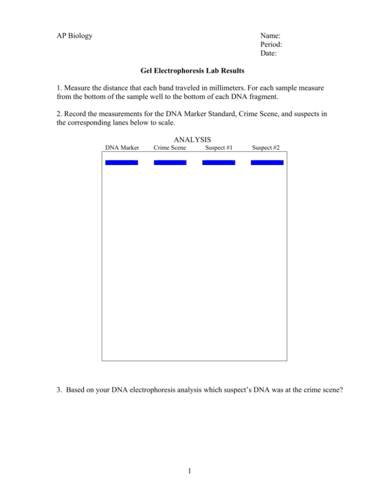
AP Biology Name: Period: Date: Gel Electrophoresis Lab Results 1. Measure the distance that each band traveled in millimeters. For each sample measure from the bottom of the sample well to the bottom of each DNA fragment. 2. Record the measurements for the DNA Marker Standard, Crime Scene, and suspects in the corresponding lanes below to scale. ANALYSIS DNA Marker Crime Scene Suspect #1 Suspect #2 3. Based on your DNA electrophoresis analysis which suspect’s DNA was at the crime scene? 1 Table 1 DNA Marker Standard (Hind III) Fragment 1 (Top) 2 3 4 5 6 7 8 Table 3 Fragment 1 (Top) 2 3 4 5 6 7 8 Distance Migrated (mm) Not Imaged Table 2 Length (bp) 23,109 9,416 6,557 4,361 2,328 2,027 564 125 Suspect 1 DNA Sample Distance Migrated (mm) Fragment 1 (Top) 2 3 4 5 6 7 8 9 10 Table 4 Length (bp) 2 Fragment 1 (Top) 2 3 4 5 6 7 8 9 10 Crime Scene DNA Sample Distance Migrated (mm) Length (bp) Suspect 2 DNA Sample Distance Migrated (mm) Length (bp) 3 Concept Questions: 1. Different bacteria contain different restriction enzymes. What is their function? 2. How would using an agarose gel higher than 8% change the results? 3. What would occur if the sample wells containing the DNA were placed closest to the red electrode instead of the black? 4. Both electrophoresis and paper chromatography are used for separating molecules. What are two similarities and two differences between these two methods? 5. Describe the purpose of each component involved in gel electrophoresis. Agarose gel- TBE buffer- Polarity of the electrophoresis chamber- Voltage- DNA samples (restriction fragments)- DNA stain- 4 6. The restriction endonuclease (restriction enzyme), EcoR1 recognizes the nucleotide sequence GAATTC (recognition site), and Hind III recognizes AAGCTT. If you add EcoR1 to a linear DNA sample, Hind III to a second sample, and both to a third, run a gel separation, the following occurs. Hind III _____ Eco R1 Double _____ _____ _____ _____ _____ _____ A student viewing the gel, draws the following map of the linear DNA: GAATTC cut site a). How did the student know to show the EcoR1 restriction enzyme cut site as identified above? AAGCTT cut site GAATTC cut site b) Why would the sample with both restriction enzymes be identified as shown above? 7. What are the odds that either recognition sequence from question #6 will appear in any random DNA? (This is a probability question. Give a numeric answer.) 8. After running a gel you are left with the following three fragments: 22kb, 18kb, 4kb. Mark the restriction sites and label the fragments on the plasmid below. What would the fragment banding on the gel look like? 5


