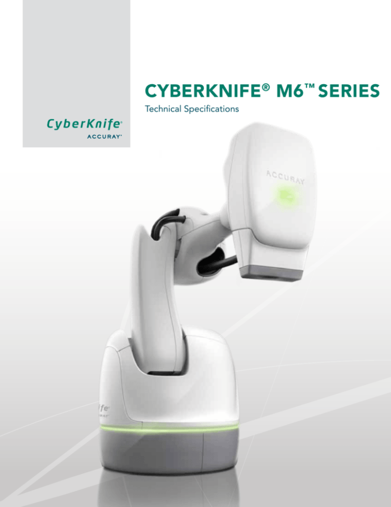
CYBERKNIFE® M6™ SERIES
Technical Specifications
System Targeting Accuracy
System targeting accuracy accounts for all components of the system. This may be understood as a combination of mechanical,
targeting, tracking and clinical accuracy, and includes error sources from the CT scan, treatment planning, patient tracking and
dose delivery systems. All these elements comprise the clinically relevant accuracy which may be termed the overall average
CyberKnife® System error.
The overall average CyberKnife System error is less than 0.95 mm RMS when a planning CT slice spacing of 1.25 mm or less
is used.
Minimum Room Size
• 15 ft 7 in (4572 mm) wide/long
• 21 ft 6 in (6553 mm) wide/long
• 9 ft 6 in (2896 mm) ceiling
Minimum Room Size
02
M6™ Series Technical Specifications
Robotic Manipulator
Four Options for
Xchange® Table
Placement
Image Detectors
Two Options for
RoboCouch®
Patient Positioning
System Placement
Room diagram with all options shown
Treatment Vault Environment
Temperature: 10˚C to 30˚C
Pressure: 103 kPa to 65 kPa
Humidity:
30% to 75% RH (non-condensing)
Mechanical Features
Robot
• 6-Axis robotic manipulator mounted on a pedestal at the head of patient area
• SmartPAD Teach Pendant with a touch screen interface
Robot Reach diagram
M6™ Series Technical Specifications
03
R o b o t i c M a n i p u l at o r
S p e c i f i c at i o n s Payload
300 kg (661 lb)
Maximum Reach
2500 mm (98 in)
Number of Axes
6
Work envelope
41 m3
Weight
1220 kb (2690 lb)
Workspace
The robotic manipulator is programmed to move in a fixed and pre-determined workspace. The workspace accounts for the
positions of objects in the treatment suite, including the treatment couch, the imaging sources and detectors, the floor and
the ceiling, and eliminates collision hazards by creating suitable paths for the robotic manipulator to move in. Additionally,
the workspace is comprised of pre-assigned points in space, termed nodes, where the manipulator is allowed to stop in
order to deliver radiation. At each node, the linac can deliver radiation from multiple beam angles. It may be noted that this
representation is conceptual as the workspace and the treatment paths adopted by the robotic manipulator are dependent
on the location of the target and patient anatomy being treated.
Workspace Geometry
04
M6™ Series Technical Specifications
Xchange® Robotic Collimator Changer
The Xchange Robotic Collimator Changer automatically changes collimator housing prior to patient treatment.
The Xchange System is compatible with the Fixed Collimator housing, the Iris™ Variable Aperture Collimator and
the InCise™ Multileaf Collimator.
Side view of Xchange Table
Top view of Xchange Table
Linear Accelerator
• Nominal source to axis (SAD) distance is 800 mm
Basic Beam Line for Fixed Collimator
M6™ Series Technical Specifications
05
Dosimetry Specification
•Chamber type
Dual sealed ion chambers segmented for symmetry monitoring
•Resolution
≥ 25 counts per MU
P h o t o n B e a m S p e c i f i c at i o n Dosimetry System
A two-channel primary/secondary dosimetry system is provided
X-ray Energy
6MV Nominal Photon Energy
Depth of Maximum Dose (Dmax)
15 mm ±2 mm
Dose Rate
1000 MU/min ±10% measured at 800 mm SAD at a depth of 15 mm in water equivalent using the 60 mm fixed collimator
Temperature And Pressure Adjustments
Within the specified operating temperature and pressure range, the dose rate and MU to dose calibration is independent of temperature and pressure
Dosimetry Linearity
Dosimetry linearity with total dose is less than ±5% or ±1 cGy, whichever is smaller over an accumulated range of 1 cGy to
100 cGy, measured at 800 mm SAD with 15 mm build up,
within the operating temperature and pressure range
Dosimetry linearity with total dose is less than ±1% or ±1 cGy, whichever is greater over an accumulated range of 100 cGy to 1000 cGy, measured at 800 mm SAD with 15 mm build-up, within the operating temperature and pressure range
Quality Index
TPR 20/10 ratio of dose rate in water
tank at 20 to 10 cm depth
Between 0.62 and 0.67 for a 60 mm fixed collimator
Leakage
Measured anywhere in the patient plane outside of the maximum useful beam
(as defined by IEC 60601-1-2) and at 1 m from the electron beam axis that is averaged
over an area no larger than 100 cm2.
Leakage in the patient plane is less than 0.2% maximum and 0.1% average
Leakage 1 m from the electron beam axis is less than 0.1%
The leakage values are given with respect to
the absorbed dose on the central axis at the
reference treatment distance of 800 mm SAD
with the 60 mm fixed collimator
Collimator Transmission
Fixed Collimator: The x-ray transmission through the blank collimator at 800 mm SAD does not exceed 0.2% of the central axis (CAX) dose rate of a 60 mm fixed collimator at 800 mm SAD
Iris™ Collimator: The x-ray transmission through an Iris
Collimator’s tungsten segments at 800 mm SAD does not exceed 0.2% of the CAX dose rate of the Iris Collimator when opened to a 60 mm field at 800 mm SAD
InCise™ Multileaf Collimator: Less than 0.5% maximum and
0.3% average at 15 mm depth and 800 mm SAD. Leaf tip
leakage (i.e., closed leaves) at full overtravel: less than 0.5% maximum. Both relative to 100 mm x 100 mm at 800 mm SAD and 15 mm depth.
06
M6™ Series Technical Specifications
Equipment Room:
Equipment Room Components
PDU (Power Distribution Unit)
Robot Controllers
Mechanical Rack including:
Chiller
Air Compressor
SF6
AMM (Advanced Magnetron Modulator)
Rack, including:
LCC (Linac Control Computer)
LPDU (Linac Power Distribution Unit)
MCC (Modulator Control Chassis)
Gun Driver
Modulator
Computer Rack including:
KVM Extender
UPS
Network switch
Network firewall
Temperature controller
Monitor and Keyboard
ELCC (E-Stop Interlock Control Chassis)
TLS (Target Locating System) Workstation
UCC (User Control Console) Workstation
Data Server
Storage Vault (option)
UCC (User Control Console) Workstation
The UCC Workstation is installed in the Equipment Room. The workstation includes mouse, keyboard and display at the Control
Console area. Power is provided to the UCC Workstation through the cabinet UPS.
UCC W o r k s t a t i o n
S p e c i f i c at i o n CPU
2 x Intel E5645 2.4 GHz CPU (6 core) for a total of
12 physical cores
Memory
12 GB DDR3 Memory
Storage
2x 300 GB SAS 2.0 15 K Drives mirrored for a total of
300 GB of storage
Graphics Card
nVidia Quadro® 2000 Graphics Card
Ethernet Port
2x Gigabit Ethernet Port
Power Supply
Dual Redundant Power Supply
M6™ Series Technical Specifications
07
CyberKnife® Data Management System (CDMS) Data Server
The CDMS data server is installed in the equipment room. The data server includes mouse, keyboard and display capabilities
through the cabinet KVM. Power is provided to the data server through the cabinet UPS (Uninterrupted Power Supply).
Notification of power events will occur over an Ethernet connection between the UPS and the data server.
D ata S e r v e r S p e c i f i c at i o n CPU
Manufacturer: Intel
Quantity: 4
RAM
4 GB
SAS Hard Disk Drive
Manufacturer: Seagate Technology
Quantity: 3
SATA Hard Disk Drive
Manufacturer: Western Digital
Quantity: 3
RAID Card
Adaptec RAID 3805
Operating System
Microsoft® Server 2003 x64 (5 CALs)
Operating System License
Microsoft® Server 2008 x64 (compatible with MS Server 2003 x64)
Database
Microsoft® SQL Server 2008 Standard x64 Edition (15 CALs)
3rd party software packages
• Windows® Server 2003 Standard x64 Edition
• SQL Server 2008 x64 Standard Edition
• SQL Server Reporting Services
• 3.5 .NET Framework
• MergeCOM-3 DICOM Libraries
Storage Vault (Option)
The Storage Vault is an optional workstation used for archiving patient data. It is housed in the equipment room.
S t o r a g e V a u lt S p e c i f i c at i o n OS
GuardianOS®
CPU
Intel® quad-core CPU
Memory
2 GB DDR3 1333 MHz Registered DIMMs
Drive Configuration
9x2 TB 7.2 K Enterprise SATA II
RAID Configuration
RAID 6, 1 Hot Swap
Network Interface
2 x Gigabit Ethernet Ports (autosensing 10/100/1000 Base-T,
dual RJ-45 network connections)
USB Interface
2 x USB 2.0 ports
08
M6™ Series Technical Specifications
MultiPlan® Treatment Planning System
The MultiPlan Treatment Planning System is a dedicated planning system for use with the CyberKnife® System. The MultiPlan
hardware is housed in the “dosimetry” or “planning” room.
M u lt i P l a n Tr e a t m e n t P l a n n i n g
S y s t e m W o r k s tat i o n S p e c i f i c at i o n CPU
Dual Intel® quad-core CPUs
Memory
24 GB DDR3
Storage
500 GB Hard drive
Graphics Card
Nvidia Quadro 4000 and Nvidia® Tesla C2075
(for M6™ Series w/MLC only)
Ethernet Port
1 Gigabit
Power Supply
>1000 W
Monitor
LCD monitor with a native resolution of 1600x1200 or 1920x1200
Operating System
Windows® 7 x64 operating system
MultiPlan MD Suite (Option)
The MultiPlan MD Suite provides remote secure access to patient record data from the CyberKnife System database. The MD Suite
workstation is housed in the remote location.
M D S u i t e W o r k s tat i o n
S p e c i f i c at i o n CPU
One Intel quad-core CPU
Memory
12 GB DDR3
Storage
500 GB Hard drive
Graphics Card
Nvidia Quadro 4000 and Nvidia Tesla C2075
(for M6™ Series w/MLC only)
Ethernet Port
1 Gigabit
Power Supply
>1000W
Monitor
LCD monitor with a native resolution of 1600 x 1200
or 1920 x 1200
Operating System
Windows 7 x64 operating system
M6™ Series Technical Specifications
09
Treatment Control Area
• Dual Monitors
Tr e a t m e n t D e l i v e r y S y s t e m
M o n i t o r S p e c i f i c at i o n Monitor Type
2x NEC® MyltiSync EA 243WM
Monitor Size 24 inches each
Resolution
1920 x 1200 each for a total workspace of 3840 x 1200
Monitor Stands
The monitors are attached to a solid frame desktop
dual monitor mount
Operator Panel, including the following:
• Indication of MV beam on
• Indication of KV image acquisition
• Indication of remote/local control
• Button to enable High Voltage
• Indication of High Voltage on
• Key switch to turn MV radiation on and off
• Emergency Stop button
• Audible sounds for KV and MV radiation
Operator Panel
10
M6™ Series Technical Specifications
Administration Workstation (Option)
The Administration Workstation provides access to a series of patient, plan and system administration applications.
This workstation, including mouse, keyboard and monitor, can be housed at a remote location.
Adm i n i s t r a t i o n W o r k s t a t i o n
S p e c i f i c at i o n CPU
Intel Core 2 Duo E6400 2.13 Ghz 2 MB Cache 106 Mhz
RAM
DIMM 1024 MB 800 MHz Unbuffered DDR2 installed in Socket
DIMM 1024 MB 800 MHz Unbuffered DDR2 installed in Socket 2
DVD Drive
Plextor® PX-820A DL Dual RW Int Drive
Hard Disk
Seagate Barracuda ES.2 900 GB 7200 RPM 32 MB Cache SATA 3.0 GB/S x 4
Video Card
ATI FireGL® V3600 256 MB PCI Express
Operating System
MS Windows 7 Professional (64 bit)
Collimation Systems
Secondary Collimation
The CyberKnife® M6™ Series uses multiple secondary collimator types to deliver beams as defined by the treatment plan.
• CyberKnife M6 FI System includes the Fixed Collimators and the Iris™ Variable Aperture Collimator
• CyberKnife M6 FM System includes the Fixed Collimators and the InCise™ Multileaf Collimator
• CyberKnife M6 FIM System includes the Fixed Collimators, the Iris Variable Aperture Collimator and the InCise
Multileaf Collimator
Fixed Collimators:
Fixed secondary collimators deliver circular field sizes of 5, 7.5, 10, 12.5, 15, 20, 25, 30, 35, 40, 50 and 60 mm diameter at 800 mm
SAD. These collimators can be changed to vary the beam size as generated by the treatment plan. For each fixed collimator, the
manipulator traverses a separate path.
F i x e d C o l l i m at o r
S p e c i f i c at i o n Collimator Transmission
The x-ray transmission through the blank collimator at a SAD of 800 mm does not exceed 0.2% of the central axis (CAX) dose rate of a 60 mm fixed collimator at 800 mm SAD
Available Apertures
Collimation sizes: 5, 7.5, 10, 12.5, 15, 20, 25, 30, 35, 40, 50 and 60 mm nominal field sizes at 800 mm SAD
M6™ Series Technical Specifications
11
Iris™ Variable Aperture Collimator
The Iris Variable Aperture Collimator creates beams with characteristics virtually identical to those of fixed collimators. It consists
of two banks of 6 tungsten segments each with each bank creating a hexagonal aperture. The two are offset by 30˚ relative to each
other resulting in a dodecahedral (12-sided) aperture when viewed from one end of the collimator to the other. The Iris Variable
Aperture Collimator replicates the existing 12 fixed collimator sizes.
Iris™ Variable Aperture Collimator
Ir i s ™ V a r i a b l e A p e r t u r e
C o l l i m at o r S p e c i f i c at i o n Circularity
The standard deviation of the radial distance from the beam
axis to the 50% dose level is less than 2% of the average
radial distance
Collimator Transmission
• Maximum: < 0.2% of the delivered dose rate
• Average: < 0.1% of the delivered dose rate
Reproducibility
• Mechanical: less than 0.1 mm
• Treatment field size: < 0.2 mm at the nominal treatment distance of 800 mm SAD
Available Apertures
• Effective collimation sizes: 5, 7.5, 10, 12.5, 15, 20, 25, 30, 35, 40, 50 and 60 mm diameter field sizes at 800 mm SAD
12
M6™ Series Technical Specifications
InCise™ Multileaf Collimator
Basic Beam Line for InCise™ Multileaf Collimator
I n C i s e ™ M u lt i l e a f C o l l i m at o r
S p e c i f i c a t i o n As defined by IEC 60976
Beam Targeting Non-Isocentric, Non-Coplanar Beam Targeting
Maximum Field Size Nominal: 120 mm (leaf motion direction) x 100 mm at
800 mm SAD
Number of Leaves
41 leaf pairs
Leaf Thickness 2.5 mm at 800 mm SAD
Leaf Tilt
Leaves tilted 0.5˚
Leaf Tip Design
3-Sided
Leaf Height
90 mm
Leaf Material Tungsten
Distal Plane of Leaves to Linac Source Distance
400 mm
Leaf Positioning Accuracy 0.5 mm at 800 mm SAD
Mechanical Accuracy
0.25 mm
Leaf Positioning Reproducibility 0.4 mm at 800 mm SAD
Mechanical Reproducibility 0.2 mm
Leaf Over-Travel
100%
Leaf Inter-Digitation Full Leaf Inter-Digitation
Transmission
Includes intra-leaf, inter-leaf and leaf tip transmission
<0.3% Average (<0.5% Maximum)
Weight 48 kg (~105 lbs)
M6™ Series Technical Specifications
13
Imaging
The CyberKnife® M6™ Series use kV X-ray imaging to provide target localization during treatment. The imaging system consists
of two X-ray sources mounted to the ceiling, and corresponding image detectors mounted in the floor. The X-ray sources are
positioned such that the generated beams intersect orthogonally and create an imaging center located 92 cm (36.22 in) from
the floor. All treatments on the CyberKnife System are based around the imaging field of view. The live images are digitized and
compared to images synthesized from the patient’s CT data (Digitally Reconstructed Radiograph, or DRR). This technique allows for
determination of intra-fraction target shifts and automatic compensation by the treatment manipulator during treatment delivery.
Imaging System Geometry
14
M6™ Series Technical Specifications
C o m pa c t X - r ay G e n e r at o r
S p e c i f i c at i o n s Constant Potential Power Rating (kw) 50.0
Radiographic kVp range 40-150 ± (5% + 1 kVp)
Resolution
1 kVp
mA Range and Stations 10, 12, 5, 16, 20, 25, 32, 40, 50, 64, 80, 100, 126, 160, 200, 250, 320, 400, 500, 640 ± (5% + 1 kVp)
Power Output 640 mA @ 78 kVp
500 mA @100 kVp
400 mA @ 125 kVp
320 mA @ 150 kVp
Exposure Time 20-640 ms + (5% + 0.1 ms)
mAs
X - r ay S o u r c e s S p e c i f i c at i o n s
0.1 – 640 mAs
Electrical
• Circuit 3-phase
• Nominal tube voltage
40 – 150 kV
• Nominal focal spot value Large focus: 1.2 mm
Small focus: 0.6 mm
• Nominal Anode input power
Large focus: 100 kW
Small focus: 40 kW
Aluminum Filter
2.5 mm min.
Firing Modes
Synchronous and Asynchronous
Collimator Type
Fixed Aperture
X - r ay D e t e c t o r S p e c i f i c at i o n s
Lower Spec Limit
Detector Type Amorphous Silicon
Number of Pixels
1024 x 1024
Pixel Pitch 400 µm
Total Area 40 x 40 cm2
MTF @ 0.25 lp/mm
80%
MTF @ 1 lp/mm
33%
DQE @ 0.25 lp/mm, 1 μGy
56%
DQE @ 1 lp/mm, 1 μGy
28%
M6™ Series Technical Specifications
15
Target Tracking
Accurate target tracking and compensating for target motion are an integral part of the CyberKnife® System and its capabilities.
The target is tracked throughout the treatment and delivery is automatically altered to compensate for any motion.
CT R e q u i r e m e n t s
f o r T a r g e t Tr a c k i n g
Maximum Slices 512
kVp
120
mAs Scanner Maximum (minimum 400)
Slice thickness Contiguous slice (no gaps); < 1.25 mm slice thickness
Target tracking and motion compensation are achieved through the use of the imaging system integrated with the treatment
delivery system. The image guidance system calculates the required offsets based on the patient’s current position. During patient
set-up, the table (the Standard Treatment Couch or the RoboCouch® Patient Positioning System) is adjusted to align the patient
with the treatment plan. During the delivery of the treatment the system automatically corrects the linac position for any calculated
offsets (within a specified tolerance).
Offset Tolerance for Intra-Fraction Robotic Corrections*
RoboCouch® System
RoboCouch System Standard Treatment
Standard Treatment
with Prostate Path
Couch
Couch with
Prostate Path
X, Y, and Z
±10 mm
±10 mm
±10 mm
±10 mm
X, Y, and Z with Synchrony®
Respiratory
Tracking System
± 25 mm
± 25 mm
± 25 mm
± 25 mm
Pitch
± 1.5˚
± 1.5˚
± 1˚
± 5˚
Roll
± 1.5˚
± 2˚
± 1˚
± 2˚
Yaw
± 1.5˚
± 3˚
± 3˚
± 3˚
*Subject to change based on target tracking method selection
Target tracking on the CyberKnife® System uses one of the following tracking methods: 6D Skull Tracking, Fiducial Tracking, Xsight®
Spine Tracking, Xsight Spine Prone Tracking, Xsight Lung Tracking, or 1-View Tracking.
16
M6™ Series Technical Specifications
Target Tracking Method
6D Skull Tracking System
The 6D Skull Tracking System allows direct tracking of the bony anatomy of the skull when treating intracranial lesions. Target
tracking and motion compensation are accomplished by using image intensity and brightness differences between the DRR
and live images.
Fiducial Tracking
For lesions that are located extracranially, target tracking can be carried out with the use of fiducials.
• General guidelines for fiducials:
o Gold Seeds or gold sphere
o Diameter: 0.7 mm to 1.2 mm
o Length: 3 mm to 6 mm
• Minimum 3 fiducials are required for 6D target tracking, including corrections for translations (x, y, z) and rotations (roll, pitch, yaw)
Xsight® Spine Tracking System
The Xsight Spine Tracking System, with the patient in the supine position, enables the tracking of skeletal structures in the cervical,
thoracic, lumbar and sacral regions of the spine without the need for implanted fiducials.
Target tracking with the Xsight Spine System is accomplished using 2D-3D registrations on a hierarchical mesh where local
displacements at each of the mesh points are estimated and combined to provide 6D corrections to the treatment manipulator.
Xsight Spine Prone Tracking System (Option)
The Xsight Spine Prone Tracking System provides support for treating spine targets with the patient in the prone position. The
tracking mode combines the Xsight Spine Tracking algorithm with the Synchrony® Respiratory Tracking System to offer continuous
real-time tracking and compensation for target motion due to respiration. In this tracking mode, the patient is first aligned using
the Xsight Spine Tracking workflow, then a Synchrony correlation model is created to compensate for target translational motion
during delivery.
Xsight Lung Tracking System (Option)
The Xsight Lung Tracking System (also called 2-View Lung Tracking) tracks tumors in the lung directly without the use of fiducials by
using image intensity differences between the lesion and the background. The patient is first aligned using the Xsight Spine Tracking
workflow, then the Xsight Lung Tracking System tracks the translational motion of the target.
Lung Optimized Treatment: 1-View Lung Tracking and 0-View Lung Tracking (Option)
Lung Optimized Treatment includes a Simulation Application and these two tracking modes: 1-View Lung Tracking and 0-View Lung
Tracking. Together, these two tracking methods provide fiducial-less treatments for lung SBRT patients, regardless of the location of
the tumor.
• The Simulation Application provides a workflow to select the most appropriate tracking mode.
• 1-View Lung Tracking is used when the treatment target is clearly visible and can be tracked in only one X-ray projection
during treatment
o Provides direct tracking of lung lesions in 2-dimensions
o Uses an ITV in the non-visible 3rd dimension
• 0-View Lung Tracking is used when the treatment target is not clearly visible in either X-ray projection, resulting in the need to
track the bony anatomy of the vertebral column during treatment
o Provides direct tracking of the spine without fiducials
o Uses an ITV in all dimensions to compensate for respiratory motion of the tumor
M6™ Series Technical Specifications
17
Motion Tracking
Motion tracking on the CyberKnife® System uses one of these methods: The Synchrony® Respiratory Tracking System or the
InTempo™ Adaptive Imaging System. Motion tracking is used in conjunction with an applicable target tracking method.
Synchrony® Respiratory Tracking System
The Synchrony Respiratory Tracking System continuously synchronizes treatment beam delivery to the motion of a target that is
moving with respiration. The Synchrony System can be used in conjunction with the following target tracking methods: Fiducial
Tracking, Xsight® Lung Tracking, Xsight Spine Prone Tracking, and 1-View Tracking.
The system operates by creating a correlation model between the patients’s breathing pattern, monitored in real-time, and the
location of the target at various points in the respiration cycle. The location of the target is determined by using X-ray imaging to
visualize the lesion while the breathing pattern is tracked and monitored using external markers (LED-based, fiber optic tracking
markers with a tracking frequency of >25 Hz) in real-time.
The system automatically determines the best correlation model type to be utilized for the treatment by choosing the one that
minimizes overall correlation error. The model is chosen from linear, curvilinear and bi-curvilinear forms. The model is based on
the latest 15 sets of X-ray images taken and is updated every time a new image is acquired.
InTempo Adaptive Imaging System
The InTempo Adaptive Imaging System is a time-based technology used to compensate for non-periodic intra-fraction motion of
the target. The InTempo System can be used in conjunction with the following target tracking methods: Fiducial Tracking, 6D Skull
Tracking and Xsight Spine Tracking.
Image Age
Image Age is the time elapsed since the most recent image acquisition. The system uses the Image Age parameter to ensure
that no treatment beam is delivered based on an image that is older than that user-specified value.
Adaptive Imaging
The user may optionally allow the system to trigger adaptive imaging in the event that the target motion is greater than a
user-defined threshold, which automatically reduces the image age to 15s.
18
M6™ Series Technical Specifications
Safety Features
• Cryptzone’s SE46 Virus Protection
o Whitelist approach instead of blacklist
o No updates
o Configuration management
o Will not be able to use any software that is not a part of the release
• Contact Detection
o Contact detection sensor at the distal end of the secondary collimator housing on the linac
o Contact detection sensor on back of robot arm
o Contact with the sensor causes an Emergency Stop (E-STOP) condition halting all motion of the system
• Safety Zones: The robot workspace also takes into consideration the position of the patient and is designed to avoid contact with
the patient. This is achieved by creation of a safety zone around the patient and the treatment couch. The safety zone consists of
two elements: fixed and dynamic.
o The fixed safety zone is rigidly attached to the imaging center and thereby the part of the patient body being treated
o The dynamic safety zone is designed to encompass the entire patient body and always lies within the fixed safety zone
o The size of the dynamic safety zone is user selectable based on individual patient sizes (Small, Medium or Large)
M6™ Series Technical Specifications
19
Network
System Interfaces
• DICOM Import/Export included:
o DICOM Image Import
o DICOM RT Structure Set Import
o DICOM Image Export
o DICOM RT Structure Set Export
o DICOM RT Dose Export
• OIS License Required to generate objects:
o DICOM RT Plan Export
o DICOM RT Beams Treatment Record Export
Network diagram
20
M6™ Series Technical Specifications
Patient Positioning Support
Two types of patient positioning support systems are available with the CyberKnife® System; the Standard Treatment Couch
and the RoboCouch® Patient Positioning System (optional).
Standard Treatment Couch
RoboCouch® System (Optional)
Payload
159 kg (350 lb)
227 kg (500 lb)
• Anterior/Posterior
28 cm
42 cm
• Right/Left
±15 cm
±18 cm
• Superior/Inferior
≥91 cm
≥100 cm
• Head Up/Head Down
(pitch)
±5˚
±5˚
• Right/Left Tilt (roll)
±5˚
±5˚
• Yaw (CW/CCW)
N/A
±5˚
Remote Workstation
Local Hand Pendant
Remote Workstation Local Hand Pendant
• Translational
0.3 mm
0.1 mm
• Rotational
0.3˚
0.1˚
Most degrees of freedom are corrected serially
All degrees of freedom are corrected
simultaneously
Range of Motion
Control
Repeatability
Motion Corrections
Point of Rotation
Fixed: determined by mechanical assembly of the actuators
Variable: all axes can move
simultaneously about a set point
in space
Standard Treatment Couch
The standard Treatment Couch is the standard patient support system of the CyberKnife System. It provides the user with flexibility
in patient positioning by providing 5 DOF motion capabilities.
M6™ Series Technical Specifications
21
RoboCouch® Robotic Patient Positioning System (option)
The RoboCouch System provides a highly flexible 6-DOF mechanism for automatically positioning the patient. The combination of
the RoboCouch System and the robotic manipulator for linac positioning enables the CyberKnife® System to deliver dose precisely,
and to the right location. The upper manipulator arm (between axes A2 and A3) integrates a contact sensor on its outer surface
and an E-STOP is triggered if an object comes in contact with it. The RoboCouch System is available with either a flat carbon fiber
couch top (standard with the RoboCouch System) or a seated load carbon fiber table top (optional). The RoboCouch System has
five rotational axes and one linear axis.
• KRC4 controller
• SmartPAD Teach Pendant with a touch screen interface
• Two possible locations in room based on the room geometry: on patient right or patient left
Seated Load Table Top (Option)
The Seated Load Table Top option is offered for the RoboCouch System. It is an articulating couch top that allows patients to be
loaded in a seated position. The seated load table top converts to a flat table top to offer flexibility in positioning and loading the
patient. The patient can be treated either in a knees-flat or a knees-elevated position.
22
M6™ Series Technical Specifications
Treatment Couch Top Specifications
Radiolucency
Maximum: <1.1 mm Aluminum equivalence at 120 kVp for the
length of at least 62 inches from the superior most point
Immobilization • Alpha Cradle®
(Compatibility)
• Vacuum Lock Bags
• Thermoplastic masks
Indexing
Compatible with CIVCO indexing systems
Flat with Standard
Flat With RoboCouch®
System
Seated Load Option
with RoboCouch System
Minimum Load Height
≤64 cm (25 in)
≤56 cm (22 in)
Seated Load: ≤ 45 cm (17.9 in)
Horizontal Load: ≤ 58 cm (23 in)
Dimensions
Length: 212 cm (83.4 in)
Width: 53 cm (21 in)
Thickness: 6.4 cm (2.5 in)
Length: 213 cm (84 in)
Width: 53 cm (21 in)
Thickness: 7.6 cm (3 in)
Length: 206 cm (81 in)
Width: 53 cm (21 in)
Thickness: 5.7 cm (2.25 in)
Regulatory Classification
The CyberKnife® System is classified as follows:
• Protection against electric shock: Class I, permanently connected
• Applied part: Patient treatment table only, Type B
• Protection against harmful ingress of water: IPXO – no protection against ingress of water
• Methods of sterilization or disinfection: Not required
• Degree of safety in the presence of flammable mixtures: Not suitable for use in the presence of flammable mixtures
• Mode of operation: Continuous
M6™ Series Technical Specifications
23
UNITED STATES
Accuray Corporate Headquarters
1310 Chesapeake Terrace
Sunnyvale, CA 94089
USA
Tel: +1.408.716.4600
Toll Free: 1.888.522.3740
Fax: +1.408.716.4601
Email: sales@accuray.com
EUROPE
ASIA
Accuray Incorporated
1240 Deming Way
Madison, WI 53717
USA
Tel: +1 608 824 2800
Fax: +1 608 824 2996
Accuray Japan K.K.
Shin Otemachi Building 7F
2-2-1 Otemachi, Chiyoda-ku
Tokyo 100-0004
Japan
Tel: +81.3.6265.1526
Fax: +81.3.3272.6166
Accuray Asia Ltd.
Suites 1702 - 1704, Tower 6
The Gateway, Harbour City
9 Canton Road, T.S.T.
Hong Kong
Tel: +852.2247.8688
Fax: +852.2175.5799
Accuray Accelerator Technology
#39 Huatai Road, Longtan
Industrial Zone,
Section 2 East, 3rd Ring Road,
Chengdu, Sichuan 610051
People's Republic of China
Accuray International Sarl
Route de la Longeraie 9 (3rd floor)
CH – 1110 Morges
Switzerland
Tel: +41.21.545.9500
Fax: +41.21.545.9501
© 2013 Accuray Incorporated. All Rights Reserved. Accuray, the stylized logo, CyberKnife, Synchrony, Xsight, Xchange, RoboCouch, TomoTherapy, TomoHD, Hi·Art, Iris, InCise, MultiPlan, M6 and InTempo are among the trademarks and/or registered trademarks of Accuray Incorporated
in the United States and other countries. TomoTherapy is a wholly owned subsidiary of Accuray. Other trademarks used and identified herein are the property of their respective owners. 501047.A

